Best Seller
beta Amyloid Antibody (MOAB-2) - BSA Free
Novus Biologicals | Catalog # NBP2-13075
Key Product Details
Species Reactivity
Validated:
Human, Mouse, Rat, Bacteria, Monkey
Cited:
Human, Mouse, Rat, Bacteria, Primate - Macaca mulatta (Rhesus Macaque)
Applications
Validated:
Immunohistochemistry, Immunohistochemistry-Paraffin, Immunohistochemistry-Frozen, Immunohistochemistry Free-Floating, Western Blot, Immunoblotting, ELISA, Immunocytochemistry/ Immunofluorescence, Immunoprecipitation, Dot Blot
Cited:
Immunohistochemistry, Immunohistochemistry-Paraffin, Immunohistochemistry-Frozen, Immunohistochemistry Free-Floating, Western Blot, ELISA, Immunocytochemistry/ Immunofluorescence, Immunoprecipitation, Cytometric Bead Assay Standard, IF/IHC
Label
Unconjugated
Antibody Source
Monoclonal Mouse IgG2B Clone # MOAB-2
Format
BSA Free
Loading...
Product Specifications
Immunogen
This beta Amyloid antibody was developed against recombinant human beta Amyloid 42.
Epitope
Residues 1-4 of beta Amyloid
Reactivity Notes
Use in Mouse reported in scientific literature (PMID:35401818). Monkey reactivity reported in scientific literature (PMID: 29241829). Use in Bacteria reported in scientific literature (PMID:32413239).
Specificity
Beta Amyloid antibody (MOAB-2) recognizes unaggregated, oligomeric and fibrillar forms of beta Amyloid 42 and unaggregated beta Amyloid 40. Does not detect APP.
Clonality
Monoclonal
Host
Mouse
Isotype
IgG2B
Theoretical MW
5 kDa.
Disclaimer note: The observed molecular weight of the protein may vary from the listed predicted molecular weight due to post translational modifications, post translation cleavages, relative charges, and other experimental factors.
Disclaimer note: The observed molecular weight of the protein may vary from the listed predicted molecular weight due to post translational modifications, post translation cleavages, relative charges, and other experimental factors.
Scientific Data Images for beta Amyloid Antibody (MOAB-2) - BSA Free

Immunohistochemical Staining of beta Amyloid in Normal and 5xFAD Mouse Brain
IHC analysis of beta Amyloid on normal mouse brain (left) and 5xFAD mouse brain (right) using DAB with hematoxylin counterstain. The MOAB-2 antibody was used at 1:20 on normal mouse brain and at 1:400 on 5xFAD mouse brain.
Western Blot Detection of beta Amyloid in Mouse Brain Homogenate
Analysis of beta Amyloid (MOAB-2) antibody in (1) 100 pmole beta Amyloid 42, (2) 5xFAD mouse brain homogenate Repetition 1 and (3) 5xFAD mouse brain homogenate Repetition 2.
Western Blot Analysis of HEK-APP SWE/BACE1 Cell Lysates Using beta Amyloid Antibody
Western blot analysis in cell lysates from HEK-APP SWE/BACE1 cells probed with an antibody against the C-terminus of APP (Lanes 1 and 2) and beta Amyloid (MOAB-2, Lanes 3 and 4). Beta Amyloid (MOAB-2) does not detect APP (from PMID: 22423893).
Immunohistochemical Staining of beta Amyloid in Paraffin Embedded Human Brain
IHC analysis of a formalin fixed paraffin embedded tissue section of human brain (Alzheimers disease, hippocampus) using 1:200 dilution of anti-beta Amyloid antibody (clone MOAB-2). The staining was developed with HRP labeled anti-mouse secondary antibody and DAB reagent, and nuclei of cells were counter-stained with hematoxylin. This beta Amyloid antibody specifically stained the cells with Abeta 42/ Abeta aggregates while the normal cells were negative for abeta peptide.
Immunocytochemistry/Immunofluorescence Staining of beta Amyloid in Mouse Subiculim
Immunofluorescent detection of beta Amyloid with MOAB-2 in the subiculim of 1-, 2- and 4- month old 5xFAD mice. Scale bar 20 um (from PMID: 22423893).
Immunohistochemical Staining of beta Amyloid in the Hippocampus of APP/PS1 Mice
beta-Amyloid-Antibody-MOAB-2-Immunohistochemistry-NBP2-13075-img0019.jpg
Immunohistochemical Analysis of beta Amyloid in Paraffin Embedded Human Brain
IHC analysis of a formalin fixed paraffin embedded tissue section of human brain (Alzheimers disease, hippocampus) using 1:40 dilution of anti-beta Amyloid antibody (clone MOAB-2). The staining was developed with HRP labeled anti-mouse secondary antibody and DAB reagent, and nuclei of cells were counter-stained with hematoxylin. This beta Amyloid antibody specifically stained the cells with Abeta 42/ Abeta aggregates while the normal cells were negative for abeta peptide.
Immunohistochemical Staining of beta Amyloid in Frozen Mouse Brain
Mouse brain (cerebral cortex). Red: MOAB-2 antibody staining, Blue: DAPI. Zeiss LSM800, 40x. Image from verified customer review. - BSA Free [NBP2-13075] -")
Immunocytochemistry/ Immunofluorescence: beta Amyloid Antibody (MOAB-2) - BSA Free [NBP2-13075] -
Immunocytochemistry/ Immunofluorescence: beta Amyloid Antibody (MOAB-2) - BSA Free [NBP2-13075] - No difference in A beta plaque load between propofol (n = 8) & vehicle (n = 6) treated APP/PS1 mice. a An example image of A beta plaque immunoreactivity in the cortex of an APP/PS1 mouse following repeated propofol exposure. b A representative image of A beta plaque immunolabeling the cortex of an APP/PS1 mouse following treatment with vehicle. c Bar graph showing the percentage of the cortex occupied by A beta plaques in propofol & vehicle treated APP/PS1 mice. d Bar graph showing the average size (μm2) of A beta plaques in the cortex of APP/PS1 in the propofol & vehicle treatment groups. e Bar graph showing the average density of A beta plaques (/μm2) in the cortex of propofol & vehicle exposed APP/PS1 mice. All data is shown as mean ± SEM. Scale bar = 500 μm Image collected & cropped by CiteAb from the following publication (https://bmcanesthesiol.biomedcentral.com/articles/10.1186/s12871-018-05…), licensed under a CC-BY license. Not internally tested by Novus Biologicals. [NBP2-13075]")
Immunohistochemistry: Mouse Monoclonal beta Amyloid Antibody (MOAB-2) [NBP2-13075]
Murine brain cryosections stained with primary MOAB-2 antibody followed by a secondary donkey anti-mouse antibody. Image from a verified customer review.Applications for beta Amyloid Antibody (MOAB-2) - BSA Free
Application
Recommended Usage
Dot Blot
reported in scientific literature (PMID 22423893)
ELISA
1:100-1:1000
Immunoblotting
reported in scientific literature (PMID 28314768)
Immunocytochemistry/ Immunofluorescence
1:200-1:500
Immunohistochemistry
1:200-1:1000
Immunohistochemistry Free-Floating
reported in scientific literature (PMID 25747037)
Immunohistochemistry-Paraffin
1:200-1:1000
Immunoprecipitation
1:200-1:1000
Western Blot
1:1000-1:5000
Application Notes
In Western blot, a band can be seen at ~4 kDa, representing the beta Amyloid monomer. Larger bands may also be seen representing the unaggregated, oligomeric, and fibrillar forms of beta Amyloid. For higher beta Amyloid yield in WB, please follow the extraction protocol described in Youmans et al, J Neurosci Methods. 2011 March 15; 196(1): 51-59 (PMID: 21219931).
Reviewed Applications
Read 3 reviews rated 4.7 using NBP2-13075 in the following applications:
Formulation, Preparation, and Storage
Purification
Protein G purified
Formulation
PBS
Format
BSA Free
Preservative
0.05% Sodium Azide
Concentration
1.0 mg/ml
Shipping
The product is shipped with polar packs. Upon receipt, store it immediately at the temperature recommended below.
Stability & Storage
Store at 4C short term. Aliquot and store at -20C long term. Avoid freeze-thaw cycles.
Background: Amyloid beta
Pyroglutamate amyloid beta peptides (pGlu-Abeta) are N-terminal truncations in which the N-terminal glutamate is cyclized to pyroglutamate resulting in pGlu-Abeta (3-40/42 and 11-40/42) (4). This pyrE modification may have a greater propensity to aggregate under physiological conditions and has been implicated as the molecular species responsible for seeding larger oligomers of amyloid beta.
References
1. Chen GF, Xu TH, Yan Y, Zhou YR, Jiang Y, Melcher K, Xu HE. (2017) Amyloid beta: structure, biology and structure-based therapeutic development. Acta Pharmacol Sin. 38(9):1205-1235. PMID: 28713158
2. De-Paula VJ1, Radanovic M, Diniz BS, Forlenza OV. (2012) Alzheimer's disease. Subcell Biochem. 65:329-52. PMID: 23225010
3. Schaich CL, Maurer MS, Nadkarni NK. (2019) Amyloidosis of the Brain and Heart: Two Sides of the Same Coin? JACC Heart Fail. 7(2):129-131. PMID: 30704604
4. He W, Barrow CJ. (1999) The A beta 3-pyroglutamyl and 11-pyroglutamyl peptides found in senile plaque have greater beta-sheet forming and aggregation propensities in vitro than full-length A beta. Biochemistry. 38(33):10871-7. PMID: 10451383
Additional Amyloid beta Products
Product Documents for beta Amyloid Antibody (MOAB-2) - BSA Free
Certificate of Analysis
To download a Certificate of Analysis, please enter a lot or batch number in the search box below.
Product Specific Notices for beta Amyloid Antibody (MOAB-2) - BSA Free
This product is for research use only and is not approved for use in humans or in clinical diagnosis. Primary Antibodies are guaranteed for 1 year from date of receipt.
Related Research Areas
Citations for beta Amyloid Antibody (MOAB-2) - BSA Free
 Powered by Bioz
Customer Reviews for beta Amyloid Antibody (MOAB-2) - BSA Free (3)
4.7 out of 5
3 Customer Ratings
Have you used beta Amyloid Antibody (MOAB-2) - BSA Free?
Submit a review and receive an Amazon gift card!
$25/€18/£15/$25CAN/¥2500 Yen for a review with an image
$10/€7/£6/$10CAN/¥1110 Yen for a review without an image
Submit a review
Customer Images
Showing
1
-
3 of
3 reviews
Showing All
Filter By:
-
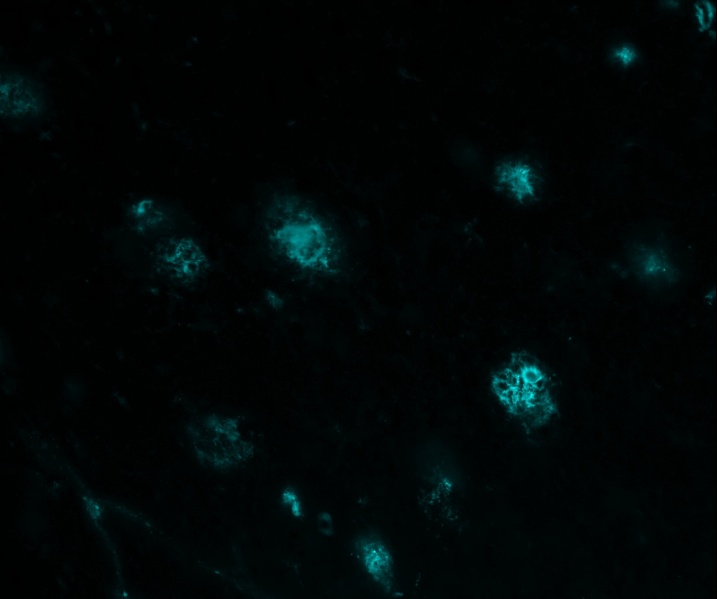
Verified Customer | Posted 10/16/2025Amyloid beta plaques in mouse brain stained with MOAB-2 (cyan)Murine brain cryosections stained with primary MOAB-2 antibody followed by a secondary donkey anti-mouse antibody

-
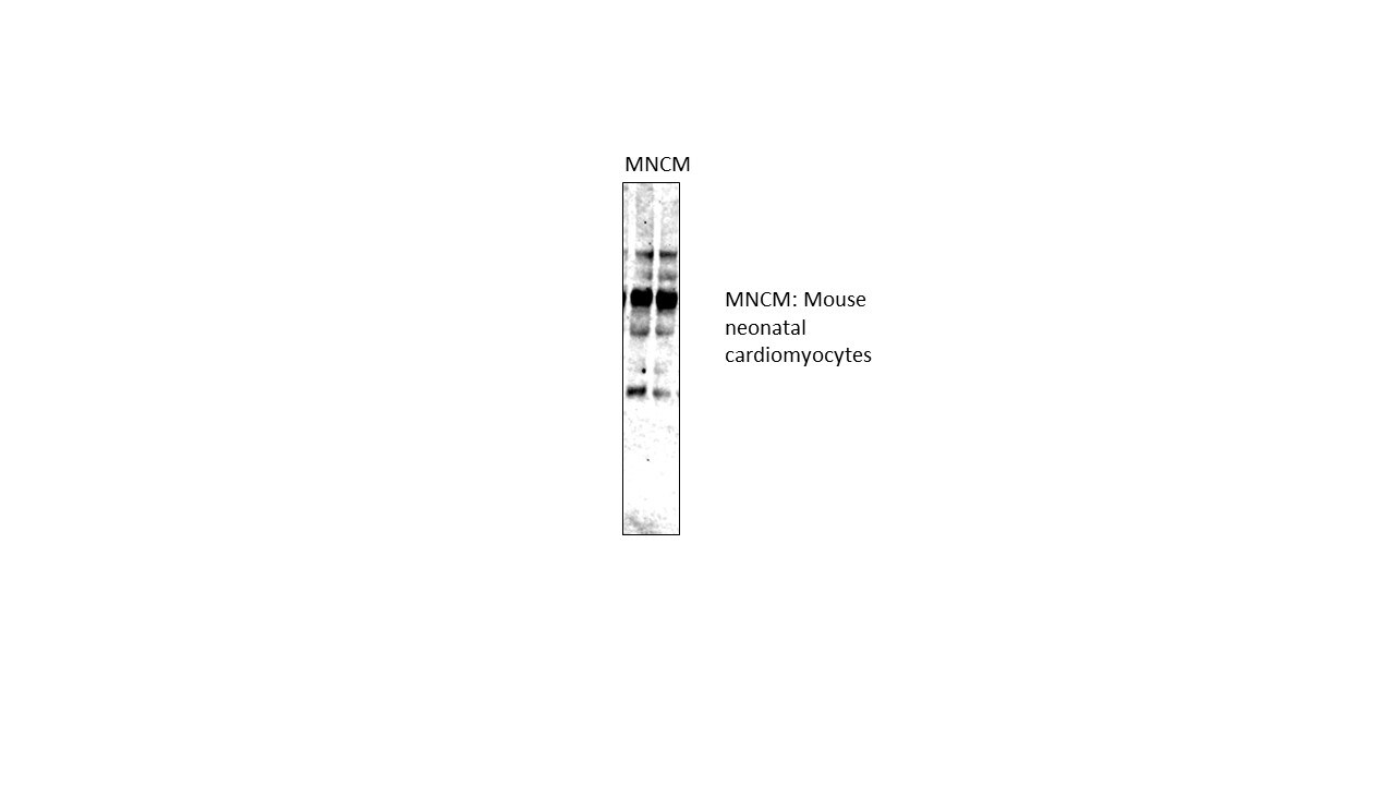
Application: Western BlotSample Tested: neonatal cardiac myocytesSpecies: MouseVerified Customer | Posted 12/11/2019Amyloid
-
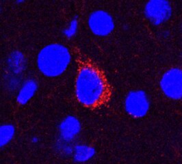
Application: Immunohistochemistry-FrozenSample Tested: Brain (cerebral cortex)Species: 5xFAD mouseVerified Customer | Posted 09/22/2018Blue: DAPI; Red: MOAB-2 Zeiss LSM 800, 40x oil objective
There are no reviews that match your criteria.
Protocols
View specific protocols for beta Amyloid Antibody (MOAB-2) - BSA Free (NBP2-13075):
Immunohistochemistry-Paraffin Embedded Sections
Antigen Unmasking:
Bring slides to a boil in 10 mM sodium citrate buffer (pH 6.0) then maintain at a sub-boiling temperature for 10 minutes. Cool slides on bench-top for 30 minutes (keep slides in the sodium citrate buffer at all times).
Staining:
1. Wash sections in deionized water three times for 5 minutes each.
2. Wash sections in PBS for 5 minutes.
3. Block each section with 100-400 ul blocking solution (1% BSA in PBS) for 1 hour at room temperature.
4. Remove blocking solution and add 100-400 ul diluted primary antibody. Incubate overnight at 4 C.
5. Remove antibody solution and wash sections in wash buffer three times for 5 minutes each.
6. Add 100-400 ul HRP polymer conjugated secondary antibody. Incubate 30 minutes at room temperature.
7. Wash sections three times in wash buffer for 5 minutes each.
8. Add 100-400 ul DAB substrate to each section and monitor staining closely.
9. As soon as the sections develop, immerse slides in deionized water.
10. Counterstain sections in hematoxylin.
11. Wash sections in deionized water two times for 5 minutes each.
12. Dehydrate sections.
13. Mount coverslips.
Western Blot Protocol
1. Perform SDS-PAGE on samples to be analyzed, loading 10-25 ug of total protein per lane.
2. Transfer proteins to PVDF membrane according to the instructions provided by the manufacturer of the membrane and transfer apparatus.
3. Stain the membrane with Ponceau S (or similar product) to assess transfer success, and mark molecular weight standards where appropriate.
4. Rinse the blot TBS -0.05% Tween 20 (TBST).
5. Block the membrane in 5% Non-fat milk in TBST (blocking buffer) for at least 1 hour.
6. Wash the membrane in TBST three times for 10 minutes each.
7. Dilute primary antibody in blocking buffer and incubate overnight at 4C with gentle rocking.
8. Wash the membrane in TBST three times for 10 minutes each.
9. Incubate the membrane in diluted HRP conjugated secondary antibody in blocking buffer (as per manufacturer's instructions) for 1 hour at room temperature.
10. Wash the blot in TBST three times for 10 minutes each (this step can be repeated as required to reduce background).
11. Apply the detection reagent of choice in accordance with the manufacturer's instructions.
Find general support by application which include: protocols, troubleshooting, illustrated assays, videos and webinars.
- Antigen Retrieval Protocol (PIER)
- Antigen Retrieval for Frozen Sections Protocol
- Appropriate Fixation of IHC/ICC Samples
- Cellular Response to Hypoxia Protocols
- Chromogenic IHC Staining of Formalin-Fixed Paraffin-Embedded (FFPE) Tissue Protocol
- Chromogenic Immunohistochemistry Staining of Frozen Tissue
- ClariTSA™ Fluorophore Kits
- Detection & Visualization of Antibody Binding
- ELISA Sample Preparation & Collection Guide
- ELISA Troubleshooting Guide
- Fluorescent IHC Staining of Frozen Tissue Protocol
- Graphic Protocol for Heat-induced Epitope Retrieval
- Graphic Protocol for the Preparation and Fluorescent IHC Staining of Frozen Tissue Sections
- Graphic Protocol for the Preparation and Fluorescent IHC Staining of Paraffin-embedded Tissue Sections
- Graphic Protocol for the Preparation of Gelatin-coated Slides for Histological Tissue Sections
- How to Run an R&D Systems DuoSet ELISA
- How to Run an R&D Systems Quantikine ELISA
- How to Run an R&D Systems Quantikine™ QuicKit™ ELISA
- ICC Cell Smear Protocol for Suspension Cells
- ICC Immunocytochemistry Protocol Videos
- ICC for Adherent Cells
- IHC Sample Preparation (Frozen sections vs Paraffin)
- Immunocytochemistry (ICC) Protocol
- Immunocytochemistry Troubleshooting
- Immunofluorescence of Organoids Embedded in Cultrex Basement Membrane Extract
- Immunofluorescent IHC Staining of Formalin-Fixed Paraffin-Embedded (FFPE) Tissue Protocol
- Immunohistochemistry (IHC) and Immunocytochemistry (ICC) Protocols
- Immunohistochemistry Frozen Troubleshooting
- Immunohistochemistry Paraffin Troubleshooting
- Immunoprecipitation Protocol
- Preparing Samples for IHC/ICC Experiments
- Preventing Non-Specific Staining (Non-Specific Binding)
- Primary Antibody Selection & Optimization
- Protocol for Heat-Induced Epitope Retrieval (HIER)
- Protocol for Making a 4% Formaldehyde Solution in PBS
- Protocol for VisUCyte™ HRP Polymer Detection Reagent
- Protocol for the Fluorescent ICC Staining of Cell Smears - Graphic
- Protocol for the Fluorescent ICC Staining of Cultured Cells on Coverslips - Graphic
- Protocol for the Preparation & Fixation of Cells on Coverslips
- Protocol for the Preparation and Chromogenic IHC Staining of Frozen Tissue Sections
- Protocol for the Preparation and Chromogenic IHC Staining of Frozen Tissue Sections - Graphic
- Protocol for the Preparation and Chromogenic IHC Staining of Paraffin-embedded Tissue Sections
- Protocol for the Preparation and Chromogenic IHC Staining of Paraffin-embedded Tissue Sections - Graphic
- Protocol for the Preparation and Fluorescent ICC Staining of Cells on Coverslips
- Protocol for the Preparation and Fluorescent ICC Staining of Non-adherent Cells
- Protocol for the Preparation and Fluorescent ICC Staining of Stem Cells on Coverslips
- Protocol for the Preparation and Fluorescent IHC Staining of Frozen Tissue Sections
- Protocol for the Preparation and Fluorescent IHC Staining of Paraffin-embedded Tissue Sections
- Protocol for the Preparation of Gelatin-coated Slides for Histological Tissue Sections
- Protocol for the Preparation of a Cell Smear for Non-adherent Cell ICC - Graphic
- Quantikine HS ELISA Kit Assay Principle, Alkaline Phosphatase
- Quantikine HS ELISA Kit Principle, Streptavidin-HRP Polymer
- R&D Systems Quality Control Western Blot Protocol
- Sandwich ELISA (Colorimetric) – Biotin/Streptavidin Detection Protocol
- Sandwich ELISA (Colorimetric) – Direct Detection Protocol
- TUNEL and Active Caspase-3 Detection by IHC/ICC Protocol
- The Importance of IHC/ICC Controls
- Troubleshooting Guide: ELISA
- Troubleshooting Guide: Immunohistochemistry
- Troubleshooting Guide: Western Blot Figures
- Western Blot Conditions
- Western Blot Protocol
- Western Blot Protocol for Cell Lysates
- Western Blot Troubleshooting
- Western Blot Troubleshooting Guide
- View all Protocols, Troubleshooting, Illustrated assays and Webinars
FAQs for beta Amyloid Antibody (MOAB-2) - BSA Free
Showing
1
-
2 of
2 FAQs
Showing All
-
Q: Does your antibody beta Amyloid Antibody (MOAB-2) # NBP2-13075 work on frozen sections for histology?
A:
This antibody has never been tested on frozen sections. It has been validated in ICC, IP and IHC on paraffin-embedded tissues, so it may work on frozen sections. While we cannot guarantee that it will work on frozen sections, if you would like to test NBP2-13075 in an untested application and share your results with us, I can recommend our Innovator's Reward Program. Our Innovator's Reward program was created to allow researchers the opportunity to try our primary antibodies in an untested species or application, without the financial risk of failure. To participate you simply submit an online review detailing your positive or negative results. In return, you receive a discount voucher for 100% of the purchase price of the reviewed product. Reviews may also be submitted by emailing innovators@novusbio.com.
-
Q: Please provide the epitope region information for NBP2-13075 Beta Amyloid Antibody.
A:
The epitope is a.a. 672 - 675 and please use https://www.uniprot.org/uniprot/P05067#sequences to check all important information.
-
Q: Does your antibody beta Amyloid Antibody (MOAB-2) # NBP2-13075 work on frozen sections for histology?
A:
This antibody has never been tested on frozen sections. It has been validated in ICC, IP and IHC on paraffin-embedded tissues, so it may work on frozen sections. While we cannot guarantee that it will work on frozen sections, if you would like to test NBP2-13075 in an untested application and share your results with us, I can recommend our Innovator's Reward Program. Our Innovator's Reward program was created to allow researchers the opportunity to try our primary antibodies in an untested species or application, without the financial risk of failure. To participate you simply submit an online review detailing your positive or negative results. In return, you receive a discount voucher for 100% of the purchase price of the reviewed product. Reviews may also be submitted by emailing innovators@novusbio.com.
-
Q: Please provide the epitope region information for NBP2-13075 Beta Amyloid Antibody.
A:
The epitope is a.a. 672 - 675 and please use https://www.uniprot.org/uniprot/P05067#sequences to check all important information.
Loading...