BMP-2 Antibody - BSA Free
Novus Biologicals | Catalog # NBP1-19751
Key Product Details
Species Reactivity
Validated:
Cited:
Applications
Validated:
Cited:
Label
Antibody Source
Format
Product Specifications
Immunogen
Reactivity Notes
Localization
Clonality
Host
Isotype
Theoretical MW
Disclaimer note: The observed molecular weight of the protein may vary from the listed predicted molecular weight due to post translational modifications, post translation cleavages, relative charges, and other experimental factors.
Scientific Data Images for BMP-2 Antibody - BSA Free
![Immunohistochemistry: BMP-2 Antibody - BSA Free [NBP1-19751]](https://resources.rndsystems.com/images/products/BMP-2-Antibody---BSA-Free-Immunohistochemistry-NBP1-19751-img0012.jpg "Immunohistochemistry: BMP-2 Antibody - BSA Free [NBP1-19751]")
Immunohistochemistry: BMP-2 Antibody - BSA Free [NBP1-19751]
BMP-2-Antibody---BSA-Free-Immunohistochemistry-NBP1-19751-img0012.jpg![Western Blot: BMP-2 AntibodyBSA Free [NBP1-19751]](https://resources.rndsystems.com/images/products/BMP-2-Antibody---BSA-Free-Western-Blot-NBP1-19751-img0008.jpg "Western Blot: BMP-2 AntibodyBSA Free [NBP1-19751]")
Western Blot: BMP-2 AntibodyBSA Free [NBP1-19751]
Western Blot: BMP-2 Antibody - BSA Free [NBP1-19751] - Extracts from HUVEC cells.![Western Blot: BMP-2 AntibodyBSA Free [NBP1-19751]](https://resources.rndsystems.com/images/products/BMP-2-Antibody---BSA-Free-Western-Blot-NBP1-19751-img0010.jpg "Western Blot: BMP-2 AntibodyBSA Free [NBP1-19751]")
Western Blot: BMP-2 AntibodyBSA Free [NBP1-19751]
BMP-2-Antibody---BSA-Free-Western-Blot-NBP1-19751-img0010.jpg![Immunohistochemistry-Paraffin: BMP-2 Antibody - BSA Free [NBP1-19751]](https://resources.rndsystems.com/images/products/BMP-2-Antibody---BSA-Free-Immunohistochemistry-Paraffin-NBP1-19751-img0007.jpg "Immunohistochemistry-Paraffin: BMP-2 Antibody - BSA Free [NBP1-19751]")
Immunohistochemistry-Paraffin: BMP-2 Antibody - BSA Free [NBP1-19751]
Immunohistochemistry-Paraffin: BMP-2 Antibody - BSA Free [NBP1-19751] - Paraffin-embedded human heart tissue.![Immunohistochemistry: BMP-2 Antibody - BSA Free [NBP1-19751]](https://resources.rndsystems.com/images/products/BMP-2-Antibody---BSA-Free-Immunohistochemistry-NBP1-19751-img0009.jpg "Immunohistochemistry: BMP-2 Antibody - BSA Free [NBP1-19751]")
Immunohistochemistry: BMP-2 Antibody - BSA Free [NBP1-19751]
Immunohistochemistry: BMP-2 Antibody - BSA Free [NBP1-19751] - Analysis of BMP2 in mouse heart using DAB with hematoxylin counterstain.![Immunohistochemistry-Paraffin: BMP-2 Antibody - BSA Free [NBP1-19751]](https://resources.rndsystems.com/images/products/BMP-2-Antibody---BSA-Free-Immunohistochemistry-Paraffin-NBP1-19751-img0011.jpg "Immunohistochemistry-Paraffin: BMP-2 Antibody - BSA Free [NBP1-19751]")
Immunohistochemistry-Paraffin: BMP-2 Antibody - BSA Free [NBP1-19751]
BMP-2-Antibody---BSA-Free-Immunohistochemistry-Paraffin-NBP1-19751-img0011.jpg
Immunohistochemistry: BMP-2 Antibody - BSA Free [NBP1-19751] -
Immunohistochemistry: BMP-2 Antibody - BSA Free [NBP1-19751] - Molecular analysis of BMP-2 expression. In order to verify the correlation between BMP-2 & satellite cells activity we performed both western blot & immune-gold analysis. In muscle biopsies of OA patients we found several large satellite cells syncytium (arrows) (a). High magnifications show immunolabeling for BMP-2 in perinuclear areas (squares) (40.000x) (b). BMP-2 molecules were expressed in the satellite cells syncytium cytoplasm (squares) & next to mitochondria (insert) (40.000 & 60.000x) (c). Numerous BMP-2 molecules were found in the fiber body (40.000x) (d). OP patients do not express BMP-2 in satellite cells (10.000x & 40.000x) ((e)-(f)). Image collected & cropped by CiteAb from the following publication (https://pubmed.ncbi.nlm.nih.gov/26101529), licensed under a CC-BY license. Not internally tested by Novus Biologicals.
Immunohistochemistry-Paraffin: BMP-2 Antibody - BSA Free [NBP1-19751] -
Immunohistochemistry-Paraffin: BMP-2 Antibody - BSA Free [NBP1-19751] - BMP-2 & satellite stem cells in muscle regenerations. ((a)-(b)) Hematoxylin & eosin sections of muscle biopsies showed a significant increase of fat tissue in OA (a) as compared to OP patients (b) (40x). (c) Image showed numerous BMP-2 positive fibers (40x). Often, in OP patients we did not observed BMP-2 expression (40x) (d). The Immunohistochemistry for myostatin was negative in OA muscle tissue (40x) (e). Muscle biopsies of OP group showed high/moderate expression of myostatin (40x) (f) inversely related to BMP-2 immunostain. Groups of satellite cells CD44 positive were focally dispersed in the tissue (200x) of OA patients (g) higher than that observed in OP patients (200x) (h). Image collected & cropped by CiteAb from the following publication (https://pubmed.ncbi.nlm.nih.gov/26101529), licensed under a CC-BY license. Not internally tested by Novus Biologicals.
Immunohistochemistry: BMP-2 Antibody - BSA Free [NBP1-19751] -
Immunohistochemistry: BMP-2 Antibody - BSA Free [NBP1-19751] - BMP-2 & satellite stem cells in muscle regenerations. ((a)-(b)) Hematoxylin & eosin sections of muscle biopsies showed a significant increase of fat tissue in OA (a) as compared to OP patients (b) (40x). (c) Image showed numerous BMP-2 positive fibers (40x). Often, in OP patients we did not observed BMP-2 expression (40x) (d). The Immunohistochemistry for myostatin was negative in OA muscle tissue (40x) (e). Muscle biopsies of OP group showed high/moderate expression of myostatin (40x) (f) inversely related to BMP-2 immunostain. Groups of satellite cells CD44 positive were focally dispersed in the tissue (200x) of OA patients (g) higher than that observed in OP patients (200x) (h). Image collected & cropped by CiteAb from the following publication (https://pubmed.ncbi.nlm.nih.gov/26101529), licensed under a CC-BY license. Not internally tested by Novus Biologicals.
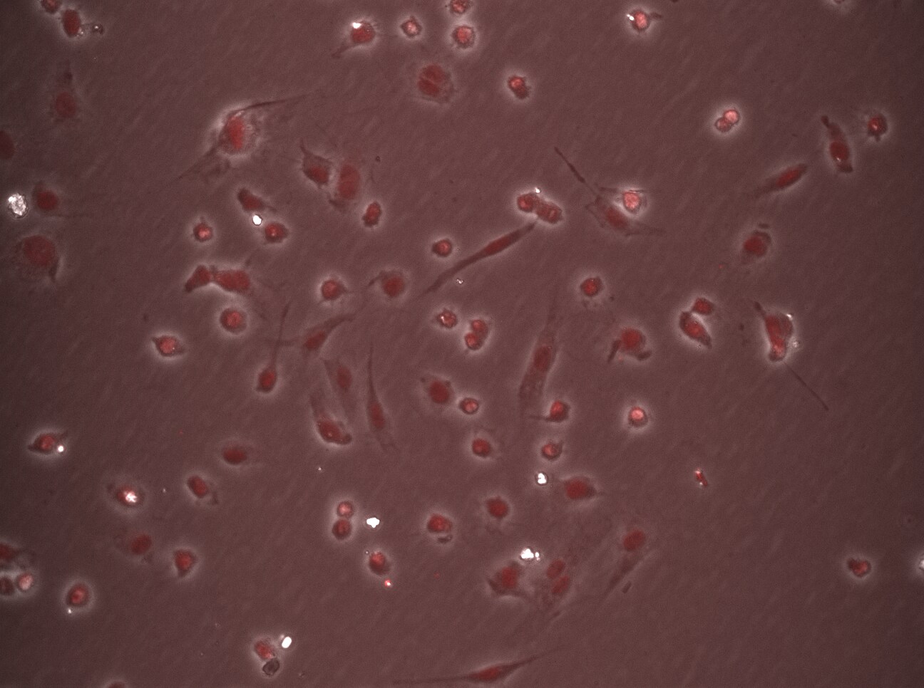
Immunocytochemistry/ Immunofluorescence: BMP-2 Antibody - BSA Free [NBP1-19751] -
Immunocytochemistry/ Immunofluorescence: BMP-2 Antibody - BSA Free [NBP1-19751] - BMD macrophages & splenic macrophages express nBMP2, which is decreased in the nuclei of nBmp2NLStm mutant macrophages. (a) BMD macrophages & (b) splenic macrophages were stained with anti-BMP2 antibody (green) & counterstained with DAPI (blue), demonstrating that nBMP2 is expressed & localized to the nucleus in wild type macrophages, & that nuclear translocation of nBMP2 is inhibited in mutant macrophages. BMP2 labeling within the cytoplasm is present in both wild type & mutant cells as expected, because the targeted mutation allows translation of nBMP2 in the cytoplasm but inhibits nuclear translocation, & it allows normal synthesis & secretion of conventional BMP2. Image collected & cropped by CiteAb from the following publication (https://pubmed.ncbi.nlm.nih.gov/30700748), licensed under a CC-BY license. Not internally tested by Novus Biologicals.
Immunohistochemistry-Paraffin: BMP-2 Antibody - BSA Free [NBP1-19751] -
Immunohistochemistry-Paraffin: BMP-2 Antibody - BSA Free [NBP1-19751] - BMP-2 & satellite stem cells in muscle regenerations. ((a)-(b)) Hematoxylin & eosin sections of muscle biopsies showed a significant increase of fat tissue in OA (a) as compared to OP patients (b) (40x). (c) Image showed numerous BMP-2 positive fibers (40x). Often, in OP patients we did not observed BMP-2 expression (40x) (d). The Immunohistochemistry for myostatin was negative in OA muscle tissue (40x) (e). Muscle biopsies of OP group showed high/moderate expression of myostatin (40x) (f) inversely related to BMP-2 immunostain. Groups of satellite cells CD44 positive were focally dispersed in the tissue (200x) of OA patients (g) higher than that observed in OP patients (200x) (h). Image collected & cropped by CiteAb from the following publication (https://pubmed.ncbi.nlm.nih.gov/26101529), licensed under a CC-BY license. Not internally tested by Novus Biologicals.
Western Blot: BMP-2 Antibody - BSA Free [NBP1-19751] -
Western Blot: BMP-2 Antibody - BSA Free [NBP1-19751] - Protein expression analysis. Immunostaining for BMP-2, myostatin, & CD44 was evaluated by counting the number of positive fibers/cells on 25-high power field (HPF), whereas western blot for BMP-2 was evaluated by lines densitometry. (a) Notably, we found that OA muscle biopsies showed a significantly higher number of BMP-2-positive fibers (293.0 ± 35.4) as compared with muscle of OP patients (162.1 ± 33.7) (p < 0.0001). (b) Western blot lines show a higher expression of BMP-2 in OA patients compared to OP group. (c) The number of myostatin positive fibers in OP patients was significantly higher compared to OA group (p < 0.0200). (d) CD44 expression shows a significantly different rate of CD44 positive cells in OA group as compared with OP (p < 0.0020). Image collected & cropped by CiteAb from the following publication (https://pubmed.ncbi.nlm.nih.gov/26101529), licensed under a CC-BY license. Not internally tested by Novus Biologicals.
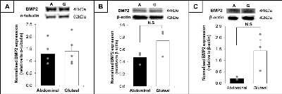
Western Blot: BMP-2 Antibody - BSA Free [NBP1-19751] -
Western Blot: BMP-2 Antibody - BSA Free [NBP1-19751] - BMP2 is a paracrine factor in WAT & regulates adipogenic differentiation in a depot-specific manner. a, b, c Western blots & summary graphs of BMP2 & alpha -tubulin expression in abdominal (A; black) & gluteal (G; white) AT (A; n = 5 women; 37–44 years, BMI 24.1–27.1 kg/m2), & BMP2 & beta -actin expression in proliferating (b) & differentiated (c) immortalised preadipocytes (n = 3). Relative protein expression data presented as mean ± SEM with individual data points overlaid (grey dots); data analysed by paired t-test. dBMP2 mRNA expression in primary abdominal (solid line) & gluteal (dashed line) preadipocytes during 3 days of proliferation (P) followed by 14 days of differentiation (d) (n = 8; 4 men, 4 women; age 32–44 years, BMI 20.5–26 kg/m2). mRNA expression normalised to PPIA. Data analysed by repeated measures ANOVA. e, f, g, h Proliferation analysis (e) in immortalised preadipocytes after 48 h culture in growth medium supplemented with BMP2 (n = 4). Triacylglycerol (TAG) accumulation (f) & PPARG2 (g) & ADIPOQ (h) mRNA expression in immortalised abdominal (black) & gluteal (white) preadipocytes after 14 day culture in BMP2-supplemented adipogenic medium (n = 3). mRNA expression normalised to 18 s. All data presented as mean fold change relative to vehicle ± SEM with individual data points overlaid (grey dots). Data analysed by ANOVA with Bonferroni post-hoc test (e, f, g, h); *p < 0.05, **p < 0.01, ***p < 0.001, compared to same depot vehicle; ‡p < 0.001, compared to same depot 5 ng/ml treatment Image collected & cropped by CiteAb from the following publication (https://pubmed.ncbi.nlm.nih.gov/31324879), licensed under a CC-BY license. Not internally tested by Novus Biologicals.Applications for BMP-2 Antibody - BSA Free
Immunocytochemistry/ Immunofluorescence
Immunohistochemistry
Immunohistochemistry-Paraffin
Western Blot
Reviewed Applications
Read 1 review rated 1 using NBP1-19751 in the following applications:
Formulation, Preparation, and Storage
Purification
Formulation
Format
Preservative
Concentration
Shipping
Stability & Storage
Background: BMP-2
Long Name
Alternate Names
Gene Symbol
UniProt
Additional BMP-2 Products
Product Documents for BMP-2 Antibody - BSA Free
Certificate of Analysis
To download a Certificate of Analysis, please enter a lot or batch number in the search box below.
Product Specific Notices for BMP-2 Antibody - BSA Free
This product is for research use only and is not approved for use in humans or in clinical diagnosis. Primary Antibodies are guaranteed for 1 year from date of receipt.
Related Research Areas
Citations for BMP-2 Antibody - BSA Free
 Powered by Bioz
Customer Reviews for BMP-2 Antibody - BSA Free (1)
Have you used BMP-2 Antibody - BSA Free?
Submit a review and receive an Amazon gift card!
$25/€18/£15/$25CAN/¥2500 Yen for a review with an image
$10/€7/£6/$10CAN/¥1110 Yen for a review without an image
Submit a review
Customer Images
-
Application: ImmunofluorescenceSample Tested: HUVEC (Human Umbilical Vein Endothelial Cells)Species: HumanVerified Customer | Posted 12/16/2017The immunofluorescence shows that cytoplasmic protein BMP-2 mainly distributed in the nucleus.Human umbilical vein endothelial cells

There are no reviews that match your criteria.
Protocols
View specific protocols for BMP-2 Antibody - BSA Free (NBP1-19751):
Immunohistochemistry-Paraffin Embedded Sections
Antigen Unmasking:
Bring slides to a boil in 10 mM sodium citrate buffer (pH 6.0) then maintain at a sub-boiling temperature for 10 minutes. Cool slides on bench-top for 30 minutes.
Staining:
1. Wash sections in deionized water three times for 5 minutes each.
2. Wash sections in wash buffer for 5 minutes.
3. Block each section with 100-400 ul blocking solution for 1 hour at room temperature.
4. Remove blocking solution and add 100-400 ul diluted primary antibody. Incubate overnight at 4C.
5. Remove antibody solution and wash sections in wash buffer three times for 5 minutes each.
6. Add 100-400 ul biotinylated diluted secondary antibody. Incubate 30 minutes at room temperature.
7. Remove secondary antibody solution and wash sections three times with wash buffer for 5 minutes each.
8. Add 100-400 ul Streptavidin-HRP reagent to each section and incubate for 30 minutes at room temperature.
9. Wash sections three times in wash buffer for 5 minutes each.
10. Add 100-400 ul DAB substrate to each section and monitor staining closely.
11. As soon as the sections develop, immerse slides in deionized water.
12. Counterstain sections in hematoxylin.
13. Wash sections in deionized water two times for 5 minutes each.
14. Dehydrate sections.
15. Mount coverslips.
Western Blot Protocol
1. Perform SDS-PAGE on samples to be analyzed, loading 40 ug of total protein per lane.
2. Transfer proteins to membrane according to the instructions provided by the manufacturer of the membrane and transfer apparatus.
3. Stain according to standard Ponceau S procedure (or similar product) to assess transfer success, and mark molecular weight standards where appropriate.
4. Rinse the blot.
5. Block the membrane using standard blocking buffer for at least 1 hour.
6. Wash the membrane in wash buffer three times for 10 minutes each.
7. Dilute primary antibody in blocking buffer and incubate 1 hour at room temperature.
8. Wash the membrane in wash buffer three times for 10 minutes each.
9. Apply the diluted HRP conjugated secondary antibody in blocking buffer (as per manufacturers instructions) and incubate 1 hour at room temperature.
10. Wash the blot in wash buffer three times for 10 minutes each (this step can be repeated as required to reduce background).
11. Apply the detection reagent of choice in accordance with the manufacturers instructions.
**Note: Tween-20 can be added to the blocking or antibody dilution buffer at a final concentration of 0.05-0.2%.
Find general support by application which include: protocols, troubleshooting, illustrated assays, videos and webinars.
- Antigen Retrieval Protocol (PIER)
- Antigen Retrieval for Frozen Sections Protocol
- Appropriate Fixation of IHC/ICC Samples
- Cellular Response to Hypoxia Protocols
- Chromogenic IHC Staining of Formalin-Fixed Paraffin-Embedded (FFPE) Tissue Protocol
- Chromogenic Immunohistochemistry Staining of Frozen Tissue
- ClariTSA™ Fluorophore Kits
- Detection & Visualization of Antibody Binding
- Fluorescent IHC Staining of Frozen Tissue Protocol
- Graphic Protocol for Heat-induced Epitope Retrieval
- Graphic Protocol for the Preparation and Fluorescent IHC Staining of Frozen Tissue Sections
- Graphic Protocol for the Preparation and Fluorescent IHC Staining of Paraffin-embedded Tissue Sections
- Graphic Protocol for the Preparation of Gelatin-coated Slides for Histological Tissue Sections
- ICC Cell Smear Protocol for Suspension Cells
- ICC Immunocytochemistry Protocol Videos
- ICC for Adherent Cells
- IHC Sample Preparation (Frozen sections vs Paraffin)
- Immunocytochemistry (ICC) Protocol
- Immunocytochemistry Troubleshooting
- Immunofluorescence of Organoids Embedded in Cultrex Basement Membrane Extract
- Immunofluorescent IHC Staining of Formalin-Fixed Paraffin-Embedded (FFPE) Tissue Protocol
- Immunohistochemistry (IHC) and Immunocytochemistry (ICC) Protocols
- Immunohistochemistry Frozen Troubleshooting
- Immunohistochemistry Paraffin Troubleshooting
- Preparing Samples for IHC/ICC Experiments
- Preventing Non-Specific Staining (Non-Specific Binding)
- Primary Antibody Selection & Optimization
- Protocol for Heat-Induced Epitope Retrieval (HIER)
- Protocol for Making a 4% Formaldehyde Solution in PBS
- Protocol for VisUCyte™ HRP Polymer Detection Reagent
- Protocol for the Fluorescent ICC Staining of Cell Smears - Graphic
- Protocol for the Fluorescent ICC Staining of Cultured Cells on Coverslips - Graphic
- Protocol for the Preparation & Fixation of Cells on Coverslips
- Protocol for the Preparation and Chromogenic IHC Staining of Frozen Tissue Sections
- Protocol for the Preparation and Chromogenic IHC Staining of Frozen Tissue Sections - Graphic
- Protocol for the Preparation and Chromogenic IHC Staining of Paraffin-embedded Tissue Sections
- Protocol for the Preparation and Chromogenic IHC Staining of Paraffin-embedded Tissue Sections - Graphic
- Protocol for the Preparation and Fluorescent ICC Staining of Cells on Coverslips
- Protocol for the Preparation and Fluorescent ICC Staining of Non-adherent Cells
- Protocol for the Preparation and Fluorescent ICC Staining of Stem Cells on Coverslips
- Protocol for the Preparation and Fluorescent IHC Staining of Frozen Tissue Sections
- Protocol for the Preparation and Fluorescent IHC Staining of Paraffin-embedded Tissue Sections
- Protocol for the Preparation of Gelatin-coated Slides for Histological Tissue Sections
- Protocol for the Preparation of a Cell Smear for Non-adherent Cell ICC - Graphic
- R&D Systems Quality Control Western Blot Protocol
- TUNEL and Active Caspase-3 Detection by IHC/ICC Protocol
- The Importance of IHC/ICC Controls
- Troubleshooting Guide: Immunohistochemistry
- Troubleshooting Guide: Western Blot Figures
- Western Blot Conditions
- Western Blot Protocol
- Western Blot Protocol for Cell Lysates
- Western Blot Troubleshooting
- Western Blot Troubleshooting Guide
- View all Protocols, Troubleshooting, Illustrated assays and Webinars
FAQs for BMP-2 Antibody - BSA Free
-
Q: I am interested in Bone Morphogenic Protein 2 (BMP-2) for clinical trial in humans. I am using this protein in stem cells culture for future use in humans. Do you have BMP-2 in pharma grade that I can use for humans?
A: Our antibodies are for research purposes only unfortunately. I am sorry we cannot be of further help.
-
Q: We onced ordered an anti BMP2 antibody (NBP1-19751) from your company, and have gotten great WB results. Your product has been included in our manuscript. Now reviewers are asking about the cross activity of this antibody with other BMPs, such as BMP6 or BMP9. Did you ever check the cross-react of the BMP2 antibody?
A: Unfortunately, we have not tested this antibody against other/recombinant BMPs so far but I can assure you that it will not be expected to react with any other BMPs. NBP1-19751 was generated against an immunogen peptide from AA250-350 of BMP2 protein [Swiss-Prot# P12643], and this sequence does not show any sequence similarity/homology with any other BMPs.
-
Q: I am interested in Bone Morphogenic Protein 2 (BMP-2) for clinical trial in humans. I am using this protein in stem cells culture for future use in humans. Do you have BMP-2 in pharma grade that I can use for humans?
A: Our antibodies are for research purposes only unfortunately. I am sorry we cannot be of further help.
-
Q: We onced ordered an anti BMP2 antibody (NBP1-19751) from your company, and have gotten great WB results. Your product has been included in our manuscript. Now reviewers are asking about the cross activity of this antibody with other BMPs, such as BMP6 or BMP9. Did you ever check the cross-react of the BMP2 antibody?
A: Unfortunately, we have not tested this antibody against other/recombinant BMPs so far but I can assure you that it will not be expected to react with any other BMPs. NBP1-19751 was generated against an immunogen peptide from AA250-350 of BMP2 protein [Swiss-Prot# P12643], and this sequence does not show any sequence similarity/homology with any other BMPs.

