BRCA1 Antibody (17F8) - Azide and BSA Free
Novus Biologicals | Catalog # NBP1-41185
![Western Blot: BRCA1 Antibody (17F8) [NBP1-41185]](https://resources.rndsystems.com/images/products/BRCA1-Antibody-17F8-Western-Blot-NBP1-41185-img0011.jpg "Western Blot: BRCA1 Antibody (17F8) [NBP1-41185]")
Loading...
Key Product Details
Validated by
Knockout/Knockdown
Species Reactivity
Validated:
Human, Mouse
Cited:
Human, Mouse
Applications
Validated:
Immunohistochemistry, Immunohistochemistry-Paraffin, Western Blot, ELISA, Immunocytochemistry/ Immunofluorescence, Immunoprecipitation, Chromatin Immunoprecipitation (ChIP)
Cited:
Knockout Validated, Immunohistochemistry-Paraffin, Western Blot, Immunocytochemistry/ Immunofluorescence
Label
Unconjugated
Antibody Source
Monoclonal Mouse IgG1 Clone # 17F8
Format
Azide and BSA Free
Loading...
Product Specifications
Immunogen
Protein fragment expressed in E. coli corresponding to amino acids 762-1315.
Reactivity Notes
Please note that this antibody is reactive to Mouse and derived from the same host, Mouse. Mouse-On-Mouse blocking reagent may be needed for IHC and ICC experiments to reduce high background signal. You can find these reagents under catalog numbers PK-2200-NB and MP-2400-NB. Please contact Technical Support if you have any questions.
Localization
Nuclear/Cytoplasmic
Specificity
This antibody does not recognize the delta exon 11 splice variant of BRCA1. In a high proportion of breast and ovarian cancer cell lines, BRCA1 aberrantly mislocates to the cytoplasm.
Clonality
Monoclonal
Host
Mouse
Isotype
IgG1
Theoretical MW
208 kDa.
Disclaimer note: The observed molecular weight of the protein may vary from the listed predicted molecular weight due to post translational modifications, post translation cleavages, relative charges, and other experimental factors.
Disclaimer note: The observed molecular weight of the protein may vary from the listed predicted molecular weight due to post translational modifications, post translation cleavages, relative charges, and other experimental factors.
Scientific Data Images for BRCA1 Antibody (17F8) - Azide and BSA Free
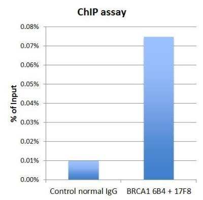
Chromatin Immunoprecipitation: BRCA1 Antibody (17F8) [NBP1-41185] - ChIP grade BRCA1 antibody 6B4 and BRCA1 antibody 17F8 were used for ChIP assay. The 6B4 and 17F8 mixture (3 microgram each), or normal mouse IgG (6 microgram) were incubated with HeLa chromatin extract (100 umicrogram each) in the ChIP assay. Enrichment of genomic DNA on a BRCA1 target gene promoter (HMGA2) was validated by a Q-PCR assay.
![Western Blot: BRCA1 Antibody (17F8) [NBP1-41185]](https://resources.rndsystems.com/images/products/BRCA1-Antibody-17F8-Western-Blot-NBP1-41185-img0012.jpg "Western Blot: BRCA1 Antibody (17F8) [NBP1-41185]")
Western Blot: BRCA1 Antibody (17F8) [NBP1-41185]
Western Blot: BRCA1 Antibody (17F8) [NBP1-41185] - ChIP grade detects BRCA1 protein by western blot analysis. Various whole cell extracts (30 ug) were separated by 5 % SDS-PAGE, and blotted with BRCA1 antibody [17F8] - ChIP grade BRCA1 diluted by 1:500![Immunoprecipitation: BRCA1 Antibody (17F8) [NBP1-41185]](https://resources.rndsystems.com/images/products/BRCA1-Antibody-17F8-Immunoprecipitation-NBP1-41185-img0006.jpg "Immunoprecipitation: BRCA1 Antibody (17F8) [NBP1-41185]")
Immunoprecipitation: BRCA1 Antibody (17F8) [NBP1-41185]
Immunoprecipitation: BRCA1 Antibody (17F8) [NBP1-41185] - BRCA1 antibody 6B4 and BRCA1 antibody 17F8 was used for IP-WB assay. 6B4 alone (4 microgram), 17F8 alone (4 microgram), 6B4 plus 17F8 (2 microgram each), and mouse control normal IgG were used in an immunoprecipitation assay with MCF7 cell extract. Immunoprecipitated BRCA1 was detected in WB using BRCA1 antibody 6B4 at 1:1000 dilution. HeLa whole cell extract (20 microgram) was used as input in the Western blot assay. [NBP1-41185] -")
Western Blot: BRCA1 Antibody (17F8) [NBP1-41185] -
Western Blot: BRCA1 Antibody (17F8) [NBP1-41185] - Wild-type (WT) and BRCA1 knockout (KO) HeLa cell extracts (30 ug) were separated by 5% SDS-PAGE, and the membrane was blotted with BRCA1 antibody [17F8] - ChIP grade diluted at 1:500. The HRP-conjugated anti-mouse IgG antibody was used to detect the primary antibody, and the signal was developed with Trident ECL plus-Enhanced.Applications for BRCA1 Antibody (17F8) - Azide and BSA Free
Application
Recommended Usage
Chromatin Immunoprecipitation (ChIP)
Assay dependent
ELISA
Assay dependent
Immunocytochemistry/ Immunofluorescence
1 ug/ml
Immunohistochemistry
Assay dependent
Immunohistochemistry-Paraffin
Assay dependent
Immunoprecipitation
Assay dependent
Western Blot
1:500-1:3000
Formulation, Preparation, and Storage
Purification
Protein G purified
Formulation
PBS
Format
Azide and BSA Free
Preservative
No Preservative
Concentration
Concentrations vary lot to lot. See vial label for concentration. If unlisted please contact technical services.
Shipping
The product is shipped with polar packs. Upon receipt, store it immediately at the temperature recommended below.
Stability & Storage
Store at 4C short term. Aliquot and store at -20C long term. Avoid freeze-thaw cycles.
Background: BRCA1
Long Name
Breast Cancer 1
Alternate Names
BRCAI, breast and ovarian cancer susceptibility protein 1, breast and ovarian cancer sususceptibility protein, breast cancer 1, early onset, breast cancer type 1 susceptibility protein, EC 6.3.2, EC 6.3.2.-, IRIS, PNCA4, PSCP, subunit 1
Entrez Gene IDs
672 (Human)
Gene Symbol
BRCA1
Additional BRCA1 Products
Product Documents for BRCA1 Antibody (17F8) - Azide and BSA Free
Certificate of Analysis
To download a Certificate of Analysis, please enter a lot or batch number in the search box below.
Product Specific Notices for BRCA1 Antibody (17F8) - Azide and BSA Free
This product is for research use only and is not approved for use in humans or in clinical diagnosis. Primary Antibodies are guaranteed for 1 year from date of receipt.
Related Research Areas
Citations for BRCA1 Antibody (17F8) - Azide and BSA Free
 Powered by Bioz
Customer Reviews for BRCA1 Antibody (17F8) - Azide and BSA Free
There are currently no reviews for this product. Be the first to review BRCA1 Antibody (17F8) - Azide and BSA Free and earn rewards!
Have you used BRCA1 Antibody (17F8) - Azide and BSA Free?
Submit a review and receive an Amazon gift card!
$25/€18/£15/$25CAN/¥2500 Yen for a review with an image
$10/€7/£6/$10CAN/¥1110 Yen for a review without an image
Submit a review
Protocols
Find general support by application which include: protocols, troubleshooting, illustrated assays, videos and webinars.
- Antigen Retrieval Protocol (PIER)
- Antigen Retrieval for Frozen Sections Protocol
- Appropriate Fixation of IHC/ICC Samples
- Cellular Response to Hypoxia Protocols
- ChIP Protocol Video
- Chromatin Immunoprecipitation (ChIP) Protocol
- Chromatin Immunoprecipitation Protocol
- Chromogenic IHC Staining of Formalin-Fixed Paraffin-Embedded (FFPE) Tissue Protocol
- Chromogenic Immunohistochemistry Staining of Frozen Tissue
- ClariTSA™ Fluorophore Kits
- Detection & Visualization of Antibody Binding
- ELISA Sample Preparation & Collection Guide
- ELISA Troubleshooting Guide
- Fluorescent IHC Staining of Frozen Tissue Protocol
- Graphic Protocol for Heat-induced Epitope Retrieval
- Graphic Protocol for the Preparation and Fluorescent IHC Staining of Frozen Tissue Sections
- Graphic Protocol for the Preparation and Fluorescent IHC Staining of Paraffin-embedded Tissue Sections
- Graphic Protocol for the Preparation of Gelatin-coated Slides for Histological Tissue Sections
- How to Run an R&D Systems DuoSet ELISA
- How to Run an R&D Systems Quantikine ELISA
- How to Run an R&D Systems Quantikine™ QuicKit™ ELISA
- ICC Cell Smear Protocol for Suspension Cells
- ICC Immunocytochemistry Protocol Videos
- ICC for Adherent Cells
- IHC Sample Preparation (Frozen sections vs Paraffin)
- Immunocytochemistry (ICC) Protocol
- Immunocytochemistry Troubleshooting
- Immunofluorescence of Organoids Embedded in Cultrex Basement Membrane Extract
- Immunofluorescent IHC Staining of Formalin-Fixed Paraffin-Embedded (FFPE) Tissue Protocol
- Immunohistochemistry (IHC) and Immunocytochemistry (ICC) Protocols
- Immunohistochemistry Frozen Troubleshooting
- Immunohistochemistry Paraffin Troubleshooting
- Immunoprecipitation Protocol
- Preparing Samples for IHC/ICC Experiments
- Preventing Non-Specific Staining (Non-Specific Binding)
- Primary Antibody Selection & Optimization
- Protocol for Heat-Induced Epitope Retrieval (HIER)
- Protocol for Making a 4% Formaldehyde Solution in PBS
- Protocol for VisUCyte™ HRP Polymer Detection Reagent
- Protocol for the Fluorescent ICC Staining of Cell Smears - Graphic
- Protocol for the Fluorescent ICC Staining of Cultured Cells on Coverslips - Graphic
- Protocol for the Preparation & Fixation of Cells on Coverslips
- Protocol for the Preparation and Chromogenic IHC Staining of Frozen Tissue Sections
- Protocol for the Preparation and Chromogenic IHC Staining of Frozen Tissue Sections - Graphic
- Protocol for the Preparation and Chromogenic IHC Staining of Paraffin-embedded Tissue Sections
- Protocol for the Preparation and Chromogenic IHC Staining of Paraffin-embedded Tissue Sections - Graphic
- Protocol for the Preparation and Fluorescent ICC Staining of Cells on Coverslips
- Protocol for the Preparation and Fluorescent ICC Staining of Non-adherent Cells
- Protocol for the Preparation and Fluorescent ICC Staining of Stem Cells on Coverslips
- Protocol for the Preparation and Fluorescent IHC Staining of Frozen Tissue Sections
- Protocol for the Preparation and Fluorescent IHC Staining of Paraffin-embedded Tissue Sections
- Protocol for the Preparation of Gelatin-coated Slides for Histological Tissue Sections
- Protocol for the Preparation of a Cell Smear for Non-adherent Cell ICC - Graphic
- Quantikine HS ELISA Kit Assay Principle, Alkaline Phosphatase
- Quantikine HS ELISA Kit Principle, Streptavidin-HRP Polymer
- R&D Systems Quality Control Western Blot Protocol
- Sandwich ELISA (Colorimetric) – Biotin/Streptavidin Detection Protocol
- Sandwich ELISA (Colorimetric) – Direct Detection Protocol
- TUNEL and Active Caspase-3 Detection by IHC/ICC Protocol
- The Importance of IHC/ICC Controls
- Troubleshooting Guide: ELISA
- Troubleshooting Guide: Immunohistochemistry
- Troubleshooting Guide: Western Blot Figures
- Western Blot Conditions
- Western Blot Protocol
- Western Blot Protocol for Cell Lysates
- Western Blot Troubleshooting
- Western Blot Troubleshooting Guide
- View all Protocols, Troubleshooting, Illustrated assays and Webinars
Loading...