Key Product Details
Species Reactivity
Validated:
Non-species specific
Cited:
Human, Mouse
Applications
Validated:
Western Blot, ELISA, Immunocytochemistry/ Immunofluorescence, Immunoprecipitation, Immunohistochemistry (Negative)
Cited:
Western Blot, Immunocytochemistry/ Immunofluorescence, Immunoprecipitation
Label
HRP
Antibody Source
Polyclonal Goat IgG
Loading...
Product Specifications
Immunogen
Goats were immunized with a synthetic peptide representing amino acid residues 410-419 (EQKLISEEDL) of human c-Myc Antibody conjugated to KLH. Antibody was isolated by affinity chromatography using the peptide immobilized on solid support.
Localization
Cytoplasmic and Nuclear
Clonality
Polyclonal
Host
Goat
Isotype
IgG
Theoretical MW
48.8 kDa.
Disclaimer note: The observed molecular weight of the protein may vary from the listed predicted molecular weight due to post translational modifications, post translation cleavages, relative charges, and other experimental factors.
Disclaimer note: The observed molecular weight of the protein may vary from the listed predicted molecular weight due to post translational modifications, post translation cleavages, relative charges, and other experimental factors.
Scientific Data Images for c-Myc Antibody [HRP]
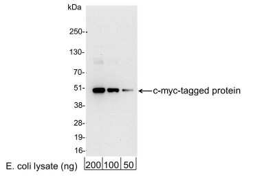
Western Blot: c-Myc Antibody [HRP] [NB600-341] - 200, 100, or 50 ng of E. coli whole cell lysate expressing a multi-tag fusion protein. Antibody used for WB at 0.4 ug/ml (1:25,000).
Applications for c-Myc Antibody [HRP]
Application
Recommended Usage
ELISA
1:10000-1:100000
Immunocytochemistry/ Immunofluorescence
1:200-1:500
Immunoprecipitation
1:10-1:500
Western Blot
1:1000-1:30000
Application Notes
IP reactivity reported in scientific literature (PMID: 22971543). ICC/IF reactivity reported in scientific literature (PMID: 27434673).
Reviewed Applications
Read 1 review rated 5 using NB600-341 in the following applications:
Formulation, Preparation, and Storage
Purification
Immunogen affinity purified
Formulation
PBS and 0.2% BSA
Preservative
0.05% Pro-Clean 400
Concentration
1.0 mg/ml
Shipping
The product is shipped with polar packs. Upon receipt, store it immediately at the temperature recommended below.
Stability & Storage
Store at 4C. Do not freeze.
Background: c-Myc
A basic Helix-Loop-Helix, Leucine Zipper domain (bHLH/LZ), designated Max, specifically associates with c-Myc, N-Myc and L-Myc proteins. The Myc-Max complex binds to DNA in a sequence-specific manner under conditions where neither Max nor Myc exhibit appreciable binding. Max can also form heterodimers with other bHLH-Zip proteins, Mad and Mxi1. c-Myc plays a role in cell cycle progression, apoptosis, cellular transformation and angiogenesis (2). Mutations, overexpression, rearrangement and translocation of this gene have been associated with a variety of cancers including B-cell Lymphomas, acute myeloid leukemia, glioblastoma, stomach adenocarcinoma, and prostate adenocarcinoma (3).
References
1. Wilkinson, D. S., Tsai, W. W., Schumacher, M. A., & Barton, M. C. (2008). Chromatin-bound p53 anchors activated Smads and the mSin3A corepressor to confer transforming-growth-factor-beta-mediated transcription repression. Mol Cell Biol, 28(6), 1988-1998. doi:10.1128/mcb.01442-07
2. Pedrosa, A. R., Bodrug, N., Gomez-Escudero, J., Carter, E. P., Reynolds, L. E., Georgiou, P. N.,... Hodivala-Dilke, K. M. (2019). Tumor Angiogenesis Is Differentially Regulated by Phosphorylation of Endothelial Cell Focal Adhesion Kinase Tyrosines-397 and -861. Cancer Res, 79(17), 4371-4386. doi:10.1158/0008-5472.Can-18-3934
3. Nagasaka, M., Tsuzuki, K., Ozeki, Y., Tokugawa, M., Ohoka, N., Inoue, Y., & Hayashi, H. (2019). Lysine-Specific Demethylase 1 (LSD1/KDM1A) Is a Novel Target Gene of c-Myc. Biol Pharm Bull, 42(3), 481-488. doi:10.1248/bpb.b18-00892
Long Name
v-Myc Avian Myelocytomatosis Viral Oncogene Homolog (Avian)
Alternate Names
cMyc, Myc, Myc2, Niard, Nird
Gene Symbol
MYC
Additional c-Myc Products
Product Documents for c-Myc Antibody [HRP]
Certificate of Analysis
To download a Certificate of Analysis, please enter a lot or batch number in the search box below.
Product Specific Notices for c-Myc Antibody [HRP]
This product is for research use only and is not approved for use in humans or in clinical diagnosis. Primary Antibodies are guaranteed for 1 year from date of receipt.
Citations for c-Myc Antibody [HRP]
 Powered by Bioz
Customer Reviews for c-Myc Antibody [HRP] (1)
5 out of 5
1 Customer Rating
Have you used c-Myc Antibody [HRP]?
Submit a review and receive an Amazon gift card!
$25/€18/£15/$25CAN/¥2500 Yen for a review with an image
$10/€7/£6/$10CAN/¥1110 Yen for a review without an image
Submit a review
Showing
1
-
1 of
1 review
Showing All
Filter By:
-
Application: ELISASample Tested: PrivateSpecies: HumanVerified Customer | Posted 06/15/2010
There are no reviews that match your criteria.
Protocols
Find general support by application which include: protocols, troubleshooting, illustrated assays, videos and webinars.
- Appropriate Fixation of IHC/ICC Samples
- Cellular Response to Hypoxia Protocols
- ClariTSA™ Fluorophore Kits
- Detection & Visualization of Antibody Binding
- ELISA Sample Preparation & Collection Guide
- ELISA Troubleshooting Guide
- How to Run an R&D Systems DuoSet ELISA
- How to Run an R&D Systems Quantikine ELISA
- How to Run an R&D Systems Quantikine™ QuicKit™ ELISA
- ICC Cell Smear Protocol for Suspension Cells
- ICC Immunocytochemistry Protocol Videos
- ICC for Adherent Cells
- Immunocytochemistry (ICC) Protocol
- Immunocytochemistry Troubleshooting
- Immunofluorescence of Organoids Embedded in Cultrex Basement Membrane Extract
- Immunohistochemistry (IHC) and Immunocytochemistry (ICC) Protocols
- Immunoprecipitation Protocol
- Preparing Samples for IHC/ICC Experiments
- Preventing Non-Specific Staining (Non-Specific Binding)
- Primary Antibody Selection & Optimization
- Protocol for VisUCyte™ HRP Polymer Detection Reagent
- Protocol for the Fluorescent ICC Staining of Cell Smears - Graphic
- Protocol for the Fluorescent ICC Staining of Cultured Cells on Coverslips - Graphic
- Protocol for the Preparation and Fluorescent ICC Staining of Cells on Coverslips
- Protocol for the Preparation and Fluorescent ICC Staining of Non-adherent Cells
- Protocol for the Preparation and Fluorescent ICC Staining of Stem Cells on Coverslips
- Protocol for the Preparation of a Cell Smear for Non-adherent Cell ICC - Graphic
- Quantikine HS ELISA Kit Assay Principle, Alkaline Phosphatase
- Quantikine HS ELISA Kit Principle, Streptavidin-HRP Polymer
- R&D Systems Quality Control Western Blot Protocol
- Sandwich ELISA (Colorimetric) – Biotin/Streptavidin Detection Protocol
- Sandwich ELISA (Colorimetric) – Direct Detection Protocol
- TUNEL and Active Caspase-3 Detection by IHC/ICC Protocol
- The Importance of IHC/ICC Controls
- Troubleshooting Guide: ELISA
- Troubleshooting Guide: Western Blot Figures
- Western Blot Conditions
- Western Blot Protocol
- Western Blot Protocol for Cell Lysates
- Western Blot Troubleshooting
- Western Blot Troubleshooting Guide
- View all Protocols, Troubleshooting, Illustrated assays and Webinars


