Best Seller
c-Myc Antibody - BSA Free
Novus Biologicals | Catalog # NB600-335
![Western Blot: c-Myc AntibodyBSA Free [NB600-335]](https://resources.rndsystems.com/images/products/c-Myc-Antibody-Western-Blot-NB600-335-img0006.jpg "Western Blot: c-Myc AntibodyBSA Free [NB600-335]")
Key Product Details
Species Reactivity
Validated:
Non-species specific
Cited:
Human, Mouse
Applications
Validated:
Immunohistochemistry, Immunohistochemistry-Paraffin, Immunohistochemistry-Frozen, Western Blot, ELISA, Immunocytochemistry/ Immunofluorescence, Immunoprecipitation
Cited:
Immunohistochemistry, Immunohistochemistry-Paraffin, Immunohistochemistry-Frozen, Western Blot, Immunocytochemistry/ Immunofluorescence, Immunoprecipitation, IF/IHC
Label
Unconjugated
Antibody Source
Polyclonal Goat IgG
Format
BSA Free
Loading...
Product Specifications
Immunogen
Goats were immunized with a synthetic peptide representing amino acid residues 410-419 (EQKLISEEDL) of human c-Myc Antibody conjugated to KLH. Antibody was isolated by affinity chromatography using the peptide immobilized on solid support.
Localization
Cytoplasmic and Nuclear
Clonality
Polyclonal
Host
Goat
Isotype
IgG
Theoretical MW
48.8 kDa.
Disclaimer note: The observed molecular weight of the protein may vary from the listed predicted molecular weight due to post translational modifications, post translation cleavages, relative charges, and other experimental factors.
Disclaimer note: The observed molecular weight of the protein may vary from the listed predicted molecular weight due to post translational modifications, post translation cleavages, relative charges, and other experimental factors.
Scientific Data Images for c-Myc Antibody - BSA Free
Western Blot: c-Myc AntibodyBSA Free [NB600-335]
Western Blot: c-Myc Antibody [NB600-335] - Detection of c-myc-tagged Protein by Western Blot. Samples: 200, 100, or 50 ng of E. coli whole cell lysate expressing a multi-tag fusion protein. Antibodies: Affinity purified, goat anti-c-myc antibody NB600-335 at 0.04 ug/ml (1:25,000). Detection: Chemiluminescence with an exposure time of 30 seconds.![Immunocytochemistry/ Immunofluorescence: c-Myc Antibody - BSA Free [NB600-335]](https://resources.rndsystems.com/images/products/c-Myc-Antibody-Immunocytochemistry-Immunofluorescence-NB600-335-img0007.jpg "Immunocytochemistry/ Immunofluorescence: c-Myc Antibody - BSA Free [NB600-335]")
Immunocytochemistry/ Immunofluorescence: c-Myc Antibody - BSA Free [NB600-335]
Immunocytochemistry/Immunofluorescence: c-Myc Antibody [NB600-335] - HeLa cells were fixed for 10 minutes using 10% formalin and then permeabilized for 5 minutes using 1X PBS + 0.5% Triton X-100. The cells were incubated with anti-c-Myc conjugated to Alexa Fluor 488 [NB600-335AF488] at 20 ug/mL for 1 hour at room temperature. Nuclei were counterstained with DAPI (Blue). Cells were imaged using a 40X objective. Image using the Alexa Fluor 488 format of this antibody.![Western Blot: c-Myc AntibodyBSA Free [NB600-335]](https://resources.rndsystems.com/images/products/c-Myc-Antibody-Western-Blot-NB600-335-img0005.jpg "Western Blot: c-Myc AntibodyBSA Free [NB600-335]")
Western Blot: c-Myc AntibodyBSA Free [NB600-335]
Western Blot: c-Myc Antibody [NB600-335] - Analysis using the HRP conjugate of NB600-335. Detection of 200, 100, or 50 ng of E. coli whole cell lysate expressing a multi-tag fusion protein. Antibody used at 0.4 ug/mL (1:25,000).
Western Blot: c-Myc Antibody - BSA Free [NB600-335] -
KLF8 regulates expression of stem cell markers in breast cancer cells. (A) Lysates from MDA-MB-231 cells stably overexpressing control or KLF8 were collected for immunoblot analysis of CSCs markers using indicated antibodies (left), and quantified graph of relative level of CSCs detected by immunoblot from MDA-MB-231 cells control or with KLF8 overexpression (right). (B) Quantified graph showing relative mRNA level of CSCs markers as detected by qRT-PCR using probes against genes in MDA-MB-231 cells stably overexpressing control or KLF8. (C) Lysates from MDA-MB-231 cells stably expressing control or KLF8 shRNA were collected for immunoblot analysis of CSCs markers using indicated antibodies (left), and quantified graph of relative level of CSCs levels (right). (D) Quantified graph showing relative mRNA level of CSCs markers as detected by qRT-PCR using probes against indicated targets in MDA-MB-231 cells stably expressing control or KLF8 shRNA. Student t test reported as mean +/- SEM, *p<0.05. Image collected and cropped by CiteAb from the following open publication (https://pubmed.ncbi.nlm.nih.gov/37152043), licensed under a CC-BY license. Not internally tested by Novus Biologicals.
Western Blot: c-Myc Antibody - BSA Free [NB600-335] -
Dynamic modeling of hBBSome and Ub‐hBBSome complexACo‐immunoprecipitation assay of BBS1 or BBS1K143R and BBS3 from lysates of HEK293 transiently expressing the indicated transgenes.BCumulative data of the experiments shown in (A). The data are expressed as mean +/- SD of three independent experiments. Student's t test, ***P < 0.001.CFull atomistic description of the homology model of the octameric complex of the BBSome human sequence (hBBSome) in the Arl6GTP‐bound (open) conformation, with the monoubiquitylation at K143 of hBBS14 alpha (Ub‐hBBSome).DFocus on the full atomistic model of hBBSome core complex components (BBS1, BBS4, BBS8, BBS9, and BBS18).ETop view of the interaction details between BBS18 alpha ‐helix and the BBS18 U‐bolt domains with the BBS8TPR and the superhelix of BBS4TPR, respectively.FTop view of the interaction details between BBS18 alpha ‐helix with the BBS1GAE domain and the superhelix of BBS4TPR.GSurface representation of the CG‐MD model of the Ub‐hBBSome, representing the starting point of the CG‐MD simulation. Residues involved in the Arl6GTP‐bound binding mode are highlighted in dark green.H, ISurface representation of the last CG‐MD frame of the wt‐hBBSome and the Ub‐hBBSome, respectively showing the progressive closure of Arl6‐binding site located between BBS1 beta prop and BBS7 beta prop. Residues involved in the Arl6GTP‐bound binding mode are highlighted in red and yellow, respectively. Image collected and cropped by CiteAb from the following open publication (https://pubmed.ncbi.nlm.nih.gov/36744302), licensed under a CC-BY license. Not internally tested by Novus Biologicals.Applications for c-Myc Antibody - BSA Free
Application
Recommended Usage
ELISA
Coating: 1:100-1:500; Primary: 1:1000-1:30000
Immunocytochemistry/ Immunofluorescence
1:100 - 1:400
Immunohistochemistry-Frozen
1:100 - 1:400
Immunoprecipitation
1 - 4 ug/mg lysate
Western Blot
1:1000 - 1:30000
Application Notes
IHC-F reactivity reported in scientific literature (PMID: 22411008). ICC/IF reactivity reported in scientific literature (PMID: 28368777). Use in IHC reported in scientific literature (PMID: 30203304). Use in IHC-P reported in scientific literature (PMID: 32159512).
Reviewed Applications
Read 1 review rated 5 using NB600-335 in the following applications:
Formulation, Preparation, and Storage
Purification
Immunogen affinity purified
Formulation
PBS
Format
BSA Free
Preservative
0.09% Sodium Azide
Concentration
1.0 mg/ml
Shipping
The product is shipped with polar packs. Upon receipt, store it immediately at the temperature recommended below.
Stability & Storage
Store at 4C. Do not freeze.
Background: c-Myc
A basic Helix-Loop-Helix, Leucine Zipper domain (bHLH/LZ), designated Max, specifically associates with c-Myc, N-Myc and L-Myc proteins. The Myc-Max complex binds to DNA in a sequence-specific manner under conditions where neither Max nor Myc exhibit appreciable binding. Max can also form heterodimers with other bHLH-Zip proteins, Mad and Mxi1. c-Myc plays a role in cell cycle progression, apoptosis, cellular transformation and angiogenesis (2). Mutations, overexpression, rearrangement and translocation of this gene have been associated with a variety of cancers including B-cell Lymphomas, acute myeloid leukemia, glioblastoma, stomach adenocarcinoma, and prostate adenocarcinoma (3).
References
1. Wilkinson, D. S., Tsai, W. W., Schumacher, M. A., & Barton, M. C. (2008). Chromatin-bound p53 anchors activated Smads and the mSin3A corepressor to confer transforming-growth-factor-beta-mediated transcription repression. Mol Cell Biol, 28(6), 1988-1998. doi:10.1128/mcb.01442-07
2. Pedrosa, A. R., Bodrug, N., Gomez-Escudero, J., Carter, E. P., Reynolds, L. E., Georgiou, P. N.,... Hodivala-Dilke, K. M. (2019). Tumor Angiogenesis Is Differentially Regulated by Phosphorylation of Endothelial Cell Focal Adhesion Kinase Tyrosines-397 and -861. Cancer Res, 79(17), 4371-4386. doi:10.1158/0008-5472.Can-18-3934
3. Nagasaka, M., Tsuzuki, K., Ozeki, Y., Tokugawa, M., Ohoka, N., Inoue, Y., & Hayashi, H. (2019). Lysine-Specific Demethylase 1 (LSD1/KDM1A) Is a Novel Target Gene of c-Myc. Biol Pharm Bull, 42(3), 481-488. doi:10.1248/bpb.b18-00892
Long Name
v-Myc Avian Myelocytomatosis Viral Oncogene Homolog (Avian)
Alternate Names
cMyc, Myc, Myc2, Niard, Nird
Gene Symbol
MYC
Additional c-Myc Products
Product Documents for c-Myc Antibody - BSA Free
Certificate of Analysis
To download a Certificate of Analysis, please enter a lot or batch number in the search box below.
Product Specific Notices for c-Myc Antibody - BSA Free
This product is for research use only and is not approved for use in humans or in clinical diagnosis. Primary Antibodies are guaranteed for 1 year from date of receipt.
Citations for c-Myc Antibody - BSA Free
 Powered by Bioz
Customer Reviews for c-Myc Antibody - BSA Free (1)
5 out of 5
1 Customer Rating
Have you used c-Myc Antibody - BSA Free?
Submit a review and receive an Amazon gift card!
$25/€18/£15/$25CAN/¥2500 Yen for a review with an image
$10/€7/£6/$10CAN/¥1110 Yen for a review without an image
Submit a review
Customer Images
Showing
1
-
1 of
1 review
Showing All
Filter By:
-
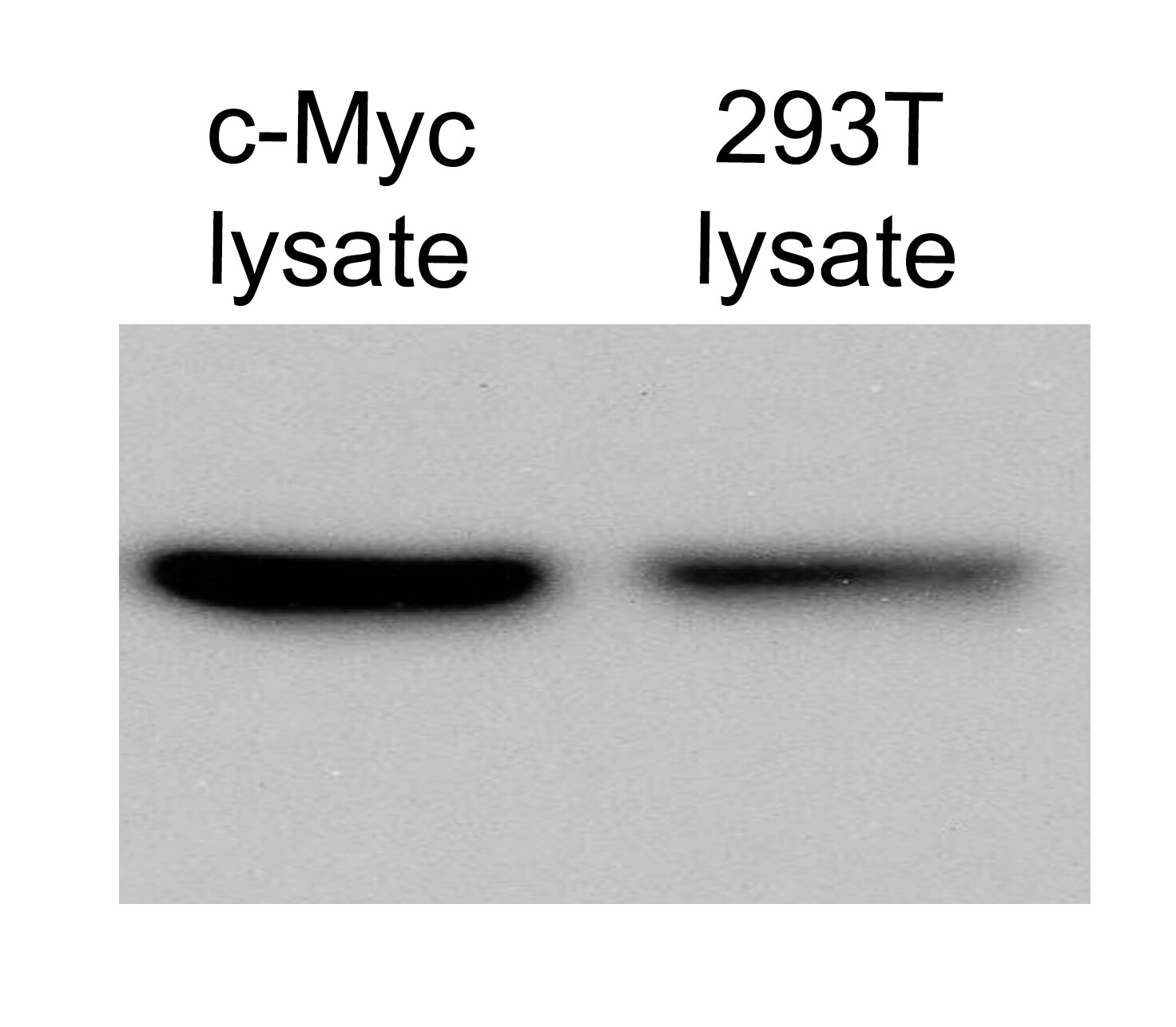
Application: Western BlotSample Tested: Whole cell lysate from 293T cells, in lysis bufferSpecies: HumanVerified Customer | Posted 12/18/2014WB for c-Myc in 293T cells

There are no reviews that match your criteria.
Protocols
Find general support by application which include: protocols, troubleshooting, illustrated assays, videos and webinars.
- Antigen Retrieval Protocol (PIER)
- Antigen Retrieval for Frozen Sections Protocol
- Appropriate Fixation of IHC/ICC Samples
- Cellular Response to Hypoxia Protocols
- Chromogenic IHC Staining of Formalin-Fixed Paraffin-Embedded (FFPE) Tissue Protocol
- Chromogenic Immunohistochemistry Staining of Frozen Tissue
- ClariTSA™ Fluorophore Kits
- Detection & Visualization of Antibody Binding
- ELISA Sample Preparation & Collection Guide
- ELISA Troubleshooting Guide
- Fluorescent IHC Staining of Frozen Tissue Protocol
- Graphic Protocol for Heat-induced Epitope Retrieval
- Graphic Protocol for the Preparation and Fluorescent IHC Staining of Frozen Tissue Sections
- Graphic Protocol for the Preparation and Fluorescent IHC Staining of Paraffin-embedded Tissue Sections
- Graphic Protocol for the Preparation of Gelatin-coated Slides for Histological Tissue Sections
- How to Run an R&D Systems DuoSet ELISA
- How to Run an R&D Systems Quantikine ELISA
- How to Run an R&D Systems Quantikine™ QuicKit™ ELISA
- ICC Cell Smear Protocol for Suspension Cells
- ICC Immunocytochemistry Protocol Videos
- ICC for Adherent Cells
- IHC Sample Preparation (Frozen sections vs Paraffin)
- Immunocytochemistry (ICC) Protocol
- Immunocytochemistry Troubleshooting
- Immunofluorescence of Organoids Embedded in Cultrex Basement Membrane Extract
- Immunofluorescent IHC Staining of Formalin-Fixed Paraffin-Embedded (FFPE) Tissue Protocol
- Immunohistochemistry (IHC) and Immunocytochemistry (ICC) Protocols
- Immunohistochemistry Frozen Troubleshooting
- Immunohistochemistry Paraffin Troubleshooting
- Immunoprecipitation Protocol
- Preparing Samples for IHC/ICC Experiments
- Preventing Non-Specific Staining (Non-Specific Binding)
- Primary Antibody Selection & Optimization
- Protocol for Heat-Induced Epitope Retrieval (HIER)
- Protocol for Making a 4% Formaldehyde Solution in PBS
- Protocol for VisUCyte™ HRP Polymer Detection Reagent
- Protocol for the Fluorescent ICC Staining of Cell Smears - Graphic
- Protocol for the Fluorescent ICC Staining of Cultured Cells on Coverslips - Graphic
- Protocol for the Preparation & Fixation of Cells on Coverslips
- Protocol for the Preparation and Chromogenic IHC Staining of Frozen Tissue Sections
- Protocol for the Preparation and Chromogenic IHC Staining of Frozen Tissue Sections - Graphic
- Protocol for the Preparation and Chromogenic IHC Staining of Paraffin-embedded Tissue Sections
- Protocol for the Preparation and Chromogenic IHC Staining of Paraffin-embedded Tissue Sections - Graphic
- Protocol for the Preparation and Fluorescent ICC Staining of Cells on Coverslips
- Protocol for the Preparation and Fluorescent ICC Staining of Non-adherent Cells
- Protocol for the Preparation and Fluorescent ICC Staining of Stem Cells on Coverslips
- Protocol for the Preparation and Fluorescent IHC Staining of Frozen Tissue Sections
- Protocol for the Preparation and Fluorescent IHC Staining of Paraffin-embedded Tissue Sections
- Protocol for the Preparation of Gelatin-coated Slides for Histological Tissue Sections
- Protocol for the Preparation of a Cell Smear for Non-adherent Cell ICC - Graphic
- Quantikine HS ELISA Kit Assay Principle, Alkaline Phosphatase
- Quantikine HS ELISA Kit Principle, Streptavidin-HRP Polymer
- R&D Systems Quality Control Western Blot Protocol
- Sandwich ELISA (Colorimetric) – Biotin/Streptavidin Detection Protocol
- Sandwich ELISA (Colorimetric) – Direct Detection Protocol
- TUNEL and Active Caspase-3 Detection by IHC/ICC Protocol
- The Importance of IHC/ICC Controls
- Troubleshooting Guide: ELISA
- Troubleshooting Guide: Immunohistochemistry
- Troubleshooting Guide: Western Blot Figures
- Western Blot Conditions
- Western Blot Protocol
- Western Blot Protocol for Cell Lysates
- Western Blot Troubleshooting
- Western Blot Troubleshooting Guide
- View all Protocols, Troubleshooting, Illustrated assays and Webinars


