Key Product Details
Species Reactivity
Human
Applications
Immunohistochemistry, Immunohistochemistry-Paraffin, Western Blot, Flow Cytometry, Immunofluorescence, Immunocytochemistry/ Immunofluorescence, Simple Western
Label
Unconjugated
Antibody Source
Monoclonal Mouse IgG1 kappa Clone # h-CALD
Loading...
Product Specifications
Immunogen
Crude human uterus extract
Localization
Cytoplasmic
Marker
Smooth Muscle Marker
Specificity
Recognizes a protein of 150kDa, which is identified as the high molecular weight variant of Caldesmon. Two closely related variants of human caldesmon have been identified which are different in their electrophoretic mobility and cellular distribution. The h-caldesmon variant (12080kDa) is found in non- muscle tissue and cells. Neither of the two variants has been detected in skeletal muscle. This monoclonal antibody recognizes only the 150kDa variant (h-caldesmon) in Western blots of human aortic media extracts and is unreactive with fibroblast extracts from cultivated human foreskin. Caldesmon is a developmentally regulated protein involved in smooth muscle and non-muscle contraction.
Clonality
Monoclonal
Host
Mouse
Isotype
IgG1 kappa
Theoretical MW
150 kDa.
Disclaimer note: The observed molecular weight of the protein may vary from the listed predicted molecular weight due to post translational modifications, post translation cleavages, relative charges, and other experimental factors.
Disclaimer note: The observed molecular weight of the protein may vary from the listed predicted molecular weight due to post translational modifications, post translation cleavages, relative charges, and other experimental factors.
Description
200ug/ml of antibody purified from Bioreactor Concentrate by Protein A or G. Prepared in 10 mM PBS with 0.05% BSA & 0.05% azide. Also available WITHOUT BSA & azide at 1.0 mg/ml. (NBP2-47816)
Antibody with azide - store at 2 to 8C. Antibody without azide - store at -20 to -80C.
Antibody with azide - store at 2 to 8C. Antibody without azide - store at -20 to -80C.
Scientific Data Images for Caldesmon/CALD1 Antibody (h-CALD)
![Western Blot: Caldesmon/CALD1 Antibody (h-CALD) [NBP2-44421]](https://resources.rndsystems.com/images/products/Caldesmon-CALD1-Antibody-h-CALD-Western-Blot-NBP2-44421-img0008.jpg "Western Blot: Caldesmon/CALD1 Antibody (h-CALD) [NBP2-44421]")
Western Blot: Caldesmon/CALD1 Antibody (h-CALD) [NBP2-44421]
Western Blot: Caldesmon/CALD1 Antibody (h-CALD) [NBP2-44421] - Western Blot Analysis of human Ovary tissue using Caldesmon/CALD1 antibody (h-CALD).![Immunohistochemistry-Paraffin: Caldesmon/CALD1 Antibody (h-CALD) [NBP2-44421]](https://resources.rndsystems.com/images/products/Caldesmon-CALD1-Antibody-h-CALD-Immunohistochemistry-Paraffin-NBP2-44421-img0007.jpg "Immunohistochemistry-Paraffin: Caldesmon/CALD1 Antibody (h-CALD) [NBP2-44421]")
Immunohistochemistry-Paraffin: Caldesmon/CALD1 Antibody (h-CALD) [NBP2-44421]
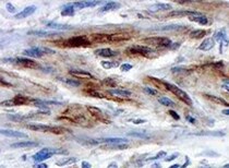
Immunohistochemistry-Paraffin: Caldesmon/CALD1 Antibody (h-CALD) [NBP2-44421] - Analysis of Human uterus tissue using Caldesmon/CALD1 antibody (h-CALD). Image from verified customer review.![Immunohistochemistry-Paraffin: Caldesmon/CALD1 Antibody (h-CALD) [NBP2-44421]](https://resources.rndsystems.com/images/products/Caldesmon-CALD1-Antibody-h-CALD-Immunohistochemistry-Paraffin-NBP2-44421-img0001.jpg "Immunohistochemistry-Paraffin: Caldesmon/CALD1 Antibody (h-CALD) [NBP2-44421]")
Immunohistochemistry-Paraffin: Caldesmon/CALD1 Antibody (h-CALD) [NBP2-44421]
Immunohistochemistry-Paraffin: Caldesmon/CALD1 Antibody (h-CALD) [NBP2-44421] - Human Uterus stained with Caldesmon Monoclonal Antibody (h-CALD).![Simple Western: Caldesmon/CALD1 Antibody (h-CALD) [NBP2-44421]](https://resources.rndsystems.com/images/products/Caldesmon-CALD1-Antibody-h-CALD-Simple-Western-NBP2-44421-img0002.jpg "Simple Western: Caldesmon/CALD1 Antibody (h-CALD) [NBP2-44421]")
Simple Western: Caldesmon/CALD1 Antibody (h-CALD) [NBP2-44421]
Simple Western: Caldesmon/CALD1 Antibody (h-CALD) [NBP2-44421] - Simple Western lane view shows a specific band for Caldesmon/CALD1 in 0.2 mg/ml of h. Aorta lysate(s). This experiment was performed under reducing conditions using the 12-230 kDa separation system.![Simple Western: Caldesmon/CALD1 Antibody (h-CALD) [NBP2-44421]](https://resources.rndsystems.com/images/products/Caldesmon-CALD1-Antibody-h-CALD-Simple-Western-NBP2-44421-img0003.jpg "Simple Western: Caldesmon/CALD1 Antibody (h-CALD) [NBP2-44421]")
Simple Western: Caldesmon/CALD1 Antibody (h-CALD) [NBP2-44421]
Simple Western: Caldesmon/CALD1 Antibody (h-CALD) [NBP2-44421] - Simple Western lane view shows a specific band for Caldesmon/CALD1 in 0.2 mg/ml of h. Uterus lysate(s). This experiment was performed under reducing conditions using the 12-230 kDa separation system.![Simple Western: Caldesmon/CALD1 Antibody (h-CALD) [NBP2-44421]](https://resources.rndsystems.com/images/products/Caldesmon-CALD1-Antibody-h-CALD-Simple-Western-NBP2-44421-img0006.jpg "Simple Western: Caldesmon/CALD1 Antibody (h-CALD) [NBP2-44421]")
Simple Western: Caldesmon/CALD1 Antibody (h-CALD) [NBP2-44421]
Simple Western: Caldesmon/CALD1 Antibody (h-CALD) [NBP2-44421] - Electropherogram image of the corresponding Simple Western lane. Caldesmon/CALD1 antibody was used at 10 ug/ml dilution of h. Aorta and h. Uterus lysates(s) respectively.Applications for Caldesmon/CALD1 Antibody (h-CALD)
Application
Recommended Usage
Flow Cytometry
1-2 ug/million cells
Immunocytochemistry/ Immunofluorescence
1-2 ug/ml
Immunofluorescence
1 - 2 ug/ml
Immunohistochemistry-Paraffin
1-2 ug/ml
Simple Western
10 ug/ml
Western Blot
2-4 ug/ml
Application Notes
Immunohistochemistry (Formalin-fixed): 1-2ug/ml for 30 minutes at RT. Staining of formalin-fixed tissues requires heating tissue sections in 10mM Tris with 1mM EDTA, pH 9.0, for 45 min at 95C followed by cooling at RT for 20 minutes.
Optimal dilution for a specific application should be determined.
In Simple Western only 10 - 15 ul of the recommended dilution is used per data point.
See Simple Western Antibody Database for Simple Western validation: Tested in h. Aorta lysate(s), h. Uterus lysate, separated by Size, antibody dilution of 10 ug/mL, apparent MW was 140-150 kDa.
Reviewed Applications
Read 1 review rated 5 using NBP2-44421 in the following applications:
Flow Cytometry Panel Builder
Bio-Techne Knows Flow Cytometry
Save time and reduce costly mistakes by quickly finding compatible reagents using the Panel Builder Tool.
Advanced Features
- Spectra Viewer - Custom analysis of spectra from multiple fluorochromes
- Spillover Popups - Visualize the spectra of individual fluorochromes
- Antigen Density Selector - Match fluorochrome brightness with antigen density

Formulation, Preparation, and Storage
Purification
Protein A or G purified
Formulation
10 mM PBS with 0.05% BSA
Preservative
0.05% Sodium Azide
Concentration
0.2 mg/ml
Shipping
The product is shipped with polar packs. Upon receipt, store it immediately at the temperature recommended below.
Stability & Storage
Store at 4C.
Background: Caldesmon/CALD1
Additional Caldesmon/CALD1 Products
Product Documents for Caldesmon/CALD1 Antibody (h-CALD)
Certificate of Analysis
To download a Certificate of Analysis, please enter a lot or batch number in the search box below.
Product Specific Notices for Caldesmon/CALD1 Antibody (h-CALD)
This product is for research use only and is not approved for use in humans or in clinical diagnosis. Primary Antibodies are guaranteed for 1 year from date of receipt.
Related Research Areas
Customer Reviews for Caldesmon/CALD1 Antibody (h-CALD) (1)
5 out of 5
1 Customer Rating
Have you used Caldesmon/CALD1 Antibody (h-CALD)?
Submit a review and receive an Amazon gift card!
$25/€18/£15/$25CAN/¥2500 Yen for a review with an image
$10/€7/£6/$10CAN/¥1110 Yen for a review without an image
Submit a review
Customer Images
Showing
1
-
1 of
1 review
Showing All
Filter By:
-
Application: Immunohistochemistry-ParaffinSample Tested: Uterus tissueSpecies: HumanVerified Customer | Posted 03/24/2022Uterus tissue

There are no reviews that match your criteria.
Protocols
Find general support by application which include: protocols, troubleshooting, illustrated assays, videos and webinars.
- 7-Amino Actinomycin D (7-AAD) Cell Viability Flow Cytometry Protocol
- Antigen Retrieval Protocol (PIER)
- Antigen Retrieval for Frozen Sections Protocol
- Appropriate Fixation of IHC/ICC Samples
- Cellular Response to Hypoxia Protocols
- Chromogenic IHC Staining of Formalin-Fixed Paraffin-Embedded (FFPE) Tissue Protocol
- Chromogenic Immunohistochemistry Staining of Frozen Tissue
- ClariTSA™ Fluorophore Kits
- Detection & Visualization of Antibody Binding
- Extracellular Membrane Flow Cytometry Protocol
- Flow Cytometry Protocol for Cell Surface Markers
- Flow Cytometry Protocol for Staining Membrane Associated Proteins
- Flow Cytometry Staining Protocols
- Flow Cytometry Troubleshooting Guide
- Fluorescent IHC Staining of Frozen Tissue Protocol
- Graphic Protocol for Heat-induced Epitope Retrieval
- Graphic Protocol for the Preparation and Fluorescent IHC Staining of Frozen Tissue Sections
- Graphic Protocol for the Preparation and Fluorescent IHC Staining of Paraffin-embedded Tissue Sections
- Graphic Protocol for the Preparation of Gelatin-coated Slides for Histological Tissue Sections
- ICC Cell Smear Protocol for Suspension Cells
- ICC Immunocytochemistry Protocol Videos
- ICC for Adherent Cells
- IHC Sample Preparation (Frozen sections vs Paraffin)
- Immunocytochemistry (ICC) Protocol
- Immunocytochemistry Troubleshooting
- Immunofluorescence of Organoids Embedded in Cultrex Basement Membrane Extract
- Immunofluorescent IHC Staining of Formalin-Fixed Paraffin-Embedded (FFPE) Tissue Protocol
- Immunohistochemistry (IHC) and Immunocytochemistry (ICC) Protocols
- Immunohistochemistry Frozen Troubleshooting
- Immunohistochemistry Paraffin Troubleshooting
- Intracellular Flow Cytometry Protocol Using Alcohol (Methanol)
- Intracellular Flow Cytometry Protocol Using Detergents
- Intracellular Nuclear Staining Flow Cytometry Protocol Using Detergents
- Intracellular Staining Flow Cytometry Protocol Using Alcohol Permeabilization
- Intracellular Staining Flow Cytometry Protocol Using Detergents to Permeabilize Cells
- Preparing Samples for IHC/ICC Experiments
- Preventing Non-Specific Staining (Non-Specific Binding)
- Primary Antibody Selection & Optimization
- Propidium Iodide Cell Viability Flow Cytometry Protocol
- Protocol for Heat-Induced Epitope Retrieval (HIER)
- Protocol for Liperfluo
- Protocol for Making a 4% Formaldehyde Solution in PBS
- Protocol for VisUCyte™ HRP Polymer Detection Reagent
- Protocol for the Characterization of Human Th22 Cells
- Protocol for the Characterization of Human Th9 Cells
- Protocol for the Fluorescent ICC Staining of Cell Smears - Graphic
- Protocol for the Fluorescent ICC Staining of Cultured Cells on Coverslips - Graphic
- Protocol for the Preparation & Fixation of Cells on Coverslips
- Protocol for the Preparation and Chromogenic IHC Staining of Frozen Tissue Sections
- Protocol for the Preparation and Chromogenic IHC Staining of Frozen Tissue Sections - Graphic
- Protocol for the Preparation and Chromogenic IHC Staining of Paraffin-embedded Tissue Sections
- Protocol for the Preparation and Chromogenic IHC Staining of Paraffin-embedded Tissue Sections - Graphic
- Protocol for the Preparation and Fluorescent ICC Staining of Cells on Coverslips
- Protocol for the Preparation and Fluorescent ICC Staining of Non-adherent Cells
- Protocol for the Preparation and Fluorescent ICC Staining of Stem Cells on Coverslips
- Protocol for the Preparation and Fluorescent IHC Staining of Frozen Tissue Sections
- Protocol for the Preparation and Fluorescent IHC Staining of Paraffin-embedded Tissue Sections
- Protocol for the Preparation of Gelatin-coated Slides for Histological Tissue Sections
- Protocol for the Preparation of a Cell Smear for Non-adherent Cell ICC - Graphic
- Protocol: Annexin V and PI Staining by Flow Cytometry
- Protocol: Annexin V and PI Staining for Apoptosis by Flow Cytometry
- R&D Systems Quality Control Western Blot Protocol
- TUNEL and Active Caspase-3 Detection by IHC/ICC Protocol
- The Importance of IHC/ICC Controls
- Troubleshooting Guide: Fluorokine Flow Cytometry Kits
- Troubleshooting Guide: Immunohistochemistry
- Troubleshooting Guide: Western Blot Figures
- Western Blot Conditions
- Western Blot Protocol
- Western Blot Protocol for Cell Lysates
- Western Blot Troubleshooting
- Western Blot Troubleshooting Guide
- View all Protocols, Troubleshooting, Illustrated assays and Webinars
Loading...