CaM Kinase II alpha Antibody
Novus Biologicals | Catalog # NBP1-51945
![Immunohistochemistry: CaM Kinase II alpha Antibody [NBP1-51945]](https://resources.rndsystems.com/images/products/CaM-Kinase-II-alpha-Antibody-Immunohistochemistry-NBP1-51945-img0008.jpg "Immunohistochemistry: CaM Kinase II alpha Antibody [NBP1-51945]")
Loading...
Key Product Details
Species Reactivity
Validated:
Human, Mouse, Rat
Cited:
Mouse
Applications
Validated:
Immunohistochemistry, Immunohistochemistry-Paraffin, Western Blot, Peptide ELISA, Flow Cytometry, Immunocytochemistry/ Immunofluorescence
Cited:
Immunocytochemistry/ Immunofluorescence
Label
Unconjugated
Antibody Source
Polyclonal Goat IgG
Loading...
Product Specifications
Immunogen
Peptide with sequence C-PRTAQSEETRVWHR, from the internal region of the protein sequence according to NP_057065.2; NP_001280099.1.
Epitope
C-PRTAQSEETRVWHR
Specificity
This antibody is expected to recognize both reported isoforms (NP_057065.2 (CAMK2A) and NP_001280099.1 (CAMK2B).
Clonality
Polyclonal
Host
Goat
Isotype
IgG
Scientific Data Images for CaM Kinase II alpha Antibody
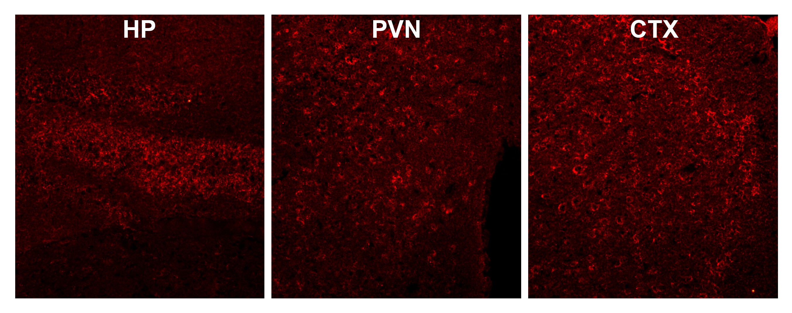
Immunohistochemistry: CaM Kinase II alpha Antibody [NBP1-51945]
Immunohistochemistry: CaM Kinase II alpha Antibody [NBP1-51945] - IHC using CamKII antibody from goat in 3% PFA perfused mouse brain. 1:100 dilution clearly provides cytosolic CamK II immunoreactivity in whole brain regions, including the hippocampus (HP), cortex (CTX) and paraventricular region (PVN). Image from verified customer review.
Flow Cytometry: CaM Kinase II alpha Antibody [NBP1-51945] -
Flow Cytometry: CaM Kinase II alpha Antibody [NBP1-51945] - Flow cytometric analysis of paraformaldehyde fixed Kelly cells (blue line), permeabilized with 0.5% Triton. Primary incubation 1hr (10ug/ml) followed by Alexa Fluor 488 secondary antibody (1ug/ml). IgG control: Unimmunized goat IgG (black line) followed by Alexa Fluor 488 secondary antibody.
Immunocytochemistry/Immunofluorescence: CaM Kinase II alpha Antibody [NBP1-51945] -
Immunocytochemistry/Immunofluorescence: CaM Kinase II alpha Antibody [NBP1-51945] - Immunofluorescence analysis of paraformaldehyde fixed U251 cells, permeabilized with 0.15% Triton. Primary incubation 1hr (10ug/ml) followed by Alexa Fluor 488 secondary antibody (2ug/ml), showing membrane staining. The nuclear stain is DAPI (blue). Negative control: Unimmunized goat IgG (10ug/ml) followed by Alexa Fluor 488 secondary antibody (2ug/ml).
Immunohistochemistry-Paraffin: CaM Kinase II alpha Antibody [NBP1-51945] -
Immunohistochemistry-Paraffin: CaM Kinase II alpha Antibody [NBP1-51945] - (3.75ug/ml) staining of paraffin embedded Human Adrenal Gland. Steamed antigen retrieval with citrate buffer pH 6, AP-staining.
Immunohistochemistry-Paraffin: CaM Kinase II alpha Antibody [NBP1-51945] -
Immunohistochemistry-Paraffin: CaM Kinase II alpha Antibody [NBP1-51945] - (3.8ug/ml) staining of paraffin embedded Human Brain Cortex. Steamed antigen retrieval with citrate buffer pH 6, AP-staining.
Western Blot: CaM Kinase II alpha Antibody [NBP1-51945] -
Western Blot: CaM Kinase II alpha Antibody [NBP1-51945] - (0.3ug/ml) staining of Human Cerebellum (A), Mouse (B) and Rat (C) Brain lysate (35ug protein in RIPA buffer). Detected by chemiluminescence
Immunocytochemistry/Immunofluorescence: CaM Kinase II alpha Antibody [NBP1-51945] -
Immunocytochemistry/Immunofluorescence: CaM Kinase II alpha Antibody [NBP1-51945] - Immunofluorescence analysis of paraformaldehyde fixed U2OS cells, permeabilized with 0.15% Triton. Primary incubation 1hr (10ug/ml) followed by Alexa Fluor 488 secondary antibody (2ug/ml), showing membrane staining. The nuclear stain is DAPI (blue). Negative control: Unimmunized goat IgG (10ug/ml) followed by Alexa Fluor 488 secondary antibody (2ug/ml).Applications for CaM Kinase II alpha Antibody
Application
Recommended Usage
Flow Cytometry
10 ug/ml
Immunocytochemistry/ Immunofluorescence
10 ug/ml
Immunohistochemistry
3 - 6 ug/ml
Immunohistochemistry-Paraffin
3 - 6 ug/ml
Peptide ELISA
Detection limit 1:64000
Western Blot
0.3-1 ug/ml
Application Notes
WB: Approx. 60kDa band observed in Human Cerebellum and approx 50+60kDa in Mouse and Rat Brain lysates, corresponding to CAMK2alpha and CAMK2beta respectively (calculated MW of 55.3kDa according to Human NP_057065.2 and 54.1kDa according to Mouse NP_803126.1 and Rat NP_037052.1 subunit Alpha and 60.4kDa according to Human NP_001280099.1, Mouse : NP_031621.3 and Rat NP_068507.2 subunit beta).. These observed molecular weights are routinely observed by other sources.and were successfully blocked by incubation with the immunizing peptide. Primary incubation 1 hour at room temperature. IHC-P: Human brain cortex shows granular staining in the periphery of pyramydal cell bodies.
Reviewed Applications
Read 1 review rated 4 using NBP1-51945 in the following applications:
Flow Cytometry Panel Builder
Bio-Techne Knows Flow Cytometry
Save time and reduce costly mistakes by quickly finding compatible reagents using the Panel Builder Tool.
Advanced Features
- Spectra Viewer - Custom analysis of spectra from multiple fluorochromes
- Spillover Popups - Visualize the spectra of individual fluorochromes
- Antigen Density Selector - Match fluorochrome brightness with antigen density

Formulation, Preparation, and Storage
Purification
Immunogen affinity purified
Formulation
Tris saline (20 mM Tris pH 7.3, 150 mM NaCl), 0.5% BSA
Preservative
0.02% Sodium Azide
Concentration
0.5 mg/ml
Shipping
The product is shipped with polar packs. Upon receipt, store it immediately at the temperature recommended below.
Stability & Storage
Store at -20C. Avoid freeze-thaw cycles.
Background: CaM Kinase II alpha
Long Name
Calcium/Calmodulin-dependent Protein Kinase II alpha
Alternate Names
CAMK2A
Gene Symbol
CAMK2A
UniProt
Additional CaM Kinase II alpha Products
Product Documents for CaM Kinase II alpha Antibody
Certificate of Analysis
To download a Certificate of Analysis, please enter a lot or batch number in the search box below.
Product Specific Notices for CaM Kinase II alpha Antibody
This product is for research use only and is not approved for use in humans or in clinical diagnosis. Primary Antibodies are guaranteed for 1 year from date of receipt.
Related Research Areas
Citations for CaM Kinase II alpha Antibody
 Powered by Bioz
Customer Reviews for CaM Kinase II alpha Antibody (1)
4 out of 5
1 Customer Rating
Have you used CaM Kinase II alpha Antibody?
Submit a review and receive an Amazon gift card!
$25/€18/£15/$25CAN/¥2500 Yen for a review with an image
$10/€7/£6/$10CAN/¥1110 Yen for a review without an image
Submit a review
Customer Images
Showing
1
-
1 of
1 review
Showing All
Filter By:
-
Application: ImmunohistochemistrySample Tested: Mouse brainSpecies: MouseVerified Customer | Posted 04/30/2018IHC using CamKII antibody from goat in 3% PFA perfused mouse brain. 1:100 dilution clearly provides cytosolic CamK II immunoreactivity in whole brain regions, including the hippocampus (HP), cortex (CTX) and paraventricular region (PVN).

There are no reviews that match your criteria.
Protocols
Find general support by application which include: protocols, troubleshooting, illustrated assays, videos and webinars.
- 7-Amino Actinomycin D (7-AAD) Cell Viability Flow Cytometry Protocol
- Antigen Retrieval Protocol (PIER)
- Antigen Retrieval for Frozen Sections Protocol
- Appropriate Fixation of IHC/ICC Samples
- Cellular Response to Hypoxia Protocols
- Chromogenic IHC Staining of Formalin-Fixed Paraffin-Embedded (FFPE) Tissue Protocol
- Chromogenic Immunohistochemistry Staining of Frozen Tissue
- ClariTSA™ Fluorophore Kits
- Detection & Visualization of Antibody Binding
- ELISA Sample Preparation & Collection Guide
- ELISA Troubleshooting Guide
- Extracellular Membrane Flow Cytometry Protocol
- Flow Cytometry Protocol for Cell Surface Markers
- Flow Cytometry Protocol for Staining Membrane Associated Proteins
- Flow Cytometry Staining Protocols
- Flow Cytometry Troubleshooting Guide
- Fluorescent IHC Staining of Frozen Tissue Protocol
- Graphic Protocol for Heat-induced Epitope Retrieval
- Graphic Protocol for the Preparation and Fluorescent IHC Staining of Frozen Tissue Sections
- Graphic Protocol for the Preparation and Fluorescent IHC Staining of Paraffin-embedded Tissue Sections
- Graphic Protocol for the Preparation of Gelatin-coated Slides for Histological Tissue Sections
- How to Run an R&D Systems DuoSet ELISA
- How to Run an R&D Systems Quantikine ELISA
- How to Run an R&D Systems Quantikine™ QuicKit™ ELISA
- ICC Cell Smear Protocol for Suspension Cells
- ICC Immunocytochemistry Protocol Videos
- ICC for Adherent Cells
- IHC Sample Preparation (Frozen sections vs Paraffin)
- Immunocytochemistry (ICC) Protocol
- Immunocytochemistry Troubleshooting
- Immunofluorescence of Organoids Embedded in Cultrex Basement Membrane Extract
- Immunofluorescent IHC Staining of Formalin-Fixed Paraffin-Embedded (FFPE) Tissue Protocol
- Immunohistochemistry (IHC) and Immunocytochemistry (ICC) Protocols
- Immunohistochemistry Frozen Troubleshooting
- Immunohistochemistry Paraffin Troubleshooting
- Intracellular Flow Cytometry Protocol Using Alcohol (Methanol)
- Intracellular Flow Cytometry Protocol Using Detergents
- Intracellular Nuclear Staining Flow Cytometry Protocol Using Detergents
- Intracellular Staining Flow Cytometry Protocol Using Alcohol Permeabilization
- Intracellular Staining Flow Cytometry Protocol Using Detergents to Permeabilize Cells
- Preparing Samples for IHC/ICC Experiments
- Preventing Non-Specific Staining (Non-Specific Binding)
- Primary Antibody Selection & Optimization
- Propidium Iodide Cell Viability Flow Cytometry Protocol
- Protocol for Heat-Induced Epitope Retrieval (HIER)
- Protocol for Liperfluo
- Protocol for Making a 4% Formaldehyde Solution in PBS
- Protocol for VisUCyte™ HRP Polymer Detection Reagent
- Protocol for the Characterization of Human Th22 Cells
- Protocol for the Characterization of Human Th9 Cells
- Protocol for the Fluorescent ICC Staining of Cell Smears - Graphic
- Protocol for the Fluorescent ICC Staining of Cultured Cells on Coverslips - Graphic
- Protocol for the Preparation & Fixation of Cells on Coverslips
- Protocol for the Preparation and Chromogenic IHC Staining of Frozen Tissue Sections
- Protocol for the Preparation and Chromogenic IHC Staining of Frozen Tissue Sections - Graphic
- Protocol for the Preparation and Chromogenic IHC Staining of Paraffin-embedded Tissue Sections
- Protocol for the Preparation and Chromogenic IHC Staining of Paraffin-embedded Tissue Sections - Graphic
- Protocol for the Preparation and Fluorescent ICC Staining of Cells on Coverslips
- Protocol for the Preparation and Fluorescent ICC Staining of Non-adherent Cells
- Protocol for the Preparation and Fluorescent ICC Staining of Stem Cells on Coverslips
- Protocol for the Preparation and Fluorescent IHC Staining of Frozen Tissue Sections
- Protocol for the Preparation and Fluorescent IHC Staining of Paraffin-embedded Tissue Sections
- Protocol for the Preparation of Gelatin-coated Slides for Histological Tissue Sections
- Protocol for the Preparation of a Cell Smear for Non-adherent Cell ICC - Graphic
- Protocol: Annexin V and PI Staining by Flow Cytometry
- Protocol: Annexin V and PI Staining for Apoptosis by Flow Cytometry
- Quantikine HS ELISA Kit Assay Principle, Alkaline Phosphatase
- Quantikine HS ELISA Kit Principle, Streptavidin-HRP Polymer
- R&D Systems Quality Control Western Blot Protocol
- Sandwich ELISA (Colorimetric) – Biotin/Streptavidin Detection Protocol
- Sandwich ELISA (Colorimetric) – Direct Detection Protocol
- TUNEL and Active Caspase-3 Detection by IHC/ICC Protocol
- The Importance of IHC/ICC Controls
- Troubleshooting Guide: ELISA
- Troubleshooting Guide: Fluorokine Flow Cytometry Kits
- Troubleshooting Guide: Immunohistochemistry
- Troubleshooting Guide: Western Blot Figures
- Western Blot Conditions
- Western Blot Protocol
- Western Blot Protocol for Cell Lysates
- Western Blot Troubleshooting
- Western Blot Troubleshooting Guide
- View all Protocols, Troubleshooting, Illustrated assays and Webinars
Loading...
Associated Pathways

