Carbonic Anhydrase II/CA2 Antibody - BSA Free
Novus Biologicals | Catalog # NB600-919
![Western Blot: Carbonic Anhydrase II/CA2 Antibody [NB600-919]](https://resources.rndsystems.com/images/products/Carbonic-Anhydrase-II-CA2-Antibody-Western-Blot-NB600-919-img0008.jpg "Western Blot: Carbonic Anhydrase II/CA2 Antibody [NB600-919]")
Key Product Details
Species Reactivity
Validated:
Cited:
Applications
Validated:
Cited:
Label
Antibody Source
Format
Product Specifications
Immunogen
Reactivity Notes
Localization
Specificity
Clonality
Host
Isotype
Description
Store vial at 4C prior to restoration. For extended storage aliquot contents and freeze at -20C or below. Avoid cycles of freezing and thawing. Centrifuge product if not completely clear after standing at room temperature. This product is stable for several weeks at 4C as an undiluted liquid. Dilute only prior to immediate use.
Scientific Data Images for Carbonic Anhydrase II/CA2 Antibody - BSA Free
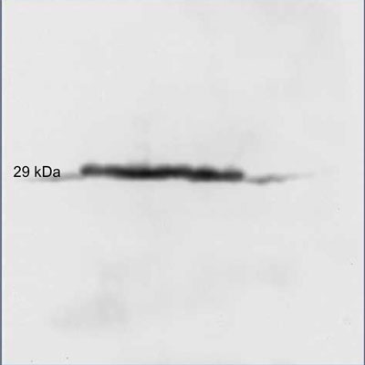
Western Blot: Carbonic Anhydrase II/CA2 Antibody [NB600-919]
Carbonic-Anhydrase-II-CA2-Antibody-Western-Blot-NB600-919-img0008.jpg![Immunohistochemistry: Carbonic Anhydrase II/CA2 Antibody [NB600-919]](https://resources.rndsystems.com/images/products/Carbonic-Anhydrase-II-CA2-Antibody-Immunohistochemistry-NB600-919-img0005.jpg "Immunohistochemistry: Carbonic Anhydrase II/CA2 Antibody [NB600-919]")
Immunohistochemistry: Carbonic Anhydrase II/CA2 Antibody [NB600-919]
Immunohistochemistry: Carbonic Anhydrase II/CA2 Antibody [NB600-919] - Staining in nucleus and cytoplasm of proximal and distal tubules, bowman's capsule and glomerular podocytes of human kidney (B) and in basal cells of retina of a mouse eye (C). Formalin fixed/paraffin embedded sections were subjected to heat induced epitope retrieval (HIER) at pH 6.2 and then incubated with rabbit anti-carbonic anhydrase II antibody at 4.0 ug/ml for 60 minutes. The reaction was developed using either MACH 1 universal HRP polymer detection (human kidney) or MACH 4 universal AP polymer detection system (mouse eye) and visualized with 3'3-diamino-benzidine substrate (DAB) or WARP RED.![Western Blot: Carbonic Anhydrase II/CA2 Antibody [NB600-919]](https://resources.rndsystems.com/images/products/Carbonic-Anhydrase-II-CA2-Antibody-Western-Blot-NB600-919-img0001.jpg "Western Blot: Carbonic Anhydrase II/CA2 Antibody [NB600-919]")
Western Blot: Carbonic Anhydrase II/CA2 Antibody [NB600-919]
Western Blot: Carbonic Anhydrase II/CA2 Antibody [NB600-919] - Detection of Carbonic Anhydrase II in mouse embryonic whole cell lysate. Image provided via product review by verified customer.![Western Blot: Carbonic Anhydrase II/CA2 Antibody [NB600-919]](https://resources.rndsystems.com/images/products/Carbonic-Anhydrase-II-CA2-Antibody-Western-Blot-NB600-919-img0002.jpg "Western Blot: Carbonic Anhydrase II/CA2 Antibody [NB600-919]")
Western Blot: Carbonic Anhydrase II/CA2 Antibody [NB600-919]
Western Blot: Carbonic Anhydrase II/CA2 Antibody [NB600-919] - analysis of Carbonic Anhydrase II protein (loading amount: 50 ng) using rabbit anti-Carbonic Anhydrase II antibody (NB600-919) at a dilution of 1:1000, incubation - overnight at 4C.![Western Blot: Carbonic Anhydrase II/CA2 Antibody [NB600-919]](https://resources.rndsystems.com/images/products/Carbonic-Anhydrase-II-CA2-Antibody-Western-Blot-NB600-919-img0004.jpg "Western Blot: Carbonic Anhydrase II/CA2 Antibody [NB600-919]")
Western Blot: Carbonic Anhydrase II/CA2 Antibody [NB600-919]
Western Blot: Carbonic Anhydrase II/CA2 Antibody [NB600-919] - Analysis using the Biotin conjugate of NB600-919. Detection of Lane 1: Carbonic Anhydrase II. Load: 50 ng per lane. Primary antibody: Rabbit anti-Carbonic Anhydrase II Antibody Biotin Conjugated at 1:1,000 overnight at 4C. Secondary antibody: HRP streptav![Western Blot: Carbonic Anhydrase II/CA2 Antibody [NB600-919]](https://resources.rndsystems.com/images/products/Carbonic-Anhydrase-II-CA2-Antibody-Western-Blot-NB600-919-img0007.jpg "Western Blot: Carbonic Anhydrase II/CA2 Antibody [NB600-919]")
Western Blot: Carbonic Anhydrase II/CA2 Antibody [NB600-919]
Western Blot: Carbonic Anhydrase II/CA2 Antibody [NB600-919] - Lane 1: Carbonic Anhydrase II. Lane 2: None. Load: 50 ng per lane. Primary antibody: Carbonic Anhydrase II primary antibody at 1:1,000 overnight at 4C. Secondary antibody: Peroxidase rabbit secondary antibody at 1:40,000 for 30 min at RT. Blocking: incubated with blocking buffer for 30 min at RT. Predicted/Observed size: 29 kDa, 29 kDa for Carbonic Anhydrase II. Other band(s): None.![Immunohistochemistry-Paraffin: Carbonic Anhydrase II/CA2 Antibody [NB600-919]](https://resources.rndsystems.com/images/products/Carbonic-Anhydrase-II-CA2-Antibody-Immunohistochemistry-Paraffin-NB600-919-img0003.jpg "Immunohistochemistry-Paraffin: Carbonic Anhydrase II/CA2 Antibody [NB600-919]")
Immunohistochemistry-Paraffin: Carbonic Anhydrase II/CA2 Antibody [NB600-919]
Immunohistochemistry-Paraffin: Carbonic Anhydrase II/CA2 Antibody [NB600-919] - Localization of CAII in mouse peritoneum. Image from verified customer review.
Carbonic Anhydrase II/CA2 Antibody
Immunohistochemistry with anti-carbonic anhydrase II antibody showing carbonic anhydrase II staining in nucleus and cytoplasm of proximal and distal tubules, bowman's capsule and glomerular podocytes of human kidney (B) and in basal cells of retina of a mouse eye (C). Formalin fixed/paraffin embedded sections were subjected to heat induced epitope retrieval (HIER) at pH 6.2 and then incubated with rabbit anti-carbonic anhydrase II antibody at 4.0 ug/ml for 60 minutes. The reaction was developed using either MACH 1 universal HRP polymer detection (human kidney) or MACH 4 universal AP polymer detection system (mouse eye) and visualized with 3'3-diamino-benzidine substrate (DAB) or WARP RED.Applications for Carbonic Anhydrase II/CA2 Antibody - BSA Free
ELISA
Immunohistochemistry
Immunohistochemistry-Frozen
Immunohistochemistry-Paraffin
Simple Western
Western Blot
Use in Immunohistochemistry-Frozen reported in scientific literature (PMID
See Simple Western Antibody Database for Simple Western validation: Tested in Blood, separated by Size, antibody dilution of 1:400
Reviewed Applications
Read 3 reviews rated 5 using NB600-919 in the following applications:
Formulation, Preparation, and Storage
Purification
Reconstitution
Formulation
Format
Preservative
Concentration
Shipping
Stability & Storage
Calculators
Background: Carbonic Anhydrase II/CA2
Additional Carbonic Anhydrase II/CA2 Products
Product Documents for Carbonic Anhydrase II/CA2 Antibody - BSA Free
Certificate of Analysis
To download a Certificate of Analysis, please enter a lot or batch number in the search box below.
Product Specific Notices for Carbonic Anhydrase II/CA2 Antibody - BSA Free
This product is for research use only and is not approved for use in humans or in clinical diagnosis. Primary Antibodies are guaranteed for 1 year from date of receipt.
Related Research Areas
Citations for Carbonic Anhydrase II/CA2 Antibody - BSA Free
 Powered by Bioz
Customer Reviews for Carbonic Anhydrase II/CA2 Antibody - BSA Free (3)
Have you used Carbonic Anhydrase II/CA2 Antibody - BSA Free?
Submit a review and receive an Amazon gift card!
$25/€18/£15/$25CAN/¥2500 Yen for a review with an image
$10/€7/£6/$10CAN/¥1110 Yen for a review without an image
Submit a review
Customer Images
-(5-mg)_NB600-919_10286.jpg)
-
Application: ImmunohistochemistrySample Tested: Mouse peritoneumSpecies: MouseVerified Customer | Posted 09/23/2014localization of CAII in mouse peritoneum

-
Application: Western BlotSample Tested: Embryonic gestational day 14.5 mouse whole embryo lysatesSpecies: MouseVerified Customer | Posted 06/24/2013Western blot analysis of mouse embryonic whole cell lysates
-
Application: Western BlotSample Tested: Detection of CA11 in human cancer cellsSpecies: HumanVerified Customer | Posted 10/06/2011
There are no reviews that match your criteria.
Protocols
Find general support by application which include: protocols, troubleshooting, illustrated assays, videos and webinars.
- Antigen Retrieval Protocol (PIER)
- Antigen Retrieval for Frozen Sections Protocol
- Appropriate Fixation of IHC/ICC Samples
- Cellular Response to Hypoxia Protocols
- Chromogenic IHC Staining of Formalin-Fixed Paraffin-Embedded (FFPE) Tissue Protocol
- Chromogenic Immunohistochemistry Staining of Frozen Tissue
- ClariTSA™ Fluorophore Kits
- Detection & Visualization of Antibody Binding
- ELISA Sample Preparation & Collection Guide
- ELISA Troubleshooting Guide
- Fluorescent IHC Staining of Frozen Tissue Protocol
- Graphic Protocol for Heat-induced Epitope Retrieval
- Graphic Protocol for the Preparation and Fluorescent IHC Staining of Frozen Tissue Sections
- Graphic Protocol for the Preparation and Fluorescent IHC Staining of Paraffin-embedded Tissue Sections
- Graphic Protocol for the Preparation of Gelatin-coated Slides for Histological Tissue Sections
- How to Run an R&D Systems DuoSet ELISA
- How to Run an R&D Systems Quantikine ELISA
- How to Run an R&D Systems Quantikine™ QuicKit™ ELISA
- IHC Sample Preparation (Frozen sections vs Paraffin)
- Immunofluorescent IHC Staining of Formalin-Fixed Paraffin-Embedded (FFPE) Tissue Protocol
- Immunohistochemistry (IHC) and Immunocytochemistry (ICC) Protocols
- Immunohistochemistry Frozen Troubleshooting
- Immunohistochemistry Paraffin Troubleshooting
- Preparing Samples for IHC/ICC Experiments
- Preventing Non-Specific Staining (Non-Specific Binding)
- Primary Antibody Selection & Optimization
- Protocol for Heat-Induced Epitope Retrieval (HIER)
- Protocol for Making a 4% Formaldehyde Solution in PBS
- Protocol for VisUCyte™ HRP Polymer Detection Reagent
- Protocol for the Preparation & Fixation of Cells on Coverslips
- Protocol for the Preparation and Chromogenic IHC Staining of Frozen Tissue Sections
- Protocol for the Preparation and Chromogenic IHC Staining of Frozen Tissue Sections - Graphic
- Protocol for the Preparation and Chromogenic IHC Staining of Paraffin-embedded Tissue Sections
- Protocol for the Preparation and Chromogenic IHC Staining of Paraffin-embedded Tissue Sections - Graphic
- Protocol for the Preparation and Fluorescent IHC Staining of Frozen Tissue Sections
- Protocol for the Preparation and Fluorescent IHC Staining of Paraffin-embedded Tissue Sections
- Protocol for the Preparation of Gelatin-coated Slides for Histological Tissue Sections
- Quantikine HS ELISA Kit Assay Principle, Alkaline Phosphatase
- Quantikine HS ELISA Kit Principle, Streptavidin-HRP Polymer
- R&D Systems Quality Control Western Blot Protocol
- Sandwich ELISA (Colorimetric) – Biotin/Streptavidin Detection Protocol
- Sandwich ELISA (Colorimetric) – Direct Detection Protocol
- TUNEL and Active Caspase-3 Detection by IHC/ICC Protocol
- The Importance of IHC/ICC Controls
- Troubleshooting Guide: ELISA
- Troubleshooting Guide: Immunohistochemistry
- Troubleshooting Guide: Western Blot Figures
- Western Blot Conditions
- Western Blot Protocol
- Western Blot Protocol for Cell Lysates
- Western Blot Troubleshooting
- Western Blot Troubleshooting Guide
- View all Protocols, Troubleshooting, Illustrated assays and Webinars
FAQs for Carbonic Anhydrase II/CA2 Antibody - BSA Free
-
Q: I am going to perform CA2 / Carbonic Anhydrase II immunohistochemistry staining for bone samples. Is this antibody better for plastic embedded bone samples? Or does it only work on paraffin embedded samples or soft tissue?
A: NB600-919 has only been tested on paraffin-embedded tissues. It will most likely work on your samples, but it has never been tested in this application specifically. From our catalog, this antibody is the best choice for your experiments.