Carbonic Anhydrase IX/CA9 Antibody (2D3) - BSA Free
Novus Biologicals | Catalog # NBP1-51691
Key Product Details
Species Reactivity
Validated:
Human, Mouse
Cited:
Human, Mouse
Applications
Validated:
Immunohistochemistry, Immunohistochemistry-Paraffin, Western Blot, ELISA, Flow Cytometry, Flow (Intracellular), Immunocytochemistry/ Immunofluorescence, CyTOF-ready
Cited:
Immunohistochemistry-Frozen, Western Blot, Immunocytochemistry/ Immunofluorescence, IF/IHC
Label
Unconjugated
Antibody Source
Monoclonal Mouse IgG1 Clone # 2D3
Format
BSA Free
Loading...
Product Specifications
Immunogen
This Carbonic Anhydrase IX/CA9 Antibody (2D3) was made to a purified recombinant fragment of human Carbonic Anhydrase IX expressed in E. coli [UniProt# Q16790].
Reactivity Notes
Mouse reactivity reported in scientific literature (PMID: 33153038).
Marker
Hypoxia Marker
Clonality
Monoclonal
Host
Mouse
Isotype
IgG1
Theoretical MW
50 kDa.
Disclaimer note: The observed molecular weight of the protein may vary from the listed predicted molecular weight due to post translational modifications, post translation cleavages, relative charges, and other experimental factors.
Disclaimer note: The observed molecular weight of the protein may vary from the listed predicted molecular weight due to post translational modifications, post translation cleavages, relative charges, and other experimental factors.
Scientific Data Images for Carbonic Anhydrase IX/CA9 Antibody (2D3) - BSA Free
![Western Blot: Carbonic Anhydrase IX/CA9 Antibody (2D3)BSA Free [NBP1-51691]](https://resources.rndsystems.com/images/products/Carbonic-Anhydrase-IX-CA9-Antibody-2D3-Western-Blot-NBP1-51691-img0006.jpg "Western Blot: Carbonic Anhydrase IX/CA9 Antibody (2D3)BSA Free [NBP1-51691]")
Western Blot: Carbonic Anhydrase IX/CA9 Antibody (2D3)BSA Free [NBP1-51691]
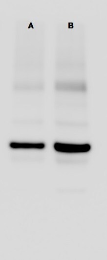
Western Blot: Carbonic Anhydrase IX/CA9 Antibody (2D3) [NBP1-51691] - Carbonic Anhydrase IX mouse antibody against Hela (1) and A549 (2) cell lysates. Bands were detected at a molecular weight of approximately 50 kDa in both cell lines.![Immunohistochemistry-Paraffin: Carbonic Anhydrase IX/CA9 Antibody (2D3) - BSA Free [NBP1-51691]](https://resources.rndsystems.com/images/products/Carbonic-Anhydrase-IX-CA9-Antibody-2D3-Immunohistochemistry-Paraffin-NBP1-51691-img0005.jpg "Immunohistochemistry-Paraffin: Carbonic Anhydrase IX/CA9 Antibody (2D3) - BSA Free [NBP1-51691]")
Immunohistochemistry-Paraffin: Carbonic Anhydrase IX/CA9 Antibody (2D3) - BSA Free [NBP1-51691]
Immunohistochemistry-Paraffin: Carbonic Anhydrase IX/CA9 Antibody (2D3) [NBP1-51691] - Paraffin-embedded lung tissues (left) and colonic tissues (right) using Carbonic Anhydrase IX mouse antibody with DAB staining.![Immunocytochemistry/ Immunofluorescence: Carbonic Anhydrase IX/CA9 Antibody (2D3) - BSA Free [NBP1-51691]](https://resources.rndsystems.com/images/products/Carbonic-Anhydrase-IX-CA9-Antibody-2D3-BSA-Free-Immunocytochemistry-Immunofluorescence-NBP1-51691-img0012.jpg "Immunocytochemistry/ Immunofluorescence: Carbonic Anhydrase IX/CA9 Antibody (2D3) - BSA Free [NBP1-51691]")
Immunocytochemistry/ Immunofluorescence: Carbonic Anhydrase IX/CA9 Antibody (2D3) - BSA Free [NBP1-51691]
Immunocytochemistry/Immunofluorescence: Carbonic Anhydrase IX/CA9 Antibody (2D3) - BSA Free [NBP1-51691] - A431 cells were fixed in 4% paraformaldehyde for 10 minutes and permeabilized in 0.05% Triton X-100 in PBS for 5 minutes. The cells were incubated with Carbonic Anhydrase IX/CA9 Antibody [2D3] (NBP1-51691) at 2ug/ml overnight at 4C and detected with an anti-mouse DyLight 488 (Green) at a 1:1000 dilution for 60 minutes. Nuclei were counterstained with DAPI (Blue). Cells were imaged using a 100X objective and digitally deconvolved.![Flow Cytometry: Carbonic Anhydrase IX/CA9 Antibody (2D3) - BSA Free [NBP1-51691]](https://resources.rndsystems.com/images/products/Carbonic-Anhydrase-IX-CA9-Antibody-2D3-Flow-Cytometry-NBP1-51691-img0008.jpg "Flow Cytometry: Carbonic Anhydrase IX/CA9 Antibody (2D3) - BSA Free [NBP1-51691]")
Flow Cytometry: Carbonic Anhydrase IX/CA9 Antibody (2D3) - BSA Free [NBP1-51691]
Flow Cytometry: Carbonic Anhydrase IX/CA9 Antibody (2D3) [NBP1-51691] - A surface stain was performed on A431 cells with Carbonic Anhydrase IX/CA9 NBP1-51691 (blue) and a matched isotype control NBP2-27287 (orange). Cells were incubated in an antibody dilution of 1 ug/mL for 20 minutes at room temperature, followed by DyLight488-conjugated anti-mouse secondary antibody.![Immunocytochemistry/ Immunofluorescence: Carbonic Anhydrase IX/CA9 Antibody (2D3) - BSA Free [NBP1-51691]](https://resources.rndsystems.com/images/products/Carbonic-Anhydrase-IX-CA9-Antibody-2D3-Immunocytochemistry-Immunofluorescence-NBP1-51691-img0011.jpg "Immunocytochemistry/ Immunofluorescence: Carbonic Anhydrase IX/CA9 Antibody (2D3) - BSA Free [NBP1-51691]")
Immunocytochemistry/ Immunofluorescence: Carbonic Anhydrase IX/CA9 Antibody (2D3) - BSA Free [NBP1-51691]
Immunocytochemistry/Immunofluorescence: Carbonic Anhydrase IX/CA9 Antibody (2D3) [NBP1-51691] - Mouse choroid stained with anti-Carbonic Anhydrase IX antibody. ICC/IF image submitted by a verified customer review.![Flow Cytometry: Carbonic Anhydrase IX/CA9 Antibody (2D3) - BSA Free [NBP1-51691]](https://resources.rndsystems.com/images/products/Carbonic-Anhydrase-IX-CA9-Antibody-2D3-Flow-Cytometry-NBP1-51691-img0007.jpg "Flow Cytometry: Carbonic Anhydrase IX/CA9 Antibody (2D3) - BSA Free [NBP1-51691]")
Flow Cytometry: Carbonic Anhydrase IX/CA9 Antibody (2D3) - BSA Free [NBP1-51691]
Flow Cytometry: Carbonic Anhydrase IX/CA9 Antibody (2D3) [NBP1-51691] - Analysis of NTERA-2 cells using Carbonic Anhydrase IX mouse mAb (green) and negative control (purple).Applications for Carbonic Anhydrase IX/CA9 Antibody (2D3) - BSA Free
Application
Recommended Usage
ELISA
1:10000
Flow (Intracellular)
1 ug/mL
Flow Cytometry
1:200 - 1:400
Immunohistochemistry
1:10 - 1:500
Immunohistochemistry-Paraffin
1:200 - 1:1000
Western Blot
1:2000
Reviewed Applications
Read 2 reviews rated 5 using NBP1-51691 in the following applications:
Flow Cytometry Panel Builder
Bio-Techne Knows Flow Cytometry
Save time and reduce costly mistakes by quickly finding compatible reagents using the Panel Builder Tool.
Advanced Features
- Spectra Viewer - Custom analysis of spectra from multiple fluorochromes
- Spillover Popups - Visualize the spectra of individual fluorochromes
- Antigen Density Selector - Match fluorochrome brightness with antigen density

Formulation, Preparation, and Storage
Purification
Protein A or G purified
Formulation
PBS
Format
BSA Free
Preservative
0.05% Sodium Azide
Concentration
1.0 mg/ml
Shipping
The product is shipped with polar packs. Upon receipt, store it immediately at the temperature recommended below.
Stability & Storage
Store at 4C short term. Aliquot and store at -20C long term. Avoid freeze-thaw cycles.
Background: Carbonic Anhydrase IX/CA9
Carbonic anhydrase IX (theoretical molecular weight 50kDa) belongs to the monomeric alpha class and is a single pass-transmembrane protein with two extracellular domains which serve catalytic and cell adhesion functions (2, 3). By cooperating with sodium bicarbonate cotransporters (NBC), lactate and proton exporting monocarboxylic acid transporters (MCT), and a sodium/hydrogen exchanger (NHE), carbonic anhydrase IX is involved in pH regulation across the cell membrane. This functional property protects cancer cells from intracellular acidification and partly explains the role of carbonic anhydrase IX in cancer cell survival and proliferation. In contrast, the pH regulating activity of carbonic anhydrase IX induces extracellular acidification, which has been implicated in epithelial to mesenchymal transition (EMT) and promoting cancer invasion. Carbonic anhydrase IX is frequently overexpressed in cancer cells (e.g., colorectal-, breast-, lung-carcinoma and brain tumors), an effect promoted by hypoxia within the tumor microenvironment (4). An exception are tumors carrying pVHL inactivating mutations, such as clear cell renal cell carcinoma (ccRCC), where HIF-alpha is stabilized due to dysfunctional proteasomal targeting and can induce HRE (Hypoxia Response Element) containing genes even under physiological normoxia (5). Carbonic anhydrase IX may be detected by immunostaining in tumors, which is found in association with necrotic tissue and metastatic cells. Because the expression of carbonic anhydrase IX correlates with both tumor grade and stage, analysis of its expression in tumors serves as a prognostic factor (4, 6).
References
1. Tripp, B. C., Smith, K., & Ferry, J. G. (2001). Carbonic Anhydrase: New Insights for an Ancient Enzyme. Journal of Biological Chemistry. https://doi.org/10.1074/jbc.R100045200
2. Nishimori, I., & Onishi, S. (2001). Carbonic anhydrase isozymes in the human pancreas. Digestive and Liver Disease. https://doi.org/10.1016/s1590-8658(01)80138-9
3. Zavadova, Z., & Zavada, J. (2005). Carbonic anhydrase IX (CA IX) mediates tumor cell interactions with microenvironment. Oncology Reports. https://doi.org/10.3892/or.13.5.977
4. Pastorekova, S., & Gillies, R. J. (2019). The role of carbonic anhydrase IX in cancer development: links to hypoxia, acidosis, and beyond. Cancer and Metastasis Reviews. https://doi.org/10.1007/s10555-019-09799-0
5. Haase, V. (2009). The VHL Tumor Suppressor: Master Regulator of HIF. Current Pharmaceutical Design. https://doi.org/10.2174/138161209789649394
6. Young, J. R., Coy, H., Kim, H. J., Douek, M., Sisk, A., Pantuck, A. J., & Raman, S. S. (2018). Association of the gross appearance of intratumoral vascularity at MDCT with the carbonic anhydrase IX score in clear cell renal cell carcinoma. American Journal of Roentgenology. https://doi.org/10.2214/AJR.18.19725
Additional Carbonic Anhydrase IX/CA9 Products
Product Documents for Carbonic Anhydrase IX/CA9 Antibody (2D3) - BSA Free
Certificate of Analysis
To download a Certificate of Analysis, please enter a lot or batch number in the search box below.
Product Specific Notices for Carbonic Anhydrase IX/CA9 Antibody (2D3) - BSA Free
This product is for research use only and is not approved for use in humans or in clinical diagnosis. Primary Antibodies are guaranteed for 1 year from date of receipt.
Related Research Areas
Citations for Carbonic Anhydrase IX/CA9 Antibody (2D3) - BSA Free
 Powered by Bioz
Customer Reviews for Carbonic Anhydrase IX/CA9 Antibody (2D3) - BSA Free (2)
5 out of 5
2 Customer Ratings
Have you used Carbonic Anhydrase IX/CA9 Antibody (2D3) - BSA Free?
Submit a review and receive an Amazon gift card!
$25/€18/£15/$25CAN/¥2500 Yen for a review with an image
$10/€7/£6/$10CAN/¥1110 Yen for a review without an image
Submit a review
Customer Images
Showing
1
-
2 of
2 reviews
Showing All
Filter By:
-
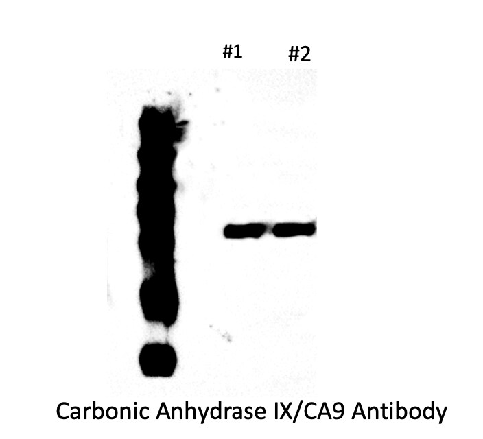
Application: Western BlotSample Tested: HEK293Species: HumanVerified Customer | Posted 07/05/2021Carbonic Anhydrase IX/CA9 Antibody #1: HEK293 #2: HEK293

-
Application: Western BlotSample Tested: MCF-7 whole cell lysateSpecies: HumanVerified Customer | Posted 11/20/2015
There are no reviews that match your criteria.
Protocols
Find general support by application which include: protocols, troubleshooting, illustrated assays, videos and webinars.
- 7-Amino Actinomycin D (7-AAD) Cell Viability Flow Cytometry Protocol
- Antigen Retrieval Protocol (PIER)
- Antigen Retrieval for Frozen Sections Protocol
- Appropriate Fixation of IHC/ICC Samples
- Cellular Response to Hypoxia Protocols
- Chromogenic IHC Staining of Formalin-Fixed Paraffin-Embedded (FFPE) Tissue Protocol
- Chromogenic Immunohistochemistry Staining of Frozen Tissue
- ClariTSA™ Fluorophore Kits
- Detection & Visualization of Antibody Binding
- ELISA Sample Preparation & Collection Guide
- ELISA Troubleshooting Guide
- Extracellular Membrane Flow Cytometry Protocol
- Flow Cytometry Protocol for Cell Surface Markers
- Flow Cytometry Protocol for Staining Membrane Associated Proteins
- Flow Cytometry Staining Protocols
- Flow Cytometry Troubleshooting Guide
- Fluorescent IHC Staining of Frozen Tissue Protocol
- Graphic Protocol for Heat-induced Epitope Retrieval
- Graphic Protocol for the Preparation and Fluorescent IHC Staining of Frozen Tissue Sections
- Graphic Protocol for the Preparation and Fluorescent IHC Staining of Paraffin-embedded Tissue Sections
- Graphic Protocol for the Preparation of Gelatin-coated Slides for Histological Tissue Sections
- How to Run an R&D Systems DuoSet ELISA
- How to Run an R&D Systems Quantikine ELISA
- How to Run an R&D Systems Quantikine™ QuicKit™ ELISA
- ICC Cell Smear Protocol for Suspension Cells
- ICC Immunocytochemistry Protocol Videos
- ICC for Adherent Cells
- IHC Sample Preparation (Frozen sections vs Paraffin)
- Immunocytochemistry (ICC) Protocol
- Immunocytochemistry Troubleshooting
- Immunofluorescence of Organoids Embedded in Cultrex Basement Membrane Extract
- Immunofluorescent IHC Staining of Formalin-Fixed Paraffin-Embedded (FFPE) Tissue Protocol
- Immunohistochemistry (IHC) and Immunocytochemistry (ICC) Protocols
- Immunohistochemistry Frozen Troubleshooting
- Immunohistochemistry Paraffin Troubleshooting
- Intracellular Flow Cytometry Protocol Using Alcohol (Methanol)
- Intracellular Flow Cytometry Protocol Using Detergents
- Intracellular Nuclear Staining Flow Cytometry Protocol Using Detergents
- Intracellular Staining Flow Cytometry Protocol Using Alcohol Permeabilization
- Intracellular Staining Flow Cytometry Protocol Using Detergents to Permeabilize Cells
- Preparing Samples for IHC/ICC Experiments
- Preventing Non-Specific Staining (Non-Specific Binding)
- Primary Antibody Selection & Optimization
- Propidium Iodide Cell Viability Flow Cytometry Protocol
- Protocol for Heat-Induced Epitope Retrieval (HIER)
- Protocol for Liperfluo
- Protocol for Making a 4% Formaldehyde Solution in PBS
- Protocol for VisUCyte™ HRP Polymer Detection Reagent
- Protocol for the Characterization of Human Th22 Cells
- Protocol for the Characterization of Human Th9 Cells
- Protocol for the Fluorescent ICC Staining of Cell Smears - Graphic
- Protocol for the Fluorescent ICC Staining of Cultured Cells on Coverslips - Graphic
- Protocol for the Preparation & Fixation of Cells on Coverslips
- Protocol for the Preparation and Chromogenic IHC Staining of Frozen Tissue Sections
- Protocol for the Preparation and Chromogenic IHC Staining of Frozen Tissue Sections - Graphic
- Protocol for the Preparation and Chromogenic IHC Staining of Paraffin-embedded Tissue Sections
- Protocol for the Preparation and Chromogenic IHC Staining of Paraffin-embedded Tissue Sections - Graphic
- Protocol for the Preparation and Fluorescent ICC Staining of Cells on Coverslips
- Protocol for the Preparation and Fluorescent ICC Staining of Non-adherent Cells
- Protocol for the Preparation and Fluorescent ICC Staining of Stem Cells on Coverslips
- Protocol for the Preparation and Fluorescent IHC Staining of Frozen Tissue Sections
- Protocol for the Preparation and Fluorescent IHC Staining of Paraffin-embedded Tissue Sections
- Protocol for the Preparation of Gelatin-coated Slides for Histological Tissue Sections
- Protocol for the Preparation of a Cell Smear for Non-adherent Cell ICC - Graphic
- Protocol: Annexin V and PI Staining by Flow Cytometry
- Protocol: Annexin V and PI Staining for Apoptosis by Flow Cytometry
- Quantikine HS ELISA Kit Assay Principle, Alkaline Phosphatase
- Quantikine HS ELISA Kit Principle, Streptavidin-HRP Polymer
- R&D Systems Quality Control Western Blot Protocol
- Sandwich ELISA (Colorimetric) – Biotin/Streptavidin Detection Protocol
- Sandwich ELISA (Colorimetric) – Direct Detection Protocol
- TUNEL and Active Caspase-3 Detection by IHC/ICC Protocol
- The Importance of IHC/ICC Controls
- Troubleshooting Guide: ELISA
- Troubleshooting Guide: Fluorokine Flow Cytometry Kits
- Troubleshooting Guide: Immunohistochemistry
- Troubleshooting Guide: Western Blot Figures
- Western Blot Conditions
- Western Blot Protocol
- Western Blot Protocol for Cell Lysates
- Western Blot Troubleshooting
- Western Blot Troubleshooting Guide
- View all Protocols, Troubleshooting, Illustrated assays and Webinars
FAQs for Carbonic Anhydrase IX/CA9 Antibody (2D3) - BSA Free
Showing
1
-
3 of
3 FAQs
Showing All
-
Q: I am looking for a primary antibody to Carbonic Anhydrase 9 and I want an antibody that recognises an extracellular epitope so that I can use it for live cell flow cytometry. Would #NBP1-51691 be a suitable candidate?
A: The immunogen of our Carbonic Anhydrase IX antibody (clone 2D3) # NBP1-51691 corresponds to peptide from amino acids region between 40-290 of human CA9, which appears to be in the extracellular domain of this protein (based on UniProt: Q16790). In our QC lab, this antibody has been validated for FLOW application, so that you may use this antibody for your current experiments.
-
Q: We are looking for the antibody available for cell sorting by CA9. The sorted cells must be cultured for the expansion after the sorting for the following experiments. Do you have the antibodies useful for this?
A:
We sell two carbonic anhydrase IX antibodies that have been validated for Flow Cytometry detection of the human protein. These have catalogue numbers NBP1-51691 and NBP1-47688 and can be seen at the following link: carbonic anhydrase IX antibodies. Since both of these products contain sodium azide as a preservative, you are unlikely to be able to culture the cells on following sorting unless you performed a buffer exchange prior to using the antibody. Unfortunately we have no direct experience with either of these products of staining and subsequently expanding cell populations.
-
Q: What is the predicted theoretical molecular weight of Carbonic Anhydrase IX/CA9 antibodies?
A: The TMW for Carbonic Anhydrase IX/CA9 antibodies is 50 kDa. Please note the observed molecular weight of the protein may vary from the listed predicted molecular weight due to post translational modifications, post translation cleavages, relative charges, and other experimental factors.
-
Q: I am looking for a primary antibody to Carbonic Anhydrase 9 and I want an antibody that recognises an extracellular epitope so that I can use it for live cell flow cytometry. Would #NBP1-51691 be a suitable candidate?
A: The immunogen of our Carbonic Anhydrase IX antibody (clone 2D3) # NBP1-51691 corresponds to peptide from amino acids region between 40-290 of human CA9, which appears to be in the extracellular domain of this protein (based on UniProt: Q16790). In our QC lab, this antibody has been validated for FLOW application, so that you may use this antibody for your current experiments.
-
Q: We are looking for the antibody available for cell sorting by CA9. The sorted cells must be cultured for the expansion after the sorting for the following experiments. Do you have the antibodies useful for this?
A:
We sell two carbonic anhydrase IX antibodies that have been validated for Flow Cytometry detection of the human protein. These have catalogue numbers NBP1-51691 and NBP1-47688 and can be seen at the following link: carbonic anhydrase IX antibodies. Since both of these products contain sodium azide as a preservative, you are unlikely to be able to culture the cells on following sorting unless you performed a buffer exchange prior to using the antibody. Unfortunately we have no direct experience with either of these products of staining and subsequently expanding cell populations.
-
Q: What is the predicted theoretical molecular weight of Carbonic Anhydrase IX/CA9 antibodies?
A: The TMW for Carbonic Anhydrase IX/CA9 antibodies is 50 kDa. Please note the observed molecular weight of the protein may vary from the listed predicted molecular weight due to post translational modifications, post translation cleavages, relative charges, and other experimental factors.
Loading...