Carbonic Anhydrase IX/CA9 Antibody - BSA Free
Novus Biologicals | Catalog # NB100-417

Key Product Details
Validated by
Knockout/Knockdown, Orthogonal Validation, Biological Validation
Species Reactivity
Validated:
Human, Mouse, Rat, Plant
Cited:
Human, Mouse, Rat, Plant
Applications
Validated:
Immunohistochemistry, Immunohistochemistry-Paraffin, Immunohistochemistry-Frozen, Western Blot, Immunoblotting, ELISA, Flow Cytometry, Dual RNAscope ISH-IHC, Immunocytochemistry/ Immunofluorescence, Simple Western, Immunoprecipitation, Chromatin Immunoprecipitation (ChIP), Proximity Ligation Assay, Gel Super Shift Assays, Microarray
Cited:
Immunohistochemistry, Immunohistochemistry-Paraffin, Immunohistochemistry-Frozen, Western Blot, ELISA, Immunofluorescence, Immunocytochemistry/ Immunofluorescence, Chemotaxis, EMSA, Proximity Ligation Assay, IF/ICC, IF/IHC, Microarray, Westen Blot
Label
Unconjugated
Antibody Source
Polyclonal Rabbit IgG
Format
BSA Free
Loading...
Product Specifications
Immunogen
This Carbonic Anhydrase IX/CA9 Antibody was made from a synthetic peptide from the C-terminal sequence of human Carbonic Anhydrase IX (within residues 400-459) [UniProt# Q16790].
Localization
Membrane.
Marker
Hypoxia Marker
Clonality
Polyclonal
Host
Rabbit
Isotype
IgG
Theoretical MW
55 kDa.
Disclaimer note: The observed molecular weight of the protein may vary from the listed predicted molecular weight due to post translational modifications, post translation cleavages, relative charges, and other experimental factors.
Disclaimer note: The observed molecular weight of the protein may vary from the listed predicted molecular weight due to post translational modifications, post translation cleavages, relative charges, and other experimental factors.
Scientific Data Images for Carbonic Anhydrase IX/CA9 Antibody - BSA Free
Immunohistological Detection of Carbonic Anhydrase IX/CA9 in SCCNij202 Tumors in Mice
Carbonic-Anhydrase-IX-CA9-Antibody-Immunohistochemistry-NB100-417-img0047.jpg
Dual RNAscope ISH-IHC Analysis of Carbonic Anhydrase IX/CA9 in Paraffin Embedded Human Stomach
Formalin-fixed paraffin-embedded tissue sections of human stomach were probed for Carbonic Anhydrase IX/CA9 mRNA (ACD RNAScope Probe, catalog # 559348; Fast Red chromogen, ACD catalog # 322750). Adjacent tissue section was processed for immunohistochemistry using Rabbit Polyclonal (Novus catalog # NB100-417) at 1:1000 dilution with overnight incubation at 4 degrees Celsius followed by incubation with anti-rabbit IgG VisUCyte HRP Polymer Antibody (Catalog # VC003) and DAB chromogen (yellow-brown). Tissue was counterstained with hematoxylin (blue). Specific staining was localized to glandular cells.
Western Blot Detection of Carbonic Anhydrase IX/CA9 in Multiple Human Cell Lysates
Analysis on various human cell lysates. Specific bands were detected for Carbonic Anhydrase IX/CA9 in HEK-293 and A498 cell lines at a molecular weight of approximately 50 kDa. WB image submitted by a verified customer review.
Immunohistological Staining of Carbonic Anhydrase IX/CA9 in Endometrial Cancer
Carbonic-Anhydrase-IX-CA9-Antibody-Immunohistochemistry-NB100-417-img0040.jpg
Western Blot Detection of Carbonic Anhydrase IX/CA9 in Hypoxic and Normoxic HeLa Cells
Hypoxias impact on proteins of the mitochondrial ISC assembly machinery. (A) Immunoblotting analyzed the total protein extracts from HeLa cells grown in normoxia (Nx, 21% O2) or hypoxia (Hx, 1% O2) conditions using VDACs poly antibody and anti-CAIX, -ISCU, -FXN, -NFS1, -HSC20 antibodies. Beta-Actin was used as loading control. Citation: Ferecatu I, Canal F, Fabbri L, Mazure NM, Bouton C, Golinelli-Cohen M-P (2018) Dysfunction in the mitochondrial Fe-S assembly machinery leads to formation of the chemoresistant truncated VDAC1 isoform without HIF-1 alpha activation. PLoS ONE 13(3): e0194782.

Detection of Carbonic Anhydrase IX/CA9 in HeLa Cell Lysate by Simple Western
Simple Western lane view shows a specific band for CAIX in 0.1 mg/mL of HeLa lysate. This experiment was performed under reducing conditions using the 12-230 kDa separation system.
Flow Cytometry of U-87 MG Cells Stained with Carbonic Anhydrase IX/CA9 Antibody
An intracellular stain was performed on U-87 MG Cells with NB100-417 and a matched isotype control. Cells were fixed with 4% PFA and then permeablized with 0.1% saponin. Cells were incubated in an antibody dilution of 2.5 ug/mL for 30 minutes at room temperature, followed by Rabbit IgG APC-conjugated Secondary Antibody, (R&D Systems, F0111).
Western Blot Detection of Carbonic Anhydrase IX/CA9 in Human Breast Cancer MCF7 Cells
Western Blot analysis of Carbonic Anhydrase IX/CA9 Antibody on human breast cancer MCF7 cells. Image from verified customer review.
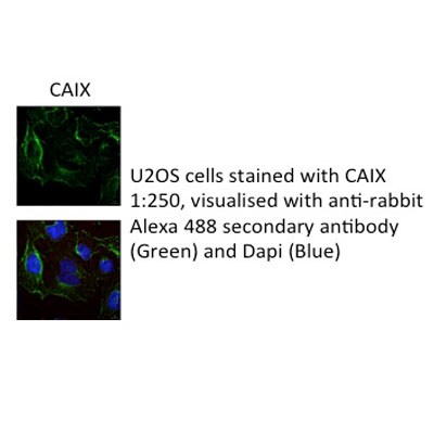
Immunocytochemistry/Immunofluorescence Staining of Carbonic Anhydrase IX/CA9 in A431 Cells
A431 cells were fixed in 4% paraformaldehyde for 10 minutes and permeabilized in 0.05% Triton X-100 in PBS for 5 minutes. The cells were incubated with anti-Carbonic Anhydrase IX/CA9 Antibody NB100-417 at 2 ug/ml overnight at 4C and detected with an anti-rabbit Dylight 488 (Green) at a 1:1000 dilution for 60 minutes. Nuclei were counterstained with DAPI (Blue). Cells were imaged using a 100X objective and digitally deconvolved.
Flow Cytometry of A431 Cells Stained with Carbonic Anhydrase IX/CA9 Antibody
An intracellular stain was performed on A431 cells with Carbonic Anhydrase IX/CA9 Antibody NB100-417 (blue) and a matched isotype control (orange). Cells were fixed with 4% PFA and then permeabilized with 0.1% saponin. Cells were incubated in an antibody dilution of 1.0 ug/mL for 30 minutes at room temperature, followed by Rabbit IgG (H+L) Cross-Adsorbed Secondary Antibody, Dylight 550 (SA5-10033, Thermo Fisher).
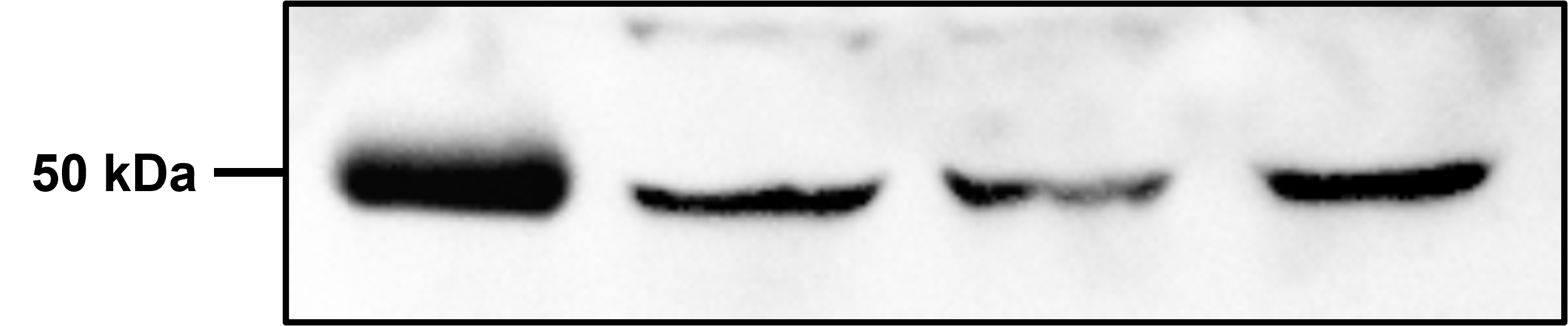
Western Blot Analysis of Carbonic Anhydrase IX/CA9 in Multiple Whole Cell Lysates
Analysis in 1) HeLa, 2) MDA-MB-231, and 3) A549 whole cell lysates. Specific bands were detected for Carbonic Anhydrase IX/CA9 at a molecular weight of 50 kDa.
Detection of Carbonic Anhydrase IX/CA9 in Rat Renal Cortex by Western Blot
Analysis on rat renal cortex. A specific band was detected at a molecular weight of approximately 50 kDa.
Staining of Carbonic Anhydrase IX/CA9 in CNHCs
Carbonic-Anhydrase-IX-CA9-Antibody-Immunocytochemistry-Immunofluorescence-NB100-417-img0039.jpg
Immunohistochemical Staining of Carbonic Anhydrase IX/CA9 in Frozen Mouse Xenografted Human Colon Carcinoma
Analysis of human colon carcinoma, xenografted in mice. IHC-Fr image submitted by a verified customer review.
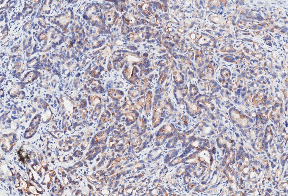
Immunohistochemical Staining of Carbonic Anhydrase IX/CA9 in Paraffin Embedded Human Breast Cancer
IHC analysis of a FFPE tissue section of human breast cancer using CAIX antibody at 1:1000 dilution. The primary antibody bound to CAIX antigens in the tissue section was detected using a HRP labeled secondary antibody and DAB reagent. Nuclei of the cells were counterstained with hematoxylin. This CAIX antibody generated an expected cytoplasmic staining of CAIX protein with an intense signal around the cellular membranes in tumor cores. The latter are more likely to be hypoxic in growing tumors which signifies that the observed CAIX staining is specific.
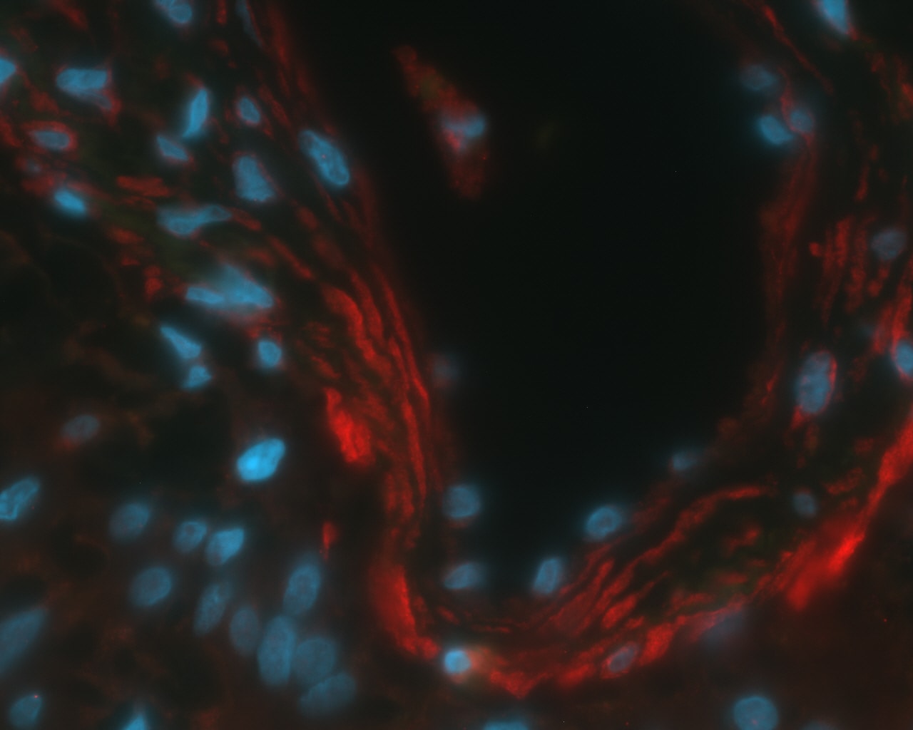
Immunohistochemical Staining of Carbonic Anhydrase IX/CA9 in Paraffin Embedded Human Stomach Tissue
Strong cytoplasmic and membranous CA9 staining in glandular cells of human stomach tissue. Citrate buffer pH 6 antigen retrieval. Antibody at 1:250 dilution and detected with an Alexa Fluor 546 labeled goat-anti-rabbit secondary antibody. Nuclei detected with DAPI. IHC-P image submitted by a verified customer review.
Immunohistological Detection of Carbonic Anhydrase IX/CA9 in Human RCC Tumor Cryosections
Immunofluorescence of human RCC tumor cryosections using NB100-417 (Panel A). Panel B shows staining with normal rabbit serum.
Immunohistochemical Staining of Carbonic Anhydrase IX/CA9 in Control and CB-PIC Treated Colorectal Cancer
Carbonic-Anhydrase-IX-CA9-Antibody-Immunohistochemistry-NB100-417-img0046.jpg
Western Blot Detection of Carbonic Anhydrase IX/CA9 in HeLa Cells Under Multiple Conditions and Treatments
Iron depletion and nitric oxide stress and Iron depletion induce the truncated VDAC1 form accumulation. (D) Total protein extracts were analyzed by immunoblotting using VDACs poly antibody and anti-HIF-1 alpha and -CAIX antibodies. (E) Immunoblotting was used to analyze total protein extracts using antibodies against the three VDAC isoforms. (F) HeLa cells were grown in hypoxia (Hx, 1% O2) conditions and transfected with iscu- or NC-siRNA for 3 days, or grown in normoxia (Nx, 21% O2), some treated with DFO for 16 h. Western Blots analyzed total proteins using VDACs poly antibody and anti-HIF-1 alpha, -CAIX, -ISCU antibodies. Citation: Ferecatu I, Canal F, Fabbri L, Mazure NM, Bouton C, Golinelli-Cohen M-P (2018) Dysfunction in the mitochondrial Fe-S assembly machinery leads to formation of the chemoresistant truncated VDAC1 isoform without HIF-1 alpha activation. PLoS ONE 13(3): e0194782.
Immunohistochemistry: Carbonic Anhydrase IX/CA9 Antibody - BSA Free [NB100-417] -
Immunohistochemical representative microphotographs representing the HIF-1 alpha, GLUT-1, and CAIX expression in endometrial cancer according to FIGO classification (IA, IB, II, IIIA, IIIC, and IV). Primary objective magnification 20x.
Immunohistochemistry: Carbonic Anhydrase IX/CA9 Antibody - BSA Free [NB100-417] -
The images are representative of immunohistochemistry staining of nuclear staining of proliferation marker Ki67, membrane protein CD 44, and CA IX. The images of scaffold culture (left panel) are compared with cell pellet paraffin section (right panel). The scaffold dissolved in xylene presented as a clear region marked ∗. All scale bars are 100 μm, magnification 40x.
Immunohistochemistry: Carbonic Anhydrase IX/CA9 Antibody - BSA Free [NB100-417] -
Chemosensitivity of the CTC SCLC lines (IC50, mean ± SD) and immunohistochemistry of sections of tumorospheres. All differences between single cells (SC) and tumorospheres (TS) are statistically significant. Immunohistochemical staining of sections of UHGc5, BHGc16 and BHGc26 CTCs was performed using antibodies directed to CD56, CHGA, CAIX and Ki67, respectively.
Western Blot: Carbonic Anhydrase IX/CA9 Antibody - BSA Free [NB100-417] -
Western Blot: Carbonic Anhydrase IX/CA9 Antibody - BSA Free [NB100-417] - Analysis of protein levels of genes with increased transcription in hypoxia, IOX2 & VH032.HIF targets were increased in hypoxia, VH298 & FG-4592. 0.05% DMSO (vehicle control), 1% O2 (hypoxia), 100 µM VH298 & 50 µM FG-4592 were introduced to (A) HeLa or HFF for 24 hours & (B) HeLa for indicated time. Protein levels were analysed by immunoblotting using antibodies against indicated proteins, with beta -Actin as loading control. The blots shown are representative of three independent experiments. * indicates longer exposure. Image collected & cropped by CiteAb from the following publication (https://pubmed.ncbi.nlm.nih.gov/30801039), licensed under a CC-BY license. Not internally tested by Novus Biologicals.
Immunohistochemistry-Paraffin: Carbonic Anhydrase IX/CA9 Antibody - BSA Free [NB100-417] -
Immunohistochemistry-Paraffin: Carbonic Anhydrase IX/CA9 Antibody - BSA Free [NB100-417] - IGF1R & CA9 expression in PDAC.(A) Representative staining of CA9 cells quantified on a scoring system of 0–3 according to staining intensity (original magnification ×200). CA9 was mainly expressed in the cell membrane of PDAC cells. (B) IGF1R was mainly expressed in the cytoplasm of PDAC cells. (C) Association between IGF1Rexpression & CA9 expression in 120 pancreas cancer. A significant correlation was found between IGF1R expression level was significantly correlated with CA9 expression in PDAC cells (p < 0.01, rs = 0.649; Spearman’s rank sum test). The size of circle indicates the number of PDAC patients. Image collected & cropped by CiteAb from the following publication (https://pubmed.ncbi.nlm.nih.gov/27487118), licensed under a CC-BY license. Not internally tested by Novus Biologicals.
Immunohistochemistry: Carbonic Anhydrase IX/CA9 Antibody - BSA Free [NB100-417] -
Immunohistochemistry: Carbonic Anhydrase IX/CA9 Antibody - BSA Free [NB100-417] - Representative human colorectal cancer specimen. Immunohistochemistry of CA9 & GGT1 in mLNs. In these cases, both CA9 & GGT1 were expressed, suggesting that GGT1 expression is related to hypoxia in mLNs. Scale bar, 500 μm. Black arrowheads: GGT of cancer cells, white arrowheads: GGT surrounding cancer cells, arrow: accumulation of GGT. mLNs: metastatic lymph nodes, GGT: gamma-glutamyl transpeptidase. Image collected & cropped by CiteAb from the following publication (https://pubmed.ncbi.nlm.nih.gov/30542087), licensed under a CC-BY license. Not internally tested by Novus Biologicals.
Immunohistochemistry: Carbonic Anhydrase IX/CA9 Antibody - BSA Free [NB100-417] -
Immunohistochemistry: Carbonic Anhydrase IX/CA9 Antibody - BSA Free [NB100-417] - Representative microscopy images of staining for hypoxia markers in prostate tissues (MO, 400×). A) HIF-1 alpha - notice the granular cytoplasmic immunoreactivity of the malignant epithelial cells. In this case, more than 50% of the glands stained. B) LOX - strong & diffuse nuclear immunoreactivity of the epithelial cells. C) CAIX - note a focal apical cytoplasmic immunoreactivity in epithelial cells. D) VEGFR2 - moderate nuclear & weak cytoplasmic expression of the epithelial cells Image collected & cropped by CiteAb from the following publication (https://pubmed.ncbi.nlm.nih.gov/28143503), licensed under a CC-BY license. Not internally tested by Novus Biologicals.
Immunohistochemistry: Carbonic Anhydrase IX/CA9 Antibody - BSA Free [NB100-417] -
Immunohistochemistry: Carbonic Anhydrase IX/CA9 Antibody - BSA Free [NB100-417] - The images are representative of immunohistochemistry staining of nuclear staining of proliferation marker Ki67, membrane protein CD 44, & CA IX. The images of scaffold culture (left panel) are compared with cell pellet paraffin section (right panel). The scaffold dissolved in xylene presented as a clear region marked ∗. All scale bars are 100 μm, magnification 40x. Image collected & cropped by CiteAb from the following publication (https://pubmed.ncbi.nlm.nih.gov/24101930), licensed under a CC-BY license. Not internally tested by Novus Biologicals.
Immunohistochemistry: Carbonic Anhydrase IX/CA9 Antibody - BSA Free [NB100-417] -
Immunohistochemistry: Carbonic Anhydrase IX/CA9 Antibody - BSA Free [NB100-417] - Hypoxia, blood vessel & proliferation staining. IHC staining in the Biopsy 23 & Biopsy 3 respectively for CA-9, EF5, CD31, Ki67 & LYVE1. 15× Magnification. Image collected & cropped by CiteAb from the following publication (http://www.mdpi.com/2072-6694/4/3/821), licensed under a CC-BY license. Not internally tested by Novus Biologicals.
Western Blot: Carbonic Anhydrase IX/CA9 Antibody - BSA Free [NB100-417] -
Western Blot: Carbonic Anhydrase IX/CA9 Antibody - BSA Free [NB100-417] - VH298 elicits a HIF-dependent hypoxic response in various cell lines.1% DMSO, hypoxia (1% O2) & indicated compounds at 100 μM were introduced to (a) HFF, HeLa, U2OS & (b) RCC4 cells expressing HA-tagged VHL (with the exception of cis-VH298 & VH298 used at 150 μM) for 24 h & (c) CTLs for 8 h. Protein levels were analysed by immunoblotting using antibodies against the indicated proteins, with beta -actin (SMC1 for CTLs) as the loading control. The blots shown are representative of three independent experiments. Image collected & cropped by CiteAb from the following publication (https://pubmed.ncbi.nlm.nih.gov/27811928), licensed under a CC-BY license. Not internally tested by Novus Biologicals.
Immunohistochemistry: Carbonic Anhydrase IX/CA9 Antibody - BSA Free [NB100-417] -
Immunohistochemistry: Carbonic Anhydrase IX/CA9 Antibody - BSA Free [NB100-417] - HE staining & immunohistochemistry (CA9 & GGT1) of fixed primary tumor & mLN specimens from orthotopic mouse model. CA9 & GGT1 were expressed inside the primary tumor & in mLNs. GGT1 was accumulated in the area of central necrosis. HE: hematoxylin-eosin. Image collected & cropped by CiteAb from the following publication (https://pubmed.ncbi.nlm.nih.gov/30542087), licensed under a CC-BY license. Not internally tested by Novus Biologicals.
Western Blot: Carbonic Anhydrase IX/CA9 Antibody - BSA Free [NB100-417] -
Western Blot: Carbonic Anhydrase IX/CA9 Antibody - BSA Free [NB100-417] - Analysis of protein levels of genes with increased transcription in hypoxia, IOX2 & VH032.HIF targets were increased in hypoxia, VH298 & FG-4592. 0.05% DMSO (vehicle control), 1% O2 (hypoxia), 100 µM VH298 & 50 µM FG-4592 were introduced to (A) HeLa or HFF for 24 hours & (B) HeLa for indicated time. Protein levels were analysed by immunoblotting using antibodies against indicated proteins, with beta -Actin as loading control. The blots shown are representative of three independent experiments. * indicates longer exposure. Image collected & cropped by CiteAb from the following publication (https://pubmed.ncbi.nlm.nih.gov/30801039), licensed under a CC-BY license. Not internally tested by Novus Biologicals.
Immunohistochemistry: Carbonic Anhydrase IX/CA9 Antibody - BSA Free [NB100-417] -
Immunohistochemistry: Carbonic Anhydrase IX/CA9 Antibody - BSA Free [NB100-417] - Comparison between primary tumor & mLNs of the mouse model. Expression of CA9 & GGT1 in mLNs was higher than that in the primary tumor at 1week after injection of cells. (A) CA9 & GGT1 expression of HT29 mLNs compared to primary tumor at 6 weeks or 1 week after injection of cells. (B) Tissue oxygen concentration (%) of rectal submucosa (SM) & nLNs (n = 3). Error bars represent SD. mNs: metastatic lymph nodes, nLNs: non metastatic lymph nodes. Image collected & cropped by CiteAb from the following publication (https://pubmed.ncbi.nlm.nih.gov/30542087), licensed under a CC-BY license. Not internally tested by Novus Biologicals.
Immunohistochemistry: Carbonic Anhydrase IX/CA9 Antibody - BSA Free [NB100-417] -
Immunohistochemistry: Carbonic Anhydrase IX/CA9 Antibody - BSA Free [NB100-417] - Chemosensitivity of the CTC SCLC lines (IC50, mean ± SD) & immunohistochemistry of sections of tumorospheres. All differences between single cells (SC) & tumorospheres (TS) are statistically significant. Immunohistochemical staining of sections of UHGc5, BHGc16 & BHGc26 CTCs was performed using antibodies directed to CD56, CHGA, CAIX & Ki67, respectively. Image collected & cropped by CiteAb from the following publication (https://www.nature.com/articles/s41598-017-05562-z), licensed under a CC-BY license. Not internally tested by Novus Biologicals.
Western Blot: Carbonic Anhydrase IX/CA9 Antibody - BSA Free [NB100-417] -
Western Blot: Carbonic Anhydrase IX/CA9 Antibody - BSA Free [NB100-417] - Analysis of protein levels of genes with increased transcription in hypoxia, IOX2 & VH032.HIF targets were increased in hypoxia, VH298 & FG-4592. 0.05% DMSO (vehicle control), 1% O2 (hypoxia), 100 µM VH298 & 50 µM FG-4592 were introduced to (A) HeLa or HFF for 24 hours & (B) HeLa for indicated time. Protein levels were analysed by immunoblotting using antibodies against indicated proteins, with beta -Actin as loading control. The blots shown are representative of three independent experiments. * indicates longer exposure. Image collected & cropped by CiteAb from the following publication (https://pubmed.ncbi.nlm.nih.gov/30801039), licensed under a CC-BY license. Not internally tested by Novus Biologicals.
Immunocytochemistry/ Immunofluorescence: Carbonic Anhydrase IX/CA9 Antibody - BSA Free [NB100-417] -
Immunocytochemistry/ Immunofluorescence: Carbonic Anhydrase IX/CA9 Antibody - BSA Free [NB100-417] - Immunocytochemical analysis of CNHCs with antibodies against the RCC marker CAIX. Clusters of CNHCs cytomorphologically classified as uncertain malignant (−UMF) with cytoplasmic positive staining with antibodies against the RCC marker CAIX (A). Clusters of CNHC-UMF & -BF without reactivity for CAIX antibodies (B & C, respectively). A single CNHC-MF with positive cytoplasmic (D) & without staining for CAIX (E). Single CAIX-negative CNHC-UMF & -BF (F & G, respectively). Image collected & cropped by CiteAb from the following publication (https://translational-medicine.biomedcentral.com/articles/10.1186/1479-…), licensed under a CC-BY license. Not internally tested by Novus Biologicals.
Western Blot: Carbonic Anhydrase IX/CA9 Antibody - BSA Free [NB100-417] -
Western Blot: Carbonic Anhydrase IX/CA9 Antibody - BSA Free [NB100-417] - GPRC5A promotes hypoxic cell survival via YAPAHypoxia induced increased total YAP expression in a panel of colorectal cancer cell lines. Actin confirmed equal loading; blots are representative of at least two independent experiments. Related to Fig 4.B–EqRT–PCR analysis revealed that hypoxia increased the expression of known YAP target genes AREG, BCL2L1 (BCL‐XL), CTGF & CYR61. Representative experiments are shown. Representative examples of n = 3 independent experiments are shown; data are presented as mean ± SD.FIncreased expression of YAP Ser397 in GPRC5A‐depleted cells was overridden by expression of constitutively active RhoA (G14V) in hypoxia. Expression of dominant negative RhoA (T19N) did not further increase YAP Ser397 phosphorylation in response to GPRC5A depletion.GDarker exposure of the GPRC5A blot in Fig 4I, confirming expression of 30–40 kDa & 80 kDa species. Note that YAP siRNA partially diminishes GPRC5A in line with the existence of positive HIF‐GPRC5A‐YAP feedback loop. Asterisk (*) indicates non‐specific band.Source data are available online for this figure. Image collected & cropped by CiteAb from the following publication (https://pubmed.ncbi.nlm.nih.gov/30143543), licensed under a CC-BY license. Not internally tested by Novus Biologicals.
Immunohistochemistry: Carbonic Anhydrase IX/CA9 Antibody - BSA Free [NB100-417] -
Immunohistochemistry: Carbonic Anhydrase IX/CA9 Antibody - BSA Free [NB100-417] - CAIX expression in PDAC & association with outcomeA. Representative images of immunohistochemical staining showing: negative expression of CAIX in both tumor cell & stromal compartments (top left), positive expression of CAIX in tumor cells but not stromal compartment (top right), positive expression of CAIX in stromal compartment but not tumor cells (bottom left), positive expression of CAIX in both tumor cell & stromal compartments (bottom right). B. Kaplan-Meier analysis of CAIX expression in the distinct tumor cell & stromal compartments & the associated overall survival. C. Representative images of immunohistochemical staining showing low stromal volume & high stromal volume & low & high microvessel density D. Representative images of immunohistochemical staining showing: positive expression of MCT4 in tumor cell compartment & positive expression of MCT4 in tumor stromal compartment. Kaplan-Meier analysis of combinatorial MCT4 & CAIX expression in the stromal compartment & the associated overall survival. Hazard ratios & p-values for each comparison are summarized in the table. Image collected & cropped by CiteAb from the following publication (https://pubmed.ncbi.nlm.nih.gov/27623078), licensed under a CC-BY license. Not internally tested by Novus Biologicals.
Immunocytochemistry/ Immunofluorescence: Carbonic Anhydrase IX/CA9 Antibody - BSA Free [NB100-417] -
Immunocytochemistry/ Immunofluorescence: Carbonic Anhydrase IX/CA9 Antibody - BSA Free [NB100-417] - Hypoxia & autophagy markers in breast cancer stem-cells(A) Co-expression of CD133 & BECLIN1 in pCR & non-pCR patients. Double IF X800. (B) Tumor cells (T) distributed around necrosis (N) express LC3B with typical cytoplasmic punctuated staining, ×1000. Double IF shows that some CD133-positive tumor cells co-express LC3B. ×400. (C) Tumor cells (T) distributed around necrosis (N) express CAIX, on their cytoplasmic membrane, ×1000. Double IF shows a co-expression of LC3B & CAIX in some tumor cells. ×400. (D) Laser microdissection of CD133-positive cells. (E) Laser-microdissected CD133-positive cells express PROMININ & not PTPRC (CD45). (F) mRNA relative quantification of HIF1A, CAIX, LC3B, BECN1, & BNIP3L show a significantly higher expression level in CD133-positive cells versus CD133-negative cells, in the 35 non-pCR biopsies. *p < 0.05. (G) Multivariate analysis of tumor cell characteristics & pCR shows an inverse correlation between pCR & cells bearing markers of stemness or autophagy. Image collected & cropped by CiteAb from the following publication (https://pubmed.ncbi.nlm.nih.gov/28445132), licensed under a CC-BY license. Not internally tested by Novus Biologicals.
Immunocytochemistry/ Immunofluorescence: Carbonic Anhydrase IX/CA9 Antibody - BSA Free [NB100-417] -
Immunocytochemistry/ Immunofluorescence: Carbonic Anhydrase IX/CA9 Antibody - BSA Free [NB100-417] - Immunofluorescent images showing CAIX expression, pimonidazole, apoptosis & proliferation in SCCNij202.CAIX is expressed in hypoxic regions as assessed by pimonidazole staining (A), but is also observed in non-pimonidazole areas (B). Yet, most of CAIX is expressed in pimonidazole positive regions (C). Proliferation (BrdUrd) & apoptosis (caspase-3) in relation to CAIX expression are shown in figure D & E respectively. Red, CAIX; Green, pimonidazole (A–B), BrdUrd (D) or caspase-3 (E); Yellow, overlap of CAIX (red) & pimonidazole (green); Light blue, vessels. Magnification 100×. Scale bars represent 100 µm. Closed circles represent CAIX expression in pimonidazole positive regions; open circles represent CAIX expression in pimonidazole negative regions. Abbrevations: 1× S4, 8 h, one i.p. injection S4, harvest after 8 hours; 1× S4, 24 h, one i.p. injection S4, harvest after 24 hours; 3× S4, one i.p. injection S4 a day for 3 days, harvest 8 hours after the last injection; 5× S4, one i.p. injection S4 a day for 5 days, harvest 8 hours after the last injection. Image collected & cropped by CiteAb from the following publication (https://pubmed.ncbi.nlm.nih.gov/25225880), licensed under a CC-BY license. Not internally tested by Novus Biologicals.
Immunohistochemistry: Carbonic Anhydrase IX/CA9 Antibody - BSA Free [NB100-417] -
Immunohistochemistry: Carbonic Anhydrase IX/CA9 Antibody - BSA Free [NB100-417] - Colorectal cancer xenograft growth was suppressed by CB-PIC(20 & 50 mg/kg body weight) in female athymic nude mice. Starting three days after SW620 cell inoculation, CB-PIC (20 & 50 mg/kg body weight) was injected in abdomen with 4% Tween 20 as vehicle once daily. (a) Body weights of mice. (b) Tumor growth in a time course. (c) Final tumor weight at termination of experiment. (d) Representative examples of immunohistochemical staining for Ki-67, CD34, TUNEL, CA IX & pAMPK alpha in tumor sections. Graphs show the Ki67 index (proliferation), CD34 index (angiogenesis), TUNEL index (apoptosis), CA IX (hypoxia region), & pAMPK alpha index in tumor sections. Values are means ± SD, n = 6. *P < 0.05 & ***P < 0.001 compared with control mice. Image collected & cropped by CiteAb from the following publication (https://pubmed.ncbi.nlm.nih.gov/23589723), licensed under a CC-BY license. Not internally tested by Novus Biologicals.
Western Blot: Carbonic Anhydrase IX/CA9 Antibody - BSA Free [NB100-417] -
Western Blot: Carbonic Anhydrase IX/CA9 Antibody - BSA Free [NB100-417] - Hypoxia induces GPRC5A directly via HIFsASILAC‐based proteomics data identify known (red) & novel (green) hypoxia‐induced proteins in SW620 cells. One‐sample t‐test was performed.BWestern blotting confirmed GPRC5A as a hypoxia‐induced protein in SILAC lysates.CValidation of GPRC5A Western blot data using siRNA. *Non‐specific band of ˜60 kDa not depleted by GPRC5A siRNA.DConfocal microscopy showing plasma membrane GPRC5A expression in hypoxic SW620 cells (scale bars: 75 μm).EWestern blotting showing GPRC5A upregulation by hypoxia in a panel of colorectal tumour cell lines.FBasal & hypoxia‐induced GPRC5A protein expression was decreased by HIF‐1/2 alpha depletion.GDepletion of HIF‐1 beta decreased GPRC5A protein upregulation in hypoxia.HHypoxia mimetic DMOG induced HIF‐1/2 alpha, CA9 & GPRC5A protein expression. Dual HIF‐1/2 alpha depletion reduced GPRC5A induction by DMOG.IqRT–PCR demonstrating that GPRC5A mRNA was upregulated by hypoxia (n = 3). GPRC5A was normalised to HPRT (error bars ± SD).JqRT–PCR demonstrating that HIF‐1/2 alpha depletion decreased GPRC5A induction during hypoxia (n = 3). GPRC5A was normalised to HPRT (error bars ± SD).KChIP‐PCR analyses identify HIF‐1 alpha binding to the GPRC5A promoter region containing a putative optimal HRE (error bars ± SD, n = 3).Data information: Asterisks (*) indicate non‐specific band. Level adjustments were made to images in Adobe Photoshop post‐acquisition for clarity (equal changes applied to the entire image). Representative examples of n = 3 independent experiments are shown.Source data are available online for this figure. Image collected & cropped by CiteAb from the following publication (https://pubmed.ncbi.nlm.nih.gov/30143543), licensed under a CC-BY license. Not internally tested by Novus Biologicals.
Immunocytochemistry/ Immunofluorescence: Carbonic Anhydrase IX/CA9 Antibody - BSA Free [NB100-417] -
Immunocytochemistry/ Immunofluorescence: Carbonic Anhydrase IX/CA9 Antibody - BSA Free [NB100-417] - Immunofluorescent images showing CAIX expression, pimonidazole, apoptosis & proliferation in SCCNij202.CAIX is expressed in hypoxic regions as assessed by pimonidazole staining (A), but is also observed in non-pimonidazole areas (B). Yet, most of CAIX is expressed in pimonidazole positive regions (C). Proliferation (BrdUrd) & apoptosis (caspase-3) in relation to CAIX expression are shown in figure D & E respectively. Red, CAIX; Green, pimonidazole (A–B), BrdUrd (D) or caspase-3 (E); Yellow, overlap of CAIX (red) & pimonidazole (green); Light blue, vessels. Magnification 100×. Scale bars represent 100 µm. Closed circles represent CAIX expression in pimonidazole positive regions; open circles represent CAIX expression in pimonidazole negative regions. Abbrevations: 1× S4, 8 h, one i.p. injection S4, harvest after 8 hours; 1× S4, 24 h, one i.p. injection S4, harvest after 24 hours; 3× S4, one i.p. injection S4 a day for 3 days, harvest 8 hours after the last injection; 5× S4, one i.p. injection S4 a day for 5 days, harvest 8 hours after the last injection. Image collected & cropped by CiteAb from the following publication (https://pubmed.ncbi.nlm.nih.gov/25225880), licensed under a CC-BY license. Not internally tested by Novus Biologicals.
Western Blot: Carbonic Anhydrase IX/CA9 Antibody - BSA Free [NB100-417] -
Western Blot: Carbonic Anhydrase IX/CA9 Antibody - BSA Free [NB100-417] - Establishment of a human protein replacement stable cell line using 47snoMEN.(a) Images of protein replacement stable cell lines (U2OSGFP–HIF-1 alpha -47PR). Expression of FP proteins was confirmed by fluorescence imaging with DFX treatment. Bar is 10 μm. (b) U2OS & U2OSGFP–HIF-1 alpha -47PR cells were exposed to 1% O2 for the indicated periods of time prior to lysis. Cell extracts were analysed by western blot using the indicated antibodies. (c) U2OS cells were transfected with 1 μg of control & HIF-1 alpha -47snoMEN vectors for 48 hours prior to lysis. DFX was added for the last 24 hours. Cell extracts were analysed by western blot using the indicated antibodies. (d) 3D culture analysis. HeLaGFP–HIF-1 alpha -47PR cells were cultured in lipidure coated plates to induce spheroid formation. HIF-1 alpha is visible in the core of the spheroid revealing hypoxia. Bar is 10 μm. Image collected & cropped by CiteAb from the following publication (https://dx.plos.org/10.1371/journal.pone.0154759), licensed under a CC-BY license. Not internally tested by Novus Biologicals.
Immunocytochemistry/ Immunofluorescence: Carbonic Anhydrase IX/CA9 Antibody - BSA Free [NB100-417] -
Immunocytochemistry/ Immunofluorescence: Carbonic Anhydrase IX/CA9 Antibody - BSA Free [NB100-417] - Immunofluorescent images showing CAIX expression, pimonidazole, apoptosis & proliferation in SCCNij202.CAIX is expressed in hypoxic regions as assessed by pimonidazole staining (A), but is also observed in non-pimonidazole areas (B). Yet, most of CAIX is expressed in pimonidazole positive regions (C). Proliferation (BrdUrd) & apoptosis (caspase-3) in relation to CAIX expression are shown in figure D & E respectively. Red, CAIX; Green, pimonidazole (A–B), BrdUrd (D) or caspase-3 (E); Yellow, overlap of CAIX (red) & pimonidazole (green); Light blue, vessels. Magnification 100×. Scale bars represent 100 µm. Closed circles represent CAIX expression in pimonidazole positive regions; open circles represent CAIX expression in pimonidazole negative regions. Abbrevations: 1× S4, 8 h, one i.p. injection S4, harvest after 8 hours; 1× S4, 24 h, one i.p. injection S4, harvest after 24 hours; 3× S4, one i.p. injection S4 a day for 3 days, harvest 8 hours after the last injection; 5× S4, one i.p. injection S4 a day for 5 days, harvest 8 hours after the last injection. Image collected & cropped by CiteAb from the following publication (https://pubmed.ncbi.nlm.nih.gov/25225880), licensed under a CC-BY license. Not internally tested by Novus Biologicals.Applications for Carbonic Anhydrase IX/CA9 Antibody - BSA Free
Application
Recommended Usage
Chromatin Immunoprecipitation (ChIP)
1:10-1:500
Dual RNAscope ISH-IHC
1:1000
ELISA
reported in scientific literature (PMID 19963243)
Flow Cytometry
1:1000
Immunocytochemistry/ Immunofluorescence
2 - 5 ug/ml
Immunohistochemistry
1:200 - 1:500
Immunohistochemistry-Frozen
1:200 - 1:500
Immunohistochemistry-Paraffin
1:200 - 1:500
Immunoprecipitation
1:10 - 1:500. Use reported in multiple pieces of scientific literature
Microarray
reported in scientific literature (PMID 31955345)
Simple Western
1:50
Western Blot
1 - 3 ug/ml
Application Notes
In Western blot a band is observed approx. 53 kDa. In Simple Western only 10 - 15 uL of the recommended dilution is used per data point.
See Simple Western Antibody Database for Simple Western validation: Tested in HeLa lysate 0.1 mg/mL, separated by Size, antibody dilution of 1:50, apparent MW was 65 kDa. Separated by Size-Wes, Sally Sue/Peggy Sue.
See Simple Western Antibody Database for Simple Western validation: Tested in HeLa lysate 0.1 mg/mL, separated by Size, antibody dilution of 1:50, apparent MW was 65 kDa. Separated by Size-Wes, Sally Sue/Peggy Sue.
Reviewed Applications
Read 7 reviews rated 4.4 using NB100-417 in the following applications:
Flow Cytometry Panel Builder
Bio-Techne Knows Flow Cytometry
Save time and reduce costly mistakes by quickly finding compatible reagents using the Panel Builder Tool.
Advanced Features
- Spectra Viewer - Custom analysis of spectra from multiple fluorochromes
- Spillover Popups - Visualize the spectra of individual fluorochromes
- Antigen Density Selector - Match fluorochrome brightness with antigen density

Formulation, Preparation, and Storage
Purification
Immunogen affinity purified
Formulation
PBS
Format
BSA Free
Preservative
0.02% Sodium Azide
Concentration
1.0 mg/ml
Shipping
The product is shipped with polar packs. Upon receipt, store it immediately at the temperature recommended below.
Stability & Storage
Store at -20C. Avoid freeze-thaw cycles.
Background: Carbonic Anhydrase IX/CA9
Carbonic anhydrase IX (theoretical molecular weight 50kDa) belongs to the monomeric alpha class and is a single pass-transmembrane protein with two extracellular domains which serve catalytic and cell adhesion functions (2, 3). By cooperating with sodium bicarbonate cotransporters (NBC), lactate and proton exporting monocarboxylic acid transporters (MCT), and a sodium/hydrogen exchanger (NHE), carbonic anhydrase IX is involved in pH regulation across the cell membrane. This functional property protects cancer cells from intracellular acidification and partly explains the role of carbonic anhydrase IX in cancer cell survival and proliferation. In contrast, the pH regulating activity of carbonic anhydrase IX induces extracellular acidification, which has been implicated in epithelial to mesenchymal transition (EMT) and promoting cancer invasion. Carbonic anhydrase IX is frequently overexpressed in cancer cells (e.g., colorectal-, breast-, lung-carcinoma and brain tumors), an effect promoted by hypoxia within the tumor microenvironment (4). An exception are tumors carrying pVHL inactivating mutations, such as clear cell renal cell carcinoma (ccRCC), where HIF-alpha is stabilized due to dysfunctional proteasomal targeting and can induce HRE (Hypoxia Response Element) containing genes even under physiological normoxia (5). Carbonic anhydrase IX may be detected by immunostaining in tumors, which is found in association with necrotic tissue and metastatic cells. Because the expression of carbonic anhydrase IX correlates with both tumor grade and stage, analysis of its expression in tumors serves as a prognostic factor (4, 6).
References
1. Tripp, B. C., Smith, K., & Ferry, J. G. (2001). Carbonic Anhydrase: New Insights for an Ancient Enzyme. Journal of Biological Chemistry. https://doi.org/10.1074/jbc.R100045200
2. Nishimori, I., & Onishi, S. (2001). Carbonic anhydrase isozymes in the human pancreas. Digestive and Liver Disease. https://doi.org/10.1016/s1590-8658(01)80138-9
3. Zavadova, Z., & Zavada, J. (2005). Carbonic anhydrase IX (CA IX) mediates tumor cell interactions with microenvironment. Oncology Reports. https://doi.org/10.3892/or.13.5.977
4. Pastorekova, S., & Gillies, R. J. (2019). The role of carbonic anhydrase IX in cancer development: links to hypoxia, acidosis, and beyond. Cancer and Metastasis Reviews. https://doi.org/10.1007/s10555-019-09799-0
5. Haase, V. (2009). The VHL Tumor Suppressor: Master Regulator of HIF. Current Pharmaceutical Design. https://doi.org/10.2174/138161209789649394
6. Young, J. R., Coy, H., Kim, H. J., Douek, M., Sisk, A., Pantuck, A. J., & Raman, S. S. (2018). Association of the gross appearance of intratumoral vascularity at MDCT with the carbonic anhydrase IX score in clear cell renal cell carcinoma. American Journal of Roentgenology. https://doi.org/10.2214/AJR.18.19725
Alternate Names
CA9, G250, MN, RCC
Gene Symbol
CA9
Additional Carbonic Anhydrase IX/CA9 Products
Product Documents for Carbonic Anhydrase IX/CA9 Antibody - BSA Free
Certificate of Analysis
To download a Certificate of Analysis, please enter a lot or batch number in the search box below.
Product Specific Notices for Carbonic Anhydrase IX/CA9 Antibody - BSA Free
This product is for research use only and is not approved for use in humans or in clinical diagnosis. Primary Antibodies are guaranteed for 1 year from date of receipt.
Related Research Areas
Citations for Carbonic Anhydrase IX/CA9 Antibody - BSA Free
 Powered by Bioz
Customer Reviews for Carbonic Anhydrase IX/CA9 Antibody - BSA Free (7)
4.4 out of 5
7 Customer Ratings
Have you used Carbonic Anhydrase IX/CA9 Antibody - BSA Free?
Submit a review and receive an Amazon gift card!
$25/€18/£15/$25CAN/¥2500 Yen for a review with an image
$10/€7/£6/$10CAN/¥1110 Yen for a review without an image
Submit a review
Customer Images
-(01-ml)_NB100-417_8936.gif)
Showing
1
-
5 of
7 reviews
Showing All
Filter By:
-
Application: Western BlotSample Tested: breast cancer MCF7 cells and Cancer Cell lysateSpecies: HumanVerified Customer | Posted 05/31/2022WB - CA9 antibody

-
Application: Immunohistochemistry-ParaffinSample Tested: Stomach tissueSpecies: HumanVerified Customer | Posted 01/27/2020Strong cytoplasmic and membranous CA9 staining in glandular cells of human stomach tissue.Used citrate pH 6 antigen retrieval buffer to prepare the tissue. Applied the antibody at 1:250 concentration and detected with an AF546 labelled goat-anti-rabbit secondary antibody. Nuclei detected with DAPI.
-
Application: Immunofluorescence in frozen tissue/Immunofluorescence in vibratome cut tissueSample Tested: P15 Mouse BrainSpecies: MouseVerified Customer | Posted 01/23/2019Image shows positive carbonic anhydrase staining in green in a P15 mouse brain cortexPFA fixed P15 mouse brains were cut 80µm using a vibratome and the sections were blocked in NGS (10%) in PBST (0.3% TritonX100) for 2 hrs at RT and incubated in primary antibody prepared in blocking buffer at 4 degrees overnight. after 3 washes in PBS (10 min each), the sections were stained in alexa-fluor conjugated secondary ab for 2 hrs at RT.
-
Application: Immunohistochemistry-ParaffinSample Tested: IHC on parrafin sections (mouse)Species: MouseVerified Customer | Posted 10/25/2017
-
Application: Western BlotSample Tested: A2780 ovarian cancer cellsSpecies: HumanVerified Customer | Posted 12/10/2014
-
Application: Western BlotSample Tested: 20 ug whole cell lysateSpecies: HumanVerified Customer | Posted 07/21/2014CA9
-
Application: ImmunocytochemistrySample Tested: Human cellsSpecies: HumanVerified Customer | Posted 12/21/2011
There are no reviews that match your criteria.
Protocols
View specific protocols for Carbonic Anhydrase IX/CA9 Antibody - BSA Free (NB100-417):
Protocol for Flow Cytometry Intracellular Staining
Sample Preparation.
1. Grow cells to 60-85% confluency. Flow cytometry requires between 2 x 10⁵ and 1 x 10⁶ cells for optimal performance.
2. If cells are adherent, harvest gently by washing once with staining buffer and then scraping. Avoid using trypsin as this can disrupt certain epitopes of interest. If enzymatic harvest is required, use Accutase, Collagenase, or TrypLE Express for a less damaging option.
3. Reserve 100 uL for counting, then transfer cell volume into a 50 mL conical tube and centrifuge for 8 minutes at 400 RCF.
a. Count cells using a hemocytometer and a 1:1 trypan blue exclusion stain to determine cell viability before starting the flow protocol. If cells appear blue, do not proceed.
4. Re-suspend cells to a concentration of 1 x 10⁶ cells/mL in staining buffer (NBP2-26247).
5. Aliquot out 100 uL samples in accordance with your experimental samples.
Tip: When cell surface and intracellular staining are required in the same sample, it is advisable that the cell surface staining be performed first since the fixation and permeablization steps might reduce the availability of surface antigens.
Intracellular Staining.
Tip: When performing intracellular staining, it is important to use appropriate fixation and permeabilization reagents based upon the target and its subcellular location. Generally, our Intracellular Flow Assay Kit (NBP2-29450) is a good place to start as it contains an optimized combination of reagents for intracellular staining as well as an inhibitor of intracellular protein transport (necessary if staining secreted proteins). Certain targets may require more gentle or transient permeabilization protocols such as the commonly employed methanol or saponin-based methods.
Protocol for Cytoplasmic Targets:
1. Fix the cells by adding 100 uL fixation solution (such as 4% PFA) to each sample for 10-15 minutes.
2. Permeabilize cells by adding 100 uL of a permeabization buffer to every 1 x 10⁶ cells present in the sample. Mix well and incubate at room temperature for 15 minutes.
a. For cytoplasmic targets, use a gentle permeabilization solution such as 1X PBS + 0.5% Saponin or 1X PBS + 0.5% Tween-20.
b. To maintain the permeabilized state throughout your experiment, use staining buffer + 0.1% of the permeabilization reagent (i.e. 0.1% Tween-20 or 0.1% Saponin).
3. Following the 15 minute incubation, add 2 mL of the staining buffer + 0.1% permeabilizer to each sample.
4. Centrifuge for 1 minute at 400 RCF.
5. Discard supernatant and re-suspend in 100 uL of staining buffer + 0.1% permeabilizer.
6. Add appropriate amount of each antibody (eg. 1 test or 1 ug per sample, as experimentally determined).
7. Mix well and incubate at room temperature for 30 minutes- 1 hour. Gently mix samples every 10-15 minutes.
8. Following the primary/conjugate incubation, add 1-2 mL/sample of staining buffer +0.1% permeabilizer and centrifuge for 1 minute at 400 RCF.
9. Wash twice by re-suspending cells in staining buffer (2 mL for tubes or 200 uL for wells) and centrifuging at 400 RCF for 5 minutes. Discard supernatant.
10. Add appropriate amount of secondary antibody (as experimentally determined) to each sample.
11. Incubate at room temperature in dark for 20 minutes.
12. Add 1-2 mL of staining buffer and centrifuge at 400 RCF for 1 minute and discard supernatant.
13. Wash twice by re-suspending cells in staining buffer (2 mL for tubes or 200 uL for wells) and centrifuging at 400 RCF for 5 minutes. Discard supernatant.
14. Resuspend in an appropriate volume of staining buffer (usually 500 uL per sample) and proceed with analysis on your flow cytometer.
Immunocytochemistry Protocol
Culture cells to appropriate density in 35 mm culture dishes or 6-well plates.
1. Remove culture medium and wash the cells briefly in PBS. Add 10% formalin to the dish and fix at room temperature for 10 minutes.
2. Remove the formalin and wash the cells in PBS.
3. Permeablize the cells with 0.1% Triton X100 or other suitable detergent for 10 min.
4. Remove the permeablization buffer and wash three times for 10 minutes each in PBS. Be sure to not let the specimen dry out.
5. To block nonspecific antibody binding, incubate in 10% normal goat serum from 1 hour to overnight at room temperature.
6. Add primary antibody at appropriate dilution and incubate overnight at 4C.
7. Remove primary antibody and replace with PBS. Wash three times for 10 minutes each.
8. Add secondary antibody at appropriate dilution. Incubate for 1 hour at room temperature.
9. Remove secondary antibody and replace with PBS. Wash three times for 10 minutes each.
10. Counter stain DNA with DAPi if required.
Antigen Unmasking:
Bring slides to a boil in 10 mM sodium citrate buffer (pH 6.0) then maintain at a sub-boiling temperature for 10 minutes. Cool slides on bench-top for 30 minutes (keep slides in the sodium citrate buffer all the time).
Staining:
1. Wash sections in deionized water three times for 5 minutes each.
2. Wash sections in PBS for 5 minutes.
3. Block each section with 100-400 ul blocking solution (1% BSA in PBS) for 1 hour at room temperature.
4. Remove blocking solution and add 100-400 ul diluted primary antibody. Incubate overnight at 4 C.
5. Remove antibody solution and wash sections in wash buffer three times for 5 minutes each.
6. Add 100-400 ul HRP polymer conjugated secondary antibody. Incubate 30 minutes at room temperature.
7. Wash sections three times in wash buffer for 5 minutes each.
8. Add 100-400 ul DAB substrate to each section and monitor staining closely.
9. As soon as the sections develop, immerse slides in deionized water.
10. Counterstain sections in hematoxylin.
11. Wash sections in deionized water two times for 5 minutes each.
12. Dehydrate sections.
13. Mount coverslips.
Carbonic Anhydrase IX/CA9 Antibody:
Western Blot Protocol
1. Perform SDS-PAGE on samples to be analyzed, loading 25 ug of total protein per lane.
2. Transfer proteins to membrane according to the instructions provided by the manufacturer of the membrane and transfer apparatus.
3. Stain according to standard Ponceau S procedure (or similar product) to assess transfer success, and mark molecular weight standards where appropriate.
4. Rinse the blot.
5. Block the membrane using standard blocking buffer for at least 1 hour.
6. Wash the membrane in wash buffer three times for 10 minutes each.
7. Dilute primary antibody in blocking buffer and incubate 1 hour at room temperature.
8. Wash the membrane in wash buffer three times for 10 minutes each.
9. Apply the diluted HRP conjugated secondary antibody in blocking buffer (as per manufacturers instructions) and incubate 1 hour at room temperature.
10. Wash the blot in wash buffer three times for 10 minutes each (this step can be repeated as required to reduce background).
11. Apply the detection reagent of choice in accordance with the manufacturers instructions.
**Note: Tween-20 can be added to the blocking or antibody dilution buffer at a final concentration of 0.05-0.2%.
Find general support by application which include: protocols, troubleshooting, illustrated assays, videos and webinars.
- 7-Amino Actinomycin D (7-AAD) Cell Viability Flow Cytometry Protocol
- Antigen Retrieval Protocol (PIER)
- Antigen Retrieval for Frozen Sections Protocol
- Appropriate Fixation of IHC/ICC Samples
- Cellular Response to Hypoxia Protocols
- ChIP Protocol Video
- Chromatin Immunoprecipitation (ChIP) Protocol
- Chromatin Immunoprecipitation Protocol
- Chromogenic IHC Staining of Formalin-Fixed Paraffin-Embedded (FFPE) Tissue Protocol
- Chromogenic Immunohistochemistry Staining of Frozen Tissue
- ClariTSA™ Fluorophore Kits
- Detection & Visualization of Antibody Binding
- ELISA Sample Preparation & Collection Guide
- ELISA Troubleshooting Guide
- Extracellular Membrane Flow Cytometry Protocol
- Flow Cytometry Protocol for Cell Surface Markers
- Flow Cytometry Protocol for Staining Membrane Associated Proteins
- Flow Cytometry Staining Protocols
- Flow Cytometry Troubleshooting Guide
- Fluorescent IHC Staining of Frozen Tissue Protocol
- Graphic Protocol for Heat-induced Epitope Retrieval
- Graphic Protocol for the Preparation and Fluorescent IHC Staining of Frozen Tissue Sections
- Graphic Protocol for the Preparation and Fluorescent IHC Staining of Paraffin-embedded Tissue Sections
- Graphic Protocol for the Preparation of Gelatin-coated Slides for Histological Tissue Sections
- How to Run an R&D Systems DuoSet ELISA
- How to Run an R&D Systems Quantikine ELISA
- How to Run an R&D Systems Quantikine™ QuicKit™ ELISA
- ICC Cell Smear Protocol for Suspension Cells
- ICC Immunocytochemistry Protocol Videos
- ICC for Adherent Cells
- IHC Sample Preparation (Frozen sections vs Paraffin)
- ISH-IHC Protocol for Chromogenic Detection on Formalin Fixed Paraffin Embedded (FFPE) Tissue
- Immunocytochemistry (ICC) Protocol
- Immunocytochemistry Troubleshooting
- Immunofluorescence of Organoids Embedded in Cultrex Basement Membrane Extract
- Immunofluorescent IHC Staining of Formalin-Fixed Paraffin-Embedded (FFPE) Tissue Protocol
- Immunohistochemistry (IHC) and Immunocytochemistry (ICC) Protocols
- Immunohistochemistry Frozen Troubleshooting
- Immunohistochemistry Paraffin Troubleshooting
- Immunoprecipitation Protocol
- Intracellular Flow Cytometry Protocol Using Alcohol (Methanol)
- Intracellular Flow Cytometry Protocol Using Detergents
- Intracellular Nuclear Staining Flow Cytometry Protocol Using Detergents
- Intracellular Staining Flow Cytometry Protocol Using Alcohol Permeabilization
- Intracellular Staining Flow Cytometry Protocol Using Detergents to Permeabilize Cells
- Preparing Samples for IHC/ICC Experiments
- Preventing Non-Specific Staining (Non-Specific Binding)
- Primary Antibody Selection & Optimization
- Propidium Iodide Cell Viability Flow Cytometry Protocol
- Protocol for Heat-Induced Epitope Retrieval (HIER)
- Protocol for Liperfluo
- Protocol for Making a 4% Formaldehyde Solution in PBS
- Protocol for VisUCyte™ HRP Polymer Detection Reagent
- Protocol for the Characterization of Human Th22 Cells
- Protocol for the Characterization of Human Th9 Cells
- Protocol for the Fluorescent ICC Staining of Cell Smears - Graphic
- Protocol for the Fluorescent ICC Staining of Cultured Cells on Coverslips - Graphic
- Protocol for the Preparation & Fixation of Cells on Coverslips
- Protocol for the Preparation and Chromogenic IHC Staining of Frozen Tissue Sections
- Protocol for the Preparation and Chromogenic IHC Staining of Frozen Tissue Sections - Graphic
- Protocol for the Preparation and Chromogenic IHC Staining of Paraffin-embedded Tissue Sections
- Protocol for the Preparation and Chromogenic IHC Staining of Paraffin-embedded Tissue Sections - Graphic
- Protocol for the Preparation and Fluorescent ICC Staining of Cells on Coverslips
- Protocol for the Preparation and Fluorescent ICC Staining of Non-adherent Cells
- Protocol for the Preparation and Fluorescent ICC Staining of Stem Cells on Coverslips
- Protocol for the Preparation and Fluorescent IHC Staining of Frozen Tissue Sections
- Protocol for the Preparation and Fluorescent IHC Staining of Paraffin-embedded Tissue Sections
- Protocol for the Preparation of Gelatin-coated Slides for Histological Tissue Sections
- Protocol for the Preparation of a Cell Smear for Non-adherent Cell ICC - Graphic
- Protocol: Annexin V and PI Staining by Flow Cytometry
- Protocol: Annexin V and PI Staining for Apoptosis by Flow Cytometry
- Quantikine HS ELISA Kit Assay Principle, Alkaline Phosphatase
- Quantikine HS ELISA Kit Principle, Streptavidin-HRP Polymer
- R&D Systems Quality Control Western Blot Protocol
- Sandwich ELISA (Colorimetric) – Biotin/Streptavidin Detection Protocol
- Sandwich ELISA (Colorimetric) – Direct Detection Protocol
- TUNEL and Active Caspase-3 Detection by IHC/ICC Protocol
- The Importance of IHC/ICC Controls
- Troubleshooting Guide: ELISA
- Troubleshooting Guide: Fluorokine Flow Cytometry Kits
- Troubleshooting Guide: Immunohistochemistry
- Troubleshooting Guide: Western Blot Figures
- Western Blot Conditions
- Western Blot Protocol
- Western Blot Protocol for Cell Lysates
- Western Blot Troubleshooting
- Western Blot Troubleshooting Guide
- View all Protocols, Troubleshooting, Illustrated assays and Webinars
FAQs for Carbonic Anhydrase IX/CA9 Antibody - BSA Free
Showing
1
-
5 of
9 FAQs
Showing All
-
Q: Could this antibody (CA9) be safely stored at -80C?
A: The recommended storage temperature for NB100-417 is -20C. It may be possible to store this antibody at -80C, but this has not been confirmed by our lab. It is most important to ensure that that antibody does not undergo too many freeze-thaw cycles.
-
Q: Could you please let me know the immunogen peptide location for this Ab?
A: The immunogen used to create this antibody was made to a cytoplasmic area of the protein.
-
Q: How many units do we need to purchase in order to stain 150 formalin fixed, paraffin embedded kidney biopsies? It depends on the amount used per slide, so could you please confirm how much antibody do you use per slide in IHC-P? Also, is the ab compatible with ENVISION FLEX+, HIGH pH, FOR AS?
A: Unfortunately there is no number that I can provide to you as it is impossible to know for sure how many slides you will be able to stain with a vial. This will depend on your experimental conditions, your samples and how highly expressed the protein is, and what dilution you end up choosing. This antibody is recommended to be used at a 1:1000 dilution for IHC staining as this has worked well for our lab. You should order a single vial and test to determine what amount will be best for your assay rather than order too much or too little without first working with the antibody. As for your second question, that is not something that our lab has tested with so I am unsure if it is compatible or not. It has a fairly normal range pH in the buffer (7-8) so most likely the functions best around that range and not at a high pH.
-
Q: I am interested in using the CA9 antibody (NB100-417) that you supply. We dilute our antibodies in 5% milk TBS-Tween and usually freeze-thaw them multiple times. Is this possible with this antibody when diluted?
A: I have checked with the lab and they have informed me that freezing is not recommended for NB100-417 under any circumstances. Unfortunately, the antibody would no longer be covered by our guarantee if it was stored at a temperature that did not match our storage instructions. Of course, you are free to test your proposed conditions if you would like, but we would not be able to accept responsibility if the antibody was damaged by this.
-
Q: I have a couple of questions regarding the Carbonic Anhydrase IX Antibody (NB100-417) listed on your website. We are wondering if this antibody is carrier-free, and if not, whether you have a carrier-free version?
A: NB100-417 is carrier protein free but does contain 0.025% sodium azide. The buffer formulation is: PBS with 0.025% sodium azide. The following products are carrier protein and azide free (catalog numbers NBP2-34411, NBP2-34408, and NBP2-33152). If you would like to use NB100-417, we do offer antibody purification kits to remove unwanted buffer ingredients.
-
Q: I'm going to use your CA-9 Antibody for paraffin-Immunohistochemistry. Do I have to do any special pretreatments?
A: We recommend antigen retrieval to ensure the epitope is not masked, and we also recommend that you add a permeablizing agent to access the epitope in the nucleus. A protocol specific for this product can be found under Product PDFs, labelled as "IHC Protocol".
-
Q: Is this antibody is carrier-free? If not, do you have a carrier-free version?
A:
NB100-417 is carrier protein free, but does contain 0.1% sodium azide. The buffer formulation is: Tris-citrate/phosphate, pH 7-8 with 0.1% sodium azide. The following products are carrier protein and azide free (catalog numbers 23300002, H00000768-B01P and H00000768-D01P). If you would like to use NB100-417, we do offer antibody purification kits to remove unwanted buffer ingredients. See this link here for those products.
-
Q: We dilute our antibodies in 5% milk TBS-Tween and usually freeze-thaw them multiple times. Is this possible with this antibody when diluted?
A: Freezing is not recommended for NB100-417 under any circumstances. Unfortunately, the antibody would no longer be covered by our guarantee if it was stored at a temperature that did not match our storage instructions. Of course, you are free to test your proposed conditions if you would like, but we would not be able to accept responsibility if the antibody was damaged by this.
-
Q: What is the predicted theoretical molecular weight of Carbonic Anhydrase IX/CA9 antibodies?
A: The TMW for Carbonic Anhydrase IX/CA9 antibodies is 50 kDa. Please note the observed molecular weight of the protein may vary from the listed predicted molecular weight due to post translational modifications, post translation cleavages, relative charges, and other experimental factors.
-
Q: Could this antibody (CA9) be safely stored at -80C?
A: The recommended storage temperature for NB100-417 is -20C. It may be possible to store this antibody at -80C, but this has not been confirmed by our lab. It is most important to ensure that that antibody does not undergo too many freeze-thaw cycles.
-
Q: Could you please let me know the immunogen peptide location for this Ab?
A: The immunogen used to create this antibody was made to a cytoplasmic area of the protein.
-
Q: How many units do we need to purchase in order to stain 150 formalin fixed, paraffin embedded kidney biopsies? It depends on the amount used per slide, so could you please confirm how much antibody do you use per slide in IHC-P? Also, is the ab compatible with ENVISION FLEX+, HIGH pH, FOR AS?
A: Unfortunately there is no number that I can provide to you as it is impossible to know for sure how many slides you will be able to stain with a vial. This will depend on your experimental conditions, your samples and how highly expressed the protein is, and what dilution you end up choosing. This antibody is recommended to be used at a 1:1000 dilution for IHC staining as this has worked well for our lab. You should order a single vial and test to determine what amount will be best for your assay rather than order too much or too little without first working with the antibody. As for your second question, that is not something that our lab has tested with so I am unsure if it is compatible or not. It has a fairly normal range pH in the buffer (7-8) so most likely the functions best around that range and not at a high pH.
-
Q: I am interested in using the CA9 antibody (NB100-417) that you supply. We dilute our antibodies in 5% milk TBS-Tween and usually freeze-thaw them multiple times. Is this possible with this antibody when diluted?
A: I have checked with the lab and they have informed me that freezing is not recommended for NB100-417 under any circumstances. Unfortunately, the antibody would no longer be covered by our guarantee if it was stored at a temperature that did not match our storage instructions. Of course, you are free to test your proposed conditions if you would like, but we would not be able to accept responsibility if the antibody was damaged by this.
-
Q: I have a couple of questions regarding the Carbonic Anhydrase IX Antibody (NB100-417) listed on your website. We are wondering if this antibody is carrier-free, and if not, whether you have a carrier-free version?
A: NB100-417 is carrier protein free but does contain 0.025% sodium azide. The buffer formulation is: PBS with 0.025% sodium azide. The following products are carrier protein and azide free (catalog numbers NBP2-34411, NBP2-34408, and NBP2-33152). If you would like to use NB100-417, we do offer antibody purification kits to remove unwanted buffer ingredients.
-
Q: I'm going to use your CA-9 Antibody for paraffin-Immunohistochemistry. Do I have to do any special pretreatments?
A: We recommend antigen retrieval to ensure the epitope is not masked, and we also recommend that you add a permeablizing agent to access the epitope in the nucleus. A protocol specific for this product can be found under Product PDFs, labelled as "IHC Protocol".
-
Q: Is this antibody is carrier-free? If not, do you have a carrier-free version?
A:
NB100-417 is carrier protein free, but does contain 0.1% sodium azide. The buffer formulation is: Tris-citrate/phosphate, pH 7-8 with 0.1% sodium azide. The following products are carrier protein and azide free (catalog numbers 23300002, H00000768-B01P and H00000768-D01P). If you would like to use NB100-417, we do offer antibody purification kits to remove unwanted buffer ingredients. See this link here for those products.
-
Q: We dilute our antibodies in 5% milk TBS-Tween and usually freeze-thaw them multiple times. Is this possible with this antibody when diluted?
A: Freezing is not recommended for NB100-417 under any circumstances. Unfortunately, the antibody would no longer be covered by our guarantee if it was stored at a temperature that did not match our storage instructions. Of course, you are free to test your proposed conditions if you would like, but we would not be able to accept responsibility if the antibody was damaged by this.
-
Q: What is the predicted theoretical molecular weight of Carbonic Anhydrase IX/CA9 antibodies?
A: The TMW for Carbonic Anhydrase IX/CA9 antibodies is 50 kDa. Please note the observed molecular weight of the protein may vary from the listed predicted molecular weight due to post translational modifications, post translation cleavages, relative charges, and other experimental factors.
Loading...