CCL2/MCP1 Antibody (2D8) - BSA Free
Novus Biologicals | Catalog # NBP2-22115
Key Product Details
Validated by
Biological Validation
Species Reactivity
Validated:
Human, Mouse, Rat, Feline, Primate
Cited:
Human, Mouse, Rat, Feline
Applications
Validated:
Immunohistochemistry, Immunohistochemistry-Paraffin, Western Blot, ELISA, Flow Cytometry, Immunocytochemistry/ Immunofluorescence
Cited:
Immunohistochemistry, Immunohistochemistry-Paraffin, Western Blot, Immunocytochemistry/ Immunofluorescence, IF/IHC, Knockdown Validated, Surface Plasmon Resonance
Label
Unconjugated
Antibody Source
Monoclonal Mouse IgG1 Clone # 2D8
Format
BSA Free
Loading...
Product Specifications
Immunogen
Full lenght human MCP1 expressed in E. coli. [Uniprot: P13500]
Reactivity Notes
Rat reactivity reported in scientific literature (PMID: 29286133).
Clonality
Monoclonal
Host
Mouse
Isotype
IgG1
Theoretical MW
11 kDa.
Disclaimer note: The observed molecular weight of the protein may vary from the listed predicted molecular weight due to post translational modifications, post translation cleavages, relative charges, and other experimental factors.
Disclaimer note: The observed molecular weight of the protein may vary from the listed predicted molecular weight due to post translational modifications, post translation cleavages, relative charges, and other experimental factors.
Scientific Data Images for CCL2/MCP1 Antibody (2D8) - BSA Free
![Western Blot: CCL2/MCP1 Antibody (2D8)BSA Free [NBP2-22115]](https://resources.rndsystems.com/images/products/CCL2-MCP1-Antibody-2D8-BSA-Free-Western-Blot-NBP2-22115-img0003.jpg "Western Blot: CCL2/MCP1 Antibody (2D8)BSA Free [NBP2-22115]")
Western Blot: CCL2/MCP1 Antibody (2D8)BSA Free [NBP2-22115]
Western Blot: CCL2/MCP1 Antibody (2D8) - BSA Free [NBP2-22115] - Analysis using MCP1 mouse mAb against A549 (1), HeLa (2), Raw264.7 (3), L1210 (4), C6 (5), and COS-7 (6) cell lysates.![Immunocytochemistry/ Immunofluorescence: CCL2/MCP1 Antibody (2D8) - BSA Free [NBP2-22115]](https://resources.rndsystems.com/images/products/CCL2-MCP1-Antibody-2D8-BSA-Free-Immunocytochemistry-Immunofluorescence-NBP2-22115-img0011.jpg "Immunocytochemistry/ Immunofluorescence: CCL2/MCP1 Antibody (2D8) - BSA Free [NBP2-22115]")
Immunocytochemistry/ Immunofluorescence: CCL2/MCP1 Antibody (2D8) - BSA Free [NBP2-22115]
Immunocytochemistry/Immunofluorescence: CCL2/MCP1 Antibody (2D8) - BSA Free [NBP2-22115] - Analysis of HepG2 cells using A) MCP1 mAb (green), DRAQ5 fluorescent DNA dye (blue), and Actin filaments have been labeled with Alexa Fluor-555 phalloidin (red). B) Staining without MCP1 mAb![Immunohistochemistry: CCL2/MCP1 Antibody (2D8) - BSA Free [NBP2-22115]](https://resources.rndsystems.com/images/products/CCL2-MCP1-Antibody-2D8-BSA-Free-Immunohistochemistry-NBP2-22115-img0009.jpg "Immunohistochemistry: CCL2/MCP1 Antibody (2D8) - BSA Free [NBP2-22115]")
Immunohistochemistry: CCL2/MCP1 Antibody (2D8) - BSA Free [NBP2-22115]
CCL2-MCP1-Antibody-2D8-BSA-Free-Immunohistochemistry-NBP2-22115-img0009.jpg![Flow Cytometry: CCL2/MCP1 Antibody (2D8) - BSA Free [NBP2-22115]](https://resources.rndsystems.com/images/products/CCL2-MCP1-Antibody-2D8-BSA-Free-Flow-Cytometry-NBP2-22115-img0004.jpg "Flow Cytometry: CCL2/MCP1 Antibody (2D8) - BSA Free [NBP2-22115]")
Flow Cytometry: CCL2/MCP1 Antibody (2D8) - BSA Free [NBP2-22115]
Flow Cytometry: CCL2/MCP1 Antibody (2D8) - BSA Free [NBP2-22115] - Analysis of A549 cells using MCP1 mouse mAb (green) and negative control (red).![Western Blot: CCL2/MCP1 Antibody (2D8)BSA Free [NBP2-22115]](https://resources.rndsystems.com/images/products/CCL2-MCP1-Antibody-2D8-BSA-Free-Western-Blot-NBP2-22115-img0002.jpg "Western Blot: CCL2/MCP1 Antibody (2D8)BSA Free [NBP2-22115]")
Western Blot: CCL2/MCP1 Antibody (2D8)BSA Free [NBP2-22115]
Western Blot: CCL2/MCP1 Antibody (2D8) - BSA Free [NBP2-22115] - Analysis using MCP1 mAb against human MCP1 (AA: 1-99) recombinant protein. (Expected MW is 36.5 kDa)![Immunocytochemistry/ Immunofluorescence: CCL2/MCP1 Antibody (2D8) - BSA Free [NBP2-22115]](https://resources.rndsystems.com/images/products/CCL2-MCP1-Antibody-2D8-BSA-Free-Immunocytochemistry-Immunofluorescence-NBP2-22115-img0010.jpg "Immunocytochemistry/ Immunofluorescence: CCL2/MCP1 Antibody (2D8) - BSA Free [NBP2-22115]")
![Immunohistochemistry: CCL2/MCP1 Antibody (2D8) - BSA Free [NBP2-22115]](https://resources.rndsystems.com/images/products/CCL2-MCP1-Antibody-2D8-BSA-Free-Immunohistochemistry-NBP2-22115-img0012.jpg "Immunohistochemistry: CCL2/MCP1 Antibody (2D8) - BSA Free [NBP2-22115]")
![Immunohistochemistry-Paraffin: CCL2/MCP1 Antibody (2D8) - BSA Free [NBP2-22115]](https://resources.rndsystems.com/images/products/CCL2-MCP1-Antibody-2D8-BSA-Free-Immunohistochemistry-Paraffin-NBP2-22115-img0007.jpg "Immunohistochemistry-Paraffin: CCL2/MCP1 Antibody (2D8) - BSA Free [NBP2-22115]")
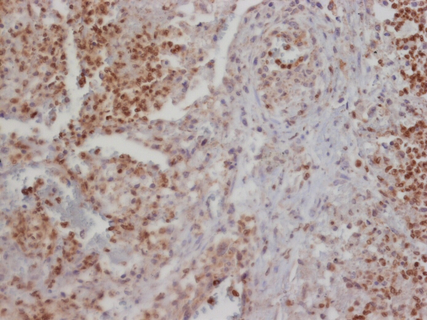
Immunohistochemistry-Paraffin: CCL2/MCP1 Antibody (2D8) - BSA Free [NBP2-22115]
Immunohistochemistry-Paraffin: CCL2/MCP1 Antibody (2D8) - BSA Free [NBP2-22115] - Analysis of paraffin-embedded liver cancer tissues using MCP1 mouse mAb with DAB staining.![ELISA: CCL2/MCP1 Antibody (2D8) - BSA Free [NBP2-22115]](https://resources.rndsystems.com/images/products/CCL2-MCP1-Antibody-2D8-BSA-Free-ELISA-NBP2-22115-img0001.jpg "ELISA: CCL2/MCP1 Antibody (2D8) - BSA Free [NBP2-22115]")
ELISA: CCL2/MCP1 Antibody (2D8) - BSA Free [NBP2-22115]
ELISA: CCL2/MCP1 Antibody (2D8) - BSA Free [NBP2-22115] - Red: Control Antigen (100ng), Purple: Antigen (10ng), Green: Antigen (50ng), Blue: Antigen (100ng). - BSA Free [NBP2-22115] -")
Immunocytochemistry/ Immunofluorescence: CCL2/MCP1 Antibody (2D8) - BSA Free [NBP2-22115] -
Immunocytochemistry/ Immunofluorescence: CCL2/MCP1 Antibody (2D8) - BSA Free [NBP2-22115] - Expression patterns of CCL2 & CCR2 in unwounded & wounded corneas of WT & MMP12 KO mice. (A) Immunofluorescence of CCL2 chemokine & (B) its receptor CCR2 in unwounded & chemically wounded (2-days after injury) WT & Mmp12−/− mouse corneas. Control images represent mouse corneas stained with secondary antibody only & without primary antibody. Nuclei were visualized by staining with DAPI (blue). Scale bars: 50 µm. A magnified image of a wounded WT cornea shows perinuclear expression of CCL2 (orange box). CCL2 staining was visualized in epithelial, stromal, & endothelial layers of wounded WT & Mmp12−/− corneas. CCR2 staining was visualized in epithelial & stromal layers of unwounded & wounded WT & Mmp12−/− corneas. Image collected & cropped by CiteAb from the following publication (https://pubmed.ncbi.nlm.nih.gov/31399604), licensed under a CC-BY license. Not internally tested by Novus Biologicals. - BSA Free [NBP2-22115] -")
Immunocytochemistry/ Immunofluorescence: CCL2/MCP1 Antibody (2D8) - BSA Free [NBP2-22115] -
Immunocytochemistry/ Immunofluorescence: CCL2/MCP1 Antibody (2D8) - BSA Free [NBP2-22115] - LIF represses CXCL9 through epigenetic silencing. a, b qRT-PCR analysis of the indicated genes in BMDMs. BMDMs were pre-incubated with 20 ng/ml LIF for 72 h & then stimulated with 5 ng/ml IFN gamma or 10 μg/ml IL4 during 6 h (a) or with 0.1, 0.5, 1 & 5 ng/ml IFN gamma for 24 h (b). c CXCL9 ELISA from BMDMs pre-incubated with 20 ng/ml LIF & then stimulated with 0.1 ng/ml IFN gamma for 24 h. d CXCL9 ELISA from human CD11b+sorted cells (77% CD11b+ CD14+, see Supplementary Fig. 7b, c) from human GBM cultured with 20 ng/ml LIF for 72 h & then with 0.1 ng/ml IFN gamma for 24 h. e ChIP of Tri-methyl-histone H3 (H3K27me3), EZH2 & acetyl-histone4 (H4ac) was performed in BMDMs treated with 20 ng/ml LIF for 72 h. Scheme shows the analysed CXCL9 promoter region. Representative data are presented as mean ± SD. f Representative images of IF of the indicated markers in human GBM organotypic slices (patients 1, 2, 3) incubated with 10 μg/ml anti-LIF for 3 days. Scale bar, 20 μm. (see Supplementary Fig. 8). Bottom, percentage of double positive cells relative to Iba1+ cells & percentage of CXCL9+ cells relative to the total number of cells. Data are mean of all patients ± SEM. Statistical analyses by Student’s t-test or Mann–Whitney T-test. *P < 0.05, **P < 0.01; ***P < 0.001; ****P < 0.0001 Image collected & cropped by CiteAb from the following publication (https://pubmed.ncbi.nlm.nih.gov/31186412), licensed under a CC-BY license. Not internally tested by Novus Biologicals. - BSA Free [NBP2-22115] -")
Immunohistochemistry: CCL2/MCP1 Antibody (2D8) - BSA Free [NBP2-22115] -
Immunohistochemistry: CCL2/MCP1 Antibody (2D8) - BSA Free [NBP2-22115] - LIF regulates CXCL9, CCL2, CD206, & CD163 in TAMs. a Differential expression analysis of isolated CD11b+ cells from anti-LIF treated ID8 mice vs. control. Volcano plot representing the genes significantly (Q-value < 0.1) overexpressed (brown) & significantly underexpressed (turquoise). Heatmap representing the expression values of the indicated genes, each column represents a sample & each row a gene. The last column represents the log2 fold change (log2 FC) of gene expression. b mRNA expression for the indicated genes in isolated CD11b+ cells from anti-LIF treated or untreated ID8 & GL261N tumors. c Percentage & mean fluorescence intensity (MFI) of CCL2 & CXCL9 in TAMs (CD11b+ Ly6G− Ly6C−) from anti-LIF treated or untreated GL261N tumors. d Representative IF images of Iba1 & the indicated markers stainings of GL261N tumors (see Supplementary Fig. 4). Scale bar, 20 μm. Bottom, percentage of double positive cells relative to the TAM marker positive cells. CXCL9 quantification is relative to the total number of cells. e Tumor growth of GL261N in WT, CXCL9−/−, & CCL2−/− mice or mice treated with the indicated antibodies is shown as total flux (p/s). f Fold change (FC) of tumor infiltrating CD8+ T cells in the indicated treatments. Data are mean ± SEM. g, h Representative IHC of the indicated markers from 20 GBM tumors. The degree of staining was quantified using H-score method. Correlations between LIF & CCL2, CD206, CD163, & CXCL9 with the R-squared coefficients (R2) were calculated (h). Statistical analyses by Mann–Whitney T-test. *P < 0.05; **P < 0.01; ***P < 0.001; ****P < 0.0001 Image collected & cropped by CiteAb from the following publication (https://pubmed.ncbi.nlm.nih.gov/31186412), licensed under a CC-BY license. Not internally tested by Novus Biologicals. - BSA Free [NBP2-22115] -")
Immunocytochemistry/ Immunofluorescence: CCL2/MCP1 Antibody (2D8) - BSA Free [NBP2-22115] -
Immunocytochemistry/ Immunofluorescence: CCL2/MCP1 Antibody (2D8) - BSA Free [NBP2-22115] - LIF regulates CXCL9, CCL2, CD206, & CD163 in TAMs. a Differential expression analysis of isolated CD11b+ cells from anti-LIF treated ID8 mice vs. control. Volcano plot representing the genes significantly (Q-value < 0.1) overexpressed (brown) & significantly underexpressed (turquoise). Heatmap representing the expression values of the indicated genes, each column represents a sample & each row a gene. The last column represents the log2 fold change (log2 FC) of gene expression. b mRNA expression for the indicated genes in isolated CD11b+ cells from anti-LIF treated or untreated ID8 & GL261N tumors. c Percentage & mean fluorescence intensity (MFI) of CCL2 & CXCL9 in TAMs (CD11b+ Ly6G− Ly6C−) from anti-LIF treated or untreated GL261N tumors. d Representative IF images of Iba1 & the indicated markers stainings of GL261N tumors (see Supplementary Fig. 4). Scale bar, 20 μm. Bottom, percentage of double positive cells relative to the TAM marker positive cells. CXCL9 quantification is relative to the total number of cells. e Tumor growth of GL261N in WT, CXCL9−/−, & CCL2−/− mice or mice treated with the indicated antibodies is shown as total flux (p/s). f Fold change (FC) of tumor infiltrating CD8+ T cells in the indicated treatments. Data are mean ± SEM. g, h Representative IHC of the indicated markers from 20 GBM tumors. The degree of staining was quantified using H-score method. Correlations between LIF & CCL2, CD206, CD163, & CXCL9 with the R-squared coefficients (R2) were calculated (h). Statistical analyses by Mann–Whitney T-test. *P < 0.05; **P < 0.01; ***P < 0.001; ****P < 0.0001 Image collected & cropped by CiteAb from the following publication (https://pubmed.ncbi.nlm.nih.gov/31186412), licensed under a CC-BY license. Not internally tested by Novus Biologicals. - BSA Free [NBP2-22115] -")
Western Blot: CCL2/MCP1 Antibody (2D8) - BSA Free [NBP2-22115] -
The production of MCP-1, VCAM-1, and ICAM-1 was significantly inhibited in Kaempferol treatment during sepsis(A) The protein expression level of MCP-1, VCAM-1, and ICAM-1 at 24 h; (B) protein expression level of MCP-1, VCAM-1, and ICAM-1 at 48 h in sham, saline-treated CLP mice, and kaempferol treated CLP mice (n = 3 per group, mean +/- S.E.M, *P<0.05, **P<0.01, ***P<0.001). Image collected and cropped by CiteAb from the following open publication (https://pubmed.ncbi.nlm.nih.gov/37440431), licensed under a CC-BY license. Not internally tested by Novus Biologicals. - BSA Free [NBP2-22115] -")
Western Blot: CCL2/MCP1 Antibody (2D8) - BSA Free [NBP2-22115] -
The production of MCP-1, VCAM-1, and ICAM-1 was significantly inhibited in Kaempferol treatment during sepsis(A) The protein expression level of MCP-1, VCAM-1, and ICAM-1 at 24 h; (B) protein expression level of MCP-1, VCAM-1, and ICAM-1 at 48 h in sham, saline-treated CLP mice, and kaempferol treated CLP mice (n = 3 per group, mean +/- S.E.M, *P<0.05, **P<0.01, ***P<0.001). Image collected and cropped by CiteAb from the following open publication (https://pubmed.ncbi.nlm.nih.gov/37440431), licensed under a CC-BY license. Not internally tested by Novus Biologicals.Applications for CCL2/MCP1 Antibody (2D8) - BSA Free
Application
Recommended Usage
ELISA
1:10000
Flow Cytometry
1:200-1:400
Immunocytochemistry/ Immunofluorescence
1:200-1:1000
Immunohistochemistry
1:200-1:1000
Immunohistochemistry-Paraffin
1:200-1:1000
Western Blot
1 - 3 ug/ml
Reviewed Applications
Read 1 review rated 3 using NBP2-22115 in the following applications:
Flow Cytometry Panel Builder
Bio-Techne Knows Flow Cytometry
Save time and reduce costly mistakes by quickly finding compatible reagents using the Panel Builder Tool.
Advanced Features
- Spectra Viewer - Custom analysis of spectra from multiple fluorochromes
- Spillover Popups - Visualize the spectra of individual fluorochromes
- Antigen Density Selector - Match fluorochrome brightness with antigen density

Formulation, Preparation, and Storage
Purification
Protein A or G purified
Formulation
PBS
Format
BSA Free
Preservative
0.02% Sodium Azide
Concentration
1.0 mg/ml
Shipping
The product is shipped with polar packs. Upon receipt, store it immediately at the temperature recommended below.
Stability & Storage
Store at 4C short term. Aliquot and store at -20C long term. Avoid freeze-thaw cycles.
Background: CCL2/MCP1
Alternate Names
C-C motif chemokine 2, chemokine (C-C motif) ligand 2, GDCF-2, HC11Monocyte secretory protein JE, MCAFMonocyte chemotactic and activating factor, MCP1 Monocyte chemotactic protein 1, MCP-1 Small-inducible cytokine A2, MGC9434, Monocyte chemoattractant protein 1, monocyte chemoattractant protein-1, SCYA2HSMCR30, small inducible cytokine A2 (monocyte chemotactic protein 1, homologous tomouse Sig-je), small inducible cytokine subfamily A (Cys-Cys), member 2, SMC-CF
Entrez Gene IDs
6347 (Human)
Gene Symbol
CCL2
UniProt
Additional CCL2/MCP1 Products
Product Documents for CCL2/MCP1 Antibody (2D8) - BSA Free
Certificate of Analysis
To download a Certificate of Analysis, please enter a lot or batch number in the search box below.
Product Specific Notices for CCL2/MCP1 Antibody (2D8) - BSA Free
This product is for research use only and is not approved for use in humans or in clinical diagnosis. Primary Antibodies are guaranteed for 1 year from date of receipt.
Citations for CCL2/MCP1 Antibody (2D8) - BSA Free
 Powered by Bioz
Customer Reviews for CCL2/MCP1 Antibody (2D8) - BSA Free (1)
3 out of 5
1 Customer Rating
Have you used CCL2/MCP1 Antibody (2D8) - BSA Free?
Submit a review and receive an Amazon gift card!
$25/€18/£15/$25CAN/¥2500 Yen for a review with an image
$10/€7/£6/$10CAN/¥1110 Yen for a review without an image
Submit a review
Customer Images
Showing
1
-
1 of
1 review
Showing All
Filter By:
-
Application: Immunohistochemistry-ParaffinSample Tested: HIV-infected Rhesus macaque adult lungSpecies: Rhesus MacaqueVerified Customer | Posted 11/25/2019Immunohistochemistry (Formalin/PFA-fixed paraffin-embedded sections)Heat-mediated Antigen Retrieval with 10mM Na Citrate buffer, pH6.0. Block with 5% Normal Goat Serum in PBS for 30min, RT Primary antibody (MCP-1, 1:500 dilution) overnight, 4 C HRP-conjugated 2nd antibody (1:500) for 30min, RT DAB staining Countersigning with hematoxylin

There are no reviews that match your criteria.
Protocols
View specific protocols for CCL2/MCP1 Antibody (2D8) - BSA Free (NBP2-22115):
Immunohistochemistry-Paraffin Embedded Sections
Antigen Unmasking:
Bring slides to a boil in 10 mM sodium citrate buffer (pH 6.0) then maintain at a sub-boiling temperature for 10 minutes. Cool slides on bench-top for 30 minutes (keep slides in the sodium citrate buffer all the time).
Staining:
1. Wash sections in deionized water three times for 5 minutes each.
2. Wash sections in PBS for 5 minutes.
3. Block each section with 100-400 ul blocking solution (1% BSA in PBS) for 1 hour at room temperature.
4. Remove blocking solution and add 100-400 ul diluted primary antibody. Incubate overnight at 4 C.
5. Remove antibody solution and wash sections in wash buffer three times for 5 minutes each.
6. Add 100-400 ul HRP polymer conjugated secondary antibody. Incubate 30 minutes at room temperature.
7. Wash sections three times in wash buffer for 5 minutes each.
8. Add 100-400 ul DAB substrate to each section and monitor staining closely.
9. As soon as the sections develop, immerse slides in deionized water.
10. Counterstain sections in hematoxylin.
11. Wash sections in deionized water two times for 5 minutes each.
12. Dehydrate sections.
13. Mount coverslips.
Find general support by application which include: protocols, troubleshooting, illustrated assays, videos and webinars.
- 7-Amino Actinomycin D (7-AAD) Cell Viability Flow Cytometry Protocol
- Antigen Retrieval Protocol (PIER)
- Antigen Retrieval for Frozen Sections Protocol
- Appropriate Fixation of IHC/ICC Samples
- Cellular Response to Hypoxia Protocols
- Chromogenic IHC Staining of Formalin-Fixed Paraffin-Embedded (FFPE) Tissue Protocol
- Chromogenic Immunohistochemistry Staining of Frozen Tissue
- ClariTSA™ Fluorophore Kits
- Detection & Visualization of Antibody Binding
- ELISA Sample Preparation & Collection Guide
- ELISA Troubleshooting Guide
- Extracellular Membrane Flow Cytometry Protocol
- Flow Cytometry Protocol for Cell Surface Markers
- Flow Cytometry Protocol for Staining Membrane Associated Proteins
- Flow Cytometry Staining Protocols
- Flow Cytometry Troubleshooting Guide
- Fluorescent IHC Staining of Frozen Tissue Protocol
- Graphic Protocol for Heat-induced Epitope Retrieval
- Graphic Protocol for the Preparation and Fluorescent IHC Staining of Frozen Tissue Sections
- Graphic Protocol for the Preparation and Fluorescent IHC Staining of Paraffin-embedded Tissue Sections
- Graphic Protocol for the Preparation of Gelatin-coated Slides for Histological Tissue Sections
- How to Run an R&D Systems DuoSet ELISA
- How to Run an R&D Systems Quantikine ELISA
- How to Run an R&D Systems Quantikine™ QuicKit™ ELISA
- ICC Cell Smear Protocol for Suspension Cells
- ICC Immunocytochemistry Protocol Videos
- ICC for Adherent Cells
- IHC Sample Preparation (Frozen sections vs Paraffin)
- Immunocytochemistry (ICC) Protocol
- Immunocytochemistry Troubleshooting
- Immunofluorescence of Organoids Embedded in Cultrex Basement Membrane Extract
- Immunofluorescent IHC Staining of Formalin-Fixed Paraffin-Embedded (FFPE) Tissue Protocol
- Immunohistochemistry (IHC) and Immunocytochemistry (ICC) Protocols
- Immunohistochemistry Frozen Troubleshooting
- Immunohistochemistry Paraffin Troubleshooting
- Intracellular Flow Cytometry Protocol Using Alcohol (Methanol)
- Intracellular Flow Cytometry Protocol Using Detergents
- Intracellular Nuclear Staining Flow Cytometry Protocol Using Detergents
- Intracellular Staining Flow Cytometry Protocol Using Alcohol Permeabilization
- Intracellular Staining Flow Cytometry Protocol Using Detergents to Permeabilize Cells
- Preparing Samples for IHC/ICC Experiments
- Preventing Non-Specific Staining (Non-Specific Binding)
- Primary Antibody Selection & Optimization
- Propidium Iodide Cell Viability Flow Cytometry Protocol
- Protocol for Heat-Induced Epitope Retrieval (HIER)
- Protocol for Liperfluo
- Protocol for Making a 4% Formaldehyde Solution in PBS
- Protocol for VisUCyte™ HRP Polymer Detection Reagent
- Protocol for the Characterization of Human Th22 Cells
- Protocol for the Characterization of Human Th9 Cells
- Protocol for the Fluorescent ICC Staining of Cell Smears - Graphic
- Protocol for the Fluorescent ICC Staining of Cultured Cells on Coverslips - Graphic
- Protocol for the Preparation & Fixation of Cells on Coverslips
- Protocol for the Preparation and Chromogenic IHC Staining of Frozen Tissue Sections
- Protocol for the Preparation and Chromogenic IHC Staining of Frozen Tissue Sections - Graphic
- Protocol for the Preparation and Chromogenic IHC Staining of Paraffin-embedded Tissue Sections
- Protocol for the Preparation and Chromogenic IHC Staining of Paraffin-embedded Tissue Sections - Graphic
- Protocol for the Preparation and Fluorescent ICC Staining of Cells on Coverslips
- Protocol for the Preparation and Fluorescent ICC Staining of Non-adherent Cells
- Protocol for the Preparation and Fluorescent ICC Staining of Stem Cells on Coverslips
- Protocol for the Preparation and Fluorescent IHC Staining of Frozen Tissue Sections
- Protocol for the Preparation and Fluorescent IHC Staining of Paraffin-embedded Tissue Sections
- Protocol for the Preparation of Gelatin-coated Slides for Histological Tissue Sections
- Protocol for the Preparation of a Cell Smear for Non-adherent Cell ICC - Graphic
- Protocol: Annexin V and PI Staining by Flow Cytometry
- Protocol: Annexin V and PI Staining for Apoptosis by Flow Cytometry
- Quantikine HS ELISA Kit Assay Principle, Alkaline Phosphatase
- Quantikine HS ELISA Kit Principle, Streptavidin-HRP Polymer
- R&D Systems Quality Control Western Blot Protocol
- Sandwich ELISA (Colorimetric) – Biotin/Streptavidin Detection Protocol
- Sandwich ELISA (Colorimetric) – Direct Detection Protocol
- TUNEL and Active Caspase-3 Detection by IHC/ICC Protocol
- The Importance of IHC/ICC Controls
- Troubleshooting Guide: ELISA
- Troubleshooting Guide: Fluorokine Flow Cytometry Kits
- Troubleshooting Guide: Immunohistochemistry
- Troubleshooting Guide: Western Blot Figures
- Western Blot Conditions
- Western Blot Protocol
- Western Blot Protocol for Cell Lysates
- Western Blot Troubleshooting
- Western Blot Troubleshooting Guide
- View all Protocols, Troubleshooting, Illustrated assays and Webinars
Loading...