CCR2 Antibody (3G7) - BSA Free
Novus Biologicals | Catalog # NBP2-35334
Key Product Details
Species Reactivity
Validated:
Human, Mouse
Cited:
Human, Mouse
Predicted:
Primate (100%). Backed by our 100% Guarantee.
Applications
Validated:
Immunohistochemistry, Immunohistochemistry-Paraffin, Western Blot, Immunocytochemistry/ Immunofluorescence
Cited:
Immunohistochemistry, Immunohistochemistry-Paraffin, Western Blot, Immunocytochemistry/ Immunofluorescence
Label
Unconjugated
Antibody Source
Monoclonal Mouse IgG2b Lambda Clone # 3G7
Format
BSA Free
Loading...
Product Specifications
Immunogen
A synthetic peptide made to an N-terminal portion of the human CD63 protein (between residues 1-100) [UniProt P41597]
Reactivity Notes
Mouse reactivity reported in scientific literature (PMID: 30213824).
Localization
Cell membrane
Clonality
Monoclonal
Host
Mouse
Isotype
IgG2b Lambda
Scientific Data Images for CCR2 Antibody (3G7) - BSA Free
![Western Blot: CCR2 Antibody (3G7)BSA Free [NBP2-35334]](https://resources.rndsystems.com/images/products/CCR2-Antibody-3G7-Western-Blot-NBP2-35334-img0007.jpg "Western Blot: CCR2 Antibody (3G7)BSA Free [NBP2-35334]")
Western Blot: CCR2 Antibody (3G7)BSA Free [NBP2-35334]
Western Blot: CCR2 Antibody (3G7) [NBP2-35334] - Western blot analysis of CCR2 [3G7] in Molt4 lysate.![Immunohistochemistry-Paraffin: CCR2 Antibody (3G7) - BSA Free [NBP2-35334]](https://resources.rndsystems.com/images/products/CCR2-Antibody-3G7-Immunohistochemistry-Paraffin-NBP2-35334-img0006.jpg "Immunohistochemistry-Paraffin: CCR2 Antibody (3G7) - BSA Free [NBP2-35334]")
Immunohistochemistry-Paraffin: CCR2 Antibody (3G7) - BSA Free [NBP2-35334]
Immunohistochemistry-Paraffin: CCR2 Antibody (3G7) [NBP2-35334] - IHC analysis of formalin-fixed paraffin-embedded tissue section of human breast using CCR2 antibody (clone 3G7) at 5 ug/ml concentration. The glandular cells in the lobules showed a specific cytoplasmic-membranous immunostaining of CCR2.![Immunohistochemistry-Paraffin: CCR2 Antibody (3G7) - BSA Free [NBP2-35334]](https://resources.rndsystems.com/images/products/CCR2-Antibody-3G7-Immunohistochemistry-Paraffin-NBP2-35334-img0003.jpg "Immunohistochemistry-Paraffin: CCR2 Antibody (3G7) - BSA Free [NBP2-35334]")
Immunohistochemistry-Paraffin: CCR2 Antibody (3G7) - BSA Free [NBP2-35334]
Immunohistochemistry-Paraffin: CCR2 Antibody (3G7) [NBP2-35334] - IHC analysis of formalin-fixed paraffin-embedded tissue section of human HCC (hepatocellular carcinoma / malignant hepatoma) using CCR2 antibody (clone 3G7) at 5 ug/ml concentration. The cancer cells depicted specific punctate to diffused cytoplasmic-membranous immuno-staining of CCR2 protein.![Immunohistochemistry-Paraffin: CCR2 Antibody (3G7) - BSA Free [NBP2-35334]](https://resources.rndsystems.com/images/products/CCR2-Antibody-3G7-Immunohistochemistry-Paraffin-NBP2-35334-img0005.jpg "Immunohistochemistry-Paraffin: CCR2 Antibody (3G7) - BSA Free [NBP2-35334]")
Immunohistochemistry-Paraffin: CCR2 Antibody (3G7) - BSA Free [NBP2-35334]
Immunohistochemistry-Paraffin: CCR2 Antibody (3G7) [NBP2-35334] - IHC analysis of formalin-fixed paraffin-embedded tissue section of human esophageal squamous cell carcinoma (SCC) using CCR2 antibody (clone 3G7) at 5 ug/ml concentration. The cancer cells depicted specific cytoplasmic-membranous immunostaining, whereas the cells of tumor stroma were largely negative for CCR2 immunopositivity.Applications for CCR2 Antibody (3G7) - BSA Free
Application
Recommended Usage
Immunocytochemistry/ Immunofluorescence
reported in scientific literature (PMID 34274535)
Immunohistochemistry
5 ug/ml
Immunohistochemistry-Paraffin
5 ug/ml
Western Blot
2 ug/ml
Reviewed Applications
Read 2 reviews rated 4 using NBP2-35334 in the following applications:
Formulation, Preparation, and Storage
Purification
Protein G purified
Formulation
PBS
Format
BSA Free
Preservative
0.05% Sodium Azide
Concentration
1.0 mg/ml
Shipping
The product is shipped with polar packs. Upon receipt, store it immediately at the temperature recommended below.
Stability & Storage
Store at 4C short term. Aliquot and store at -20C long term. Avoid freeze-thaw cycles.
Background: CCR2
Alternate Names
CC-CKR-2, CCR2, CCR2B, CD192, CKR2, CKR2A, CMKBR2, FLJ78302, MCP-1-R
Entrez Gene IDs
729230 (Human)
Gene Symbol
CCR2
UniProt
Additional CCR2 Products
Product Documents for CCR2 Antibody (3G7) - BSA Free
Certificate of Analysis
To download a Certificate of Analysis, please enter a lot or batch number in the search box below.
Product Specific Notices for CCR2 Antibody (3G7) - BSA Free
This product is for research use only and is not approved for use in humans or in clinical diagnosis. Primary Antibodies are guaranteed for 1 year from date of receipt.
Citations for CCR2 Antibody (3G7) - BSA Free
 Powered by Bioz
Customer Reviews for CCR2 Antibody (3G7) - BSA Free (2)
4 out of 5
2 Customer Ratings
Have you used CCR2 Antibody (3G7) - BSA Free?
Submit a review and receive an Amazon gift card!
$25/€18/£15/$25CAN/¥2500 Yen for a review with an image
$10/€7/£6/$10CAN/¥1110 Yen for a review without an image
Submit a review
Customer Images
Showing
1
-
2 of
2 reviews
Showing All
Filter By:
-
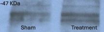
Application: Western BlotSample Tested: Liver tissueSpecies: MouseVerified Customer | Posted 11/12/2024Superior CCR2 AntibodyMice liver homogenate from sham and treated groups were resolved in SDS-PAGE and immunobloted with this antibody at 1:1000 dilution.

-
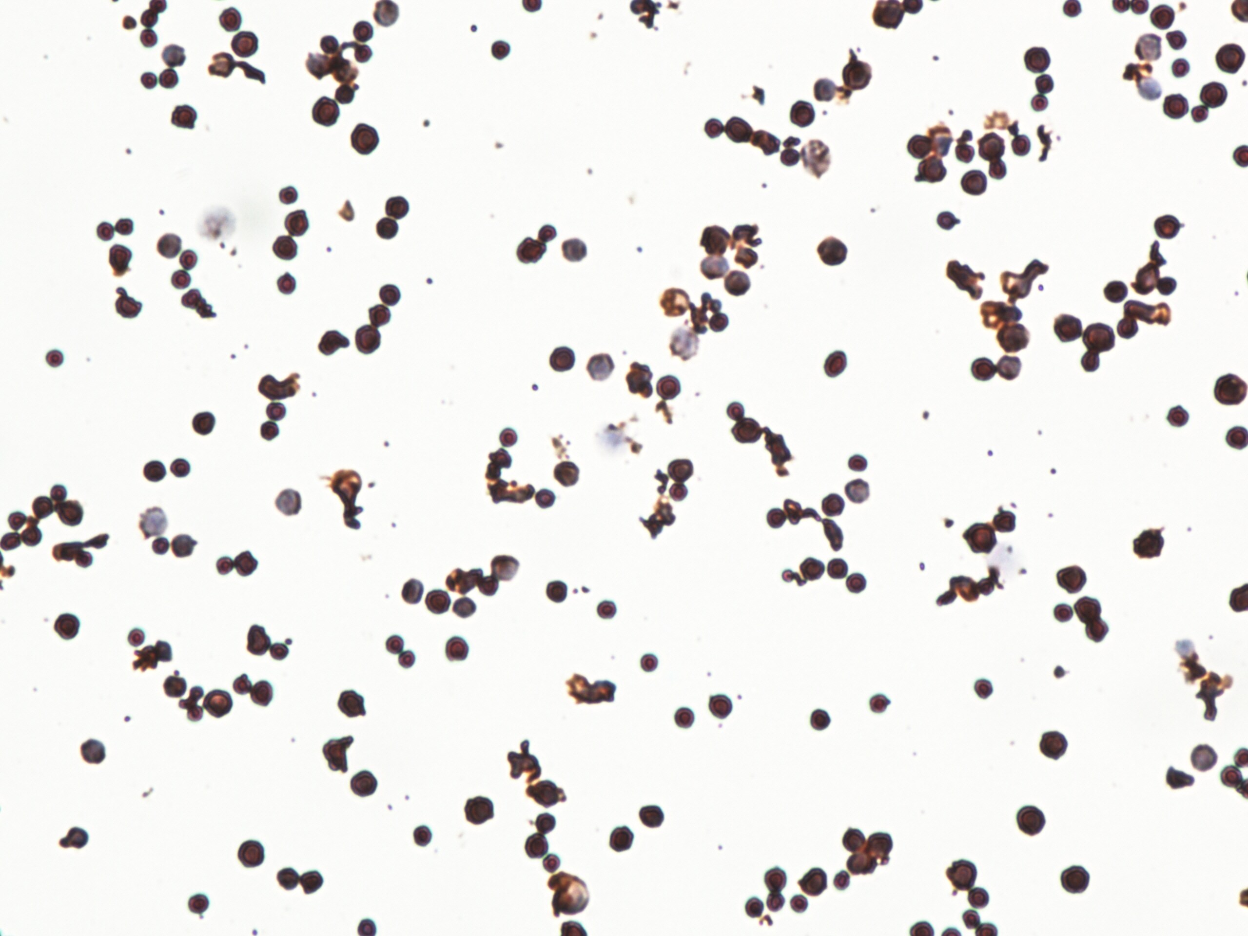
Application: ImmunocytochemistrySample Tested: rabbit spleen isolated primary cellsSpecies: OtherVerified Customer | Posted 02/02/2015rabbit spleen isolated primary cells IHC with CCR2
There are no reviews that match your criteria.
Protocols
View specific protocols for CCR2 Antibody (3G7) - BSA Free (NBP2-35334):
1. Deparaffinize the tissue sections by immersing the slides in Xylene with two changes for 10 min each. Sections should not get dried at any stage from this point.
2. Rehydrate the tissue sections by immersing the slides in decreasing grades of ethanol as follows:
a. Immerse in 100% ethanol with 2 changes for 5 minutes each
b. Immerse in 95% ethanol with 2 changes for 5 minutes each
c. Immerse in 90% ethanol for 5 minutes
d. Immerse in 70% ethanol for 5 minutes
e. Immerse in 50% ethanol for 5 minutes
f. Immerse in distilled water for 5 minutes
3. Antigen Retrieval (Microwave Method):
a. Immerse the slides in a microwave compatible tray containing 10 mM Sodium Citrate buffer (pH 6.0) with 0.05% Tween 20.
b. Boil the slides and maintain the sub-boiling temperature for 5 minutes in the microwave. Thereafter, take out the tray very carefully and cool it at room temperature (RT) for about 30 minutes.
c. Wash the slides 3 times, 3 minutes each by immersing them in TBST (Tris Buffered Saline having 0.05% Tween 20).
4. Quenching of Endogenous Peroxidase:
a. Incubate the slides in 3% hydrogen peroxide prepared in methanol for 15 minutes (at RT, in dark conditions).
b. Wash the slides in TBST 3 times, 3 minutes each.
5. Protein Blocking:
a. Incubate the sections with background sniper solution at RT for 15 minutes (Biocare Medicals, USA).
b. Wash the sections 3 times, 3 min each by immersing the slides in TBST.
6. Primary Antibody:
a. Dilute the primary antibody at 5ug/ml concentration using PBS as a diluent.
b. Incubate the sections with diluted primary antibody for 90 minutes at RT in a humidified chamber.
c. Thereafter, wash the slides 4 times, 5 minutes each with TBST.
7. Probe (Secondary Reagent):
a. Incubate with MACH 1 Mouse probe for 15 minutes at RT.
b. Incubate for 30 min at room temperature with HRP-Polymer (Biocare Medical, USA).
c. Wash the slides with TBST 4 times, 5 minutes each
8. Chromogen:
a. Mix 32ul of DAB Chromogen with 1 ml of DAB substrate buffer (Biocare Medical, USA).
b. Apply 200ul DAB mixture/section and incubate at RT in dark conditions (few seconds - 5 minutes).
c. As soon as an appropriate color develops, rinse the slides with deionized water (2-3 brief rinses).
9. Counter stain:
a. Counter stain with Hematoxylin for 30 seconds (Vector Labs, USA).
b. Wash in deionized water for 1-2 minutes to clear the extra stain.
c. Incubate the slides in bluing solution or Scott's water twice for 2 minutes each time.
10. Dehydrate the sections in increasing grades of alcohols:
a. 50% alcohol for 1 minute
b. 70% for 1 minute
c. 90% for 1 minute
d. 95% for 1 minute
e. 100% for 1 minute
f. Xylene with 2 changes for 2 minutes each
11. Mount with DPX mount and cover-slip glass (Fisher Scientific, USA), carefully not allowing any air bubbles to enter.
NOTE:- This protocol is provided as a reference tool only. Depending upon the type of tissues /tissue processing and reagents employed, the end user will need to optimize the final conditions for achieving an expected staining.
Western Blot Protocol
1. Perform SDS-PAGE on samples to be analyzed, loading 25 ug of total protein per lane.
2. Transfer proteins to membrane according to the instructions provided by the manufacturer of the membrane and transfer apparatus.
3. Stain according to standard Ponceau S procedure (or similar product) to assess transfer success, and mark molecular weight standards where appropriate.
4. Rinse the blot.
5. Block the membrane using standard blocking buffer for at least 1 hour.
6. Wash the membrane in wash buffer three times for 10 minutes each.
7. Dilute anti-CCR2 primary antibody in blocking buffer and incubate 1 hour at room temperature.
8. Wash the membrane in wash buffer three times for 10 minutes each.
9. Apply the diluted HRP conjugated secondary antibody in blocking buffer (as per manufacturers instructions) and incubate 1 hour at room temperature.
10. Wash the blot in wash buffer three times for 10 minutes each (this step can be repeated as required to reduce background).
11. Apply the detection reagent of choice in accordance with the manufacturers instructions.
Note: Tween-20 can be added to the blocking or antibody dilution buffer at a final concentration of 0.05-0.2%.
Find general support by application which include: protocols, troubleshooting, illustrated assays, videos and webinars.
- Antigen Retrieval Protocol (PIER)
- Antigen Retrieval for Frozen Sections Protocol
- Appropriate Fixation of IHC/ICC Samples
- Cellular Response to Hypoxia Protocols
- Chromogenic IHC Staining of Formalin-Fixed Paraffin-Embedded (FFPE) Tissue Protocol
- Chromogenic Immunohistochemistry Staining of Frozen Tissue
- ClariTSA™ Fluorophore Kits
- Detection & Visualization of Antibody Binding
- Fluorescent IHC Staining of Frozen Tissue Protocol
- Graphic Protocol for Heat-induced Epitope Retrieval
- Graphic Protocol for the Preparation and Fluorescent IHC Staining of Frozen Tissue Sections
- Graphic Protocol for the Preparation and Fluorescent IHC Staining of Paraffin-embedded Tissue Sections
- Graphic Protocol for the Preparation of Gelatin-coated Slides for Histological Tissue Sections
- ICC Cell Smear Protocol for Suspension Cells
- ICC Immunocytochemistry Protocol Videos
- ICC for Adherent Cells
- IHC Sample Preparation (Frozen sections vs Paraffin)
- Immunocytochemistry (ICC) Protocol
- Immunocytochemistry Troubleshooting
- Immunofluorescence of Organoids Embedded in Cultrex Basement Membrane Extract
- Immunofluorescent IHC Staining of Formalin-Fixed Paraffin-Embedded (FFPE) Tissue Protocol
- Immunohistochemistry (IHC) and Immunocytochemistry (ICC) Protocols
- Immunohistochemistry Frozen Troubleshooting
- Immunohistochemistry Paraffin Troubleshooting
- Preparing Samples for IHC/ICC Experiments
- Preventing Non-Specific Staining (Non-Specific Binding)
- Primary Antibody Selection & Optimization
- Protocol for Heat-Induced Epitope Retrieval (HIER)
- Protocol for Making a 4% Formaldehyde Solution in PBS
- Protocol for VisUCyte™ HRP Polymer Detection Reagent
- Protocol for the Fluorescent ICC Staining of Cell Smears - Graphic
- Protocol for the Fluorescent ICC Staining of Cultured Cells on Coverslips - Graphic
- Protocol for the Preparation & Fixation of Cells on Coverslips
- Protocol for the Preparation and Chromogenic IHC Staining of Frozen Tissue Sections
- Protocol for the Preparation and Chromogenic IHC Staining of Frozen Tissue Sections - Graphic
- Protocol for the Preparation and Chromogenic IHC Staining of Paraffin-embedded Tissue Sections
- Protocol for the Preparation and Chromogenic IHC Staining of Paraffin-embedded Tissue Sections - Graphic
- Protocol for the Preparation and Fluorescent ICC Staining of Cells on Coverslips
- Protocol for the Preparation and Fluorescent ICC Staining of Non-adherent Cells
- Protocol for the Preparation and Fluorescent ICC Staining of Stem Cells on Coverslips
- Protocol for the Preparation and Fluorescent IHC Staining of Frozen Tissue Sections
- Protocol for the Preparation and Fluorescent IHC Staining of Paraffin-embedded Tissue Sections
- Protocol for the Preparation of Gelatin-coated Slides for Histological Tissue Sections
- Protocol for the Preparation of a Cell Smear for Non-adherent Cell ICC - Graphic
- R&D Systems Quality Control Western Blot Protocol
- TUNEL and Active Caspase-3 Detection by IHC/ICC Protocol
- The Importance of IHC/ICC Controls
- Troubleshooting Guide: Immunohistochemistry
- Troubleshooting Guide: Western Blot Figures
- Western Blot Conditions
- Western Blot Protocol
- Western Blot Protocol for Cell Lysates
- Western Blot Troubleshooting
- Western Blot Troubleshooting Guide
- View all Protocols, Troubleshooting, Illustrated assays and Webinars


