Key Product Details
Validated by
Orthogonal Validation
Species Reactivity
Human
Applications
Multiplex Immunofluorescence, Immunohistochemistry, Immunohistochemistry-Paraffin, Western Blot, COMET
Label
Unconjugated
Antibody Source
Monoclonal Mouse IgG1 Clone # CL1831
Format
BSA Free
Loading...
Product Specifications
Immunogen
This antibody was developed using a recombinant protein derived from P20702, with the exact immunogen sequence remaining proprietary.
Reactivity Notes
Mouse reactivity reported from a verified customer review.
Marker
Dendritic Cell Marker
Clonality
Monoclonal
Host
Mouse
Isotype
IgG1
Scientific Data Images for CD11c Antibody (CL1831) - BSA Free

Detection of CD11c in Mouse Spleen via seqIF™ staining on COMET™
CD11c was detected in immersion fixed paraffin-embedded sections of mouse Spleen using Mouse anti-CD11c, Monoclonal Antibody (Catalog #NBP2-34491) at 1:200 dilution at 37 ° Celsius for 4 minutes. Before incubation with the primary antibody, tissue underwent an all-in-one dewaxing and antigen retrieval preprocessing using PreTreatment Module (PT Module) and Dewax and HIER Buffer H (pH 9; Epredia Catalog # TA-999-DHBH). Tissue was stained using the Alexa Fluor™ 647 Goat anti-Mouse IgG Secondary Antibody at 1:200 at 37 ° Celsius for 2 minutes. (Yellow; Lunaphore Catalog # DR647MS) and counterstained with DAPI (blue; Lunaphore Catalog # DR100). Specific staining was localized to the cytoplasm. Protocol available in COMET™ Panel Builder.![Immunohistochemistry-Paraffin: CD11c Antibody (CL1831) [NBP2-34491]](https://resources.rndsystems.com/images/products/CD11c-Antibody-CL1831-Immunohistochemistry-Paraffin-NBP2-34491-img0016.jpg "Immunohistochemistry-Paraffin: CD11c Antibody (CL1831) [NBP2-34491]")
![Western Blot: CD11c Antibody (CL1831) [NBP2-34491]](https://resources.rndsystems.com/images/products/CD11c-Antibody-CL1831-Western-Blot-NBP2-34491-img0001.jpg "Western Blot: CD11c Antibody (CL1831) [NBP2-34491]")
Western Blot: CD11c Antibody (CL1831) [NBP2-34491]
Western Blot: CD11c Antibody (CL1831) [NBP2-34491] - Lane 1: Marker [kDa]. Lane 2: Human tonsil.![Immunohistochemistry-Paraffin: CD11c Antibody (CL1831) [NBP2-34491]](https://resources.rndsystems.com/images/products/CD11c-Antibody-CL1831-Immunohistochemistry-Paraffin-NBP2-34491-img0017.jpg "Immunohistochemistry-Paraffin: CD11c Antibody (CL1831) [NBP2-34491]")
Immunohistochemistry-Paraffin: CD11c Antibody (CL1831) [NBP2-34491]
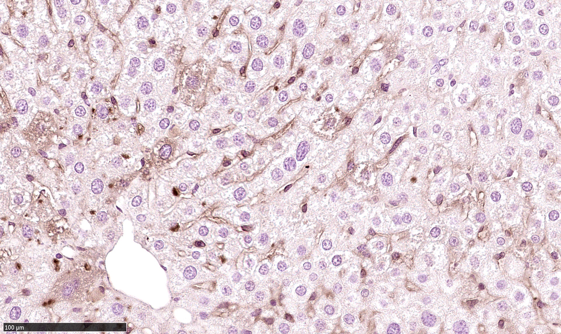
Immunohistochemistry-Paraffin: CD11c Antibody (CL1831) [NBP2-34491] - CD11c Positive cells in mouse liver tissue. Primary antibody dilution: 1:100. Citrate buffer antigen retrieval. Overnight incubation RT. Image from verified customer review.![Immunohistochemistry-Paraffin: CD11c Antibody (CL1831) [NBP2-34491]](https://resources.rndsystems.com/images/products/CD11c-Antibody-CL1831-Immunohistochemistry-Paraffin-NBP2-34491-img0012.jpg "Immunohistochemistry-Paraffin: CD11c Antibody (CL1831) [NBP2-34491]")
Immunohistochemistry-Paraffin: CD11c Antibody (CL1831) [NBP2-34491]
Immunohistochemistry-Paraffin: CD11c Antibody (CL1831) [NBP2-34491] - Staining of human tonsil shows strong membranous positivity in a subset of germinal and non-germinal center cells.![Immunohistochemistry-Paraffin: CD11c Antibody (CL1831) [NBP2-34491]](https://resources.rndsystems.com/images/products/CD11c-Antibody-CL1831-Immunohistochemistry-Paraffin-NBP2-34491-img0013.jpg "Immunohistochemistry-Paraffin: CD11c Antibody (CL1831) [NBP2-34491]")
Immunohistochemistry-Paraffin: CD11c Antibody (CL1831) [NBP2-34491]
Immunohistochemistry-Paraffin: CD11c Antibody (CL1831) [NBP2-34491] - Staining of human small intestine shows moderate positivity in a subset of lymphoid cells.![Immunohistochemistry-Paraffin: CD11c Antibody (CL1831) [NBP2-34491]](https://resources.rndsystems.com/images/products/CD11c-Antibody-CL1831-Immunohistochemistry-Paraffin-NBP2-34491-img0014.jpg "Immunohistochemistry-Paraffin: CD11c Antibody (CL1831) [NBP2-34491]")
Immunohistochemistry-Paraffin: CD11c Antibody (CL1831) [NBP2-34491]
Immunohistochemistry-Paraffin: CD11c Antibody (CL1831) [NBP2-34491] - Staining of human fallopian tube shows moderate membranous positivity in a subset of lymphoid cells.![Immunohistochemistry-Paraffin: CD11c Antibody (CL1831) [NBP2-34491]](https://resources.rndsystems.com/images/products/CD11c-Antibody-CL1831-Immunohistochemistry-Paraffin-NBP2-34491-img0015.jpg "Immunohistochemistry-Paraffin: CD11c Antibody (CL1831) [NBP2-34491]")
Immunohistochemistry-Paraffin: CD11c Antibody (CL1831) [NBP2-34491]
Immunohistochemistry-Paraffin: CD11c Antibody (CL1831) [NBP2-34491] - Staining of human kidney shows no positivity as expected.Applications for CD11c Antibody (CL1831) - BSA Free
Application
Recommended Usage
Immunohistochemistry
1:200 - 1:500
Immunohistochemistry-Paraffin
1:200 - 1:500
Western Blot
1 ug/ml
Application Notes
For IHC-Paraffin, HIER pH 6 retrieval is recommended.
Reviewed Applications
Read 1 review rated 4 using NBP2-34491 in the following applications:
Formulation, Preparation, and Storage
Purification
Protein A purified
Formulation
PBS (pH 7.2) and 40% Glycerol
Format
BSA Free
Preservative
0.02% Sodium Azide
Concentration
1 mg/ml
Shipping
The product is shipped with polar packs. Upon receipt, store it immediately at the temperature recommended below.
Stability & Storage
Store at 4C short term. Aliquot and store at -20C long term. Avoid freeze-thaw cycles.
Background: CD11c
Alternate Names
CD11c, Integrin alpha X, ITGAX, p150,95 alpha
Gene Symbol
ITGAX
UniProt
Additional CD11c Products
Product Documents for CD11c Antibody (CL1831) - BSA Free
Certificate of Analysis
To download a Certificate of Analysis, please enter a lot or batch number in the search box below.
Product Specific Notices for CD11c Antibody (CL1831) - BSA Free
This product is for research use only and is not approved for use in humans or in clinical diagnosis. Primary Antibodies are guaranteed for 1 year from date of receipt.
Related Research Areas
Customer Reviews for CD11c Antibody (CL1831) - BSA Free (1)
4 out of 5
1 Customer Rating
Have you used CD11c Antibody (CL1831) - BSA Free?
Submit a review and receive an Amazon gift card!
$25/€18/£15/$25CAN/¥2500 Yen for a review with an image
$10/€7/£6/$10CAN/¥1110 Yen for a review without an image
Submit a review
Customer Images
Showing
1
-
1 of
1 review
Showing All
Filter By:
-
Application: Immunohistochemistry-ParaffinSample Tested: Liver and Liver tissueSpecies: MouseVerified Customer | Posted 02/07/2022Cd11c Positive cells in mouse liver1:100 dilution Citrate buffer antigen retrieval Overnight incubation RT
 Bio-Techne ResponseThis review was submitted through the legacy Novus Innovators Program, reflecting a new species or application tested on a primary antibody.
There are no reviews that match your criteria.
Protocols
Find general support by application which include: protocols, troubleshooting, illustrated assays, videos and webinars.
- Antigen Retrieval Protocol (PIER)
- Antigen Retrieval for Frozen Sections Protocol
- Appropriate Fixation of IHC/ICC Samples
- Cellular Response to Hypoxia Protocols
- Chromogenic IHC Staining of Formalin-Fixed Paraffin-Embedded (FFPE) Tissue Protocol
- Chromogenic Immunohistochemistry Staining of Frozen Tissue
- ClariTSA™ Fluorophore Kits
- Detection & Visualization of Antibody Binding
- Fluorescent IHC Staining of Frozen Tissue Protocol
- Graphic Protocol for Heat-induced Epitope Retrieval
- Graphic Protocol for the Preparation and Fluorescent IHC Staining of Frozen Tissue Sections
- Graphic Protocol for the Preparation and Fluorescent IHC Staining of Paraffin-embedded Tissue Sections
- Graphic Protocol for the Preparation of Gelatin-coated Slides for Histological Tissue Sections
- IHC Sample Preparation (Frozen sections vs Paraffin)
- Immunofluorescent IHC Staining of Formalin-Fixed Paraffin-Embedded (FFPE) Tissue Protocol
- Immunohistochemistry (IHC) and Immunocytochemistry (ICC) Protocols
- Immunohistochemistry Frozen Troubleshooting
- Immunohistochemistry Paraffin Troubleshooting
- Preparing Samples for IHC/ICC Experiments
- Preventing Non-Specific Staining (Non-Specific Binding)
- Primary Antibody Selection & Optimization
- Protocol for Heat-Induced Epitope Retrieval (HIER)
- Protocol for Making a 4% Formaldehyde Solution in PBS
- Protocol for VisUCyte™ HRP Polymer Detection Reagent
- Protocol for the Preparation & Fixation of Cells on Coverslips
- Protocol for the Preparation and Chromogenic IHC Staining of Frozen Tissue Sections
- Protocol for the Preparation and Chromogenic IHC Staining of Frozen Tissue Sections - Graphic
- Protocol for the Preparation and Chromogenic IHC Staining of Paraffin-embedded Tissue Sections
- Protocol for the Preparation and Chromogenic IHC Staining of Paraffin-embedded Tissue Sections - Graphic
- Protocol for the Preparation and Fluorescent IHC Staining of Frozen Tissue Sections
- Protocol for the Preparation and Fluorescent IHC Staining of Paraffin-embedded Tissue Sections
- Protocol for the Preparation of Gelatin-coated Slides for Histological Tissue Sections
- R&D Systems Quality Control Western Blot Protocol
- TUNEL and Active Caspase-3 Detection by IHC/ICC Protocol
- The Importance of IHC/ICC Controls
- Troubleshooting Guide: Immunohistochemistry
- Troubleshooting Guide: Western Blot Figures
- Western Blot Conditions
- Western Blot Protocol
- Western Blot Protocol for Cell Lysates
- Western Blot Troubleshooting
- Western Blot Troubleshooting Guide
- View all Protocols, Troubleshooting, Illustrated assays and Webinars
Loading...
Associated Pathways

