CD133 Antibody - BSA Free
Novus Biologicals | Catalog # NB120-16518
![Western Blot: CD133 AntibodyBSA Free [NB120-16518]](https://resources.rndsystems.com/images/products/CD133-Antibody---BSA-Free-Western-Blot-NB120-16518-img0003.jpg "Western Blot: CD133 AntibodyBSA Free [NB120-16518]")
Key Product Details
Species Reactivity
Validated:
Human, Mouse, Rat, Porcine
Cited:
Human, Mouse, Rat, Porcine
Applications
Validated:
Immunohistochemistry, Immunohistochemistry-Paraffin, Immunohistochemistry-Frozen, Western Blot, ELISA, Flow Cytometry, Immunocytochemistry/ Immunofluorescence, Chromatin Immunoprecipitation (ChIP)
Cited:
Immunohistochemistry, Immunohistochemistry-Paraffin, Immunohistochemistry-Frozen, Western Blot, ELISA, Flow Cytometry, Immunocytochemistry/ Immunofluorescence, IF/IHC
Label
Unconjugated
Antibody Source
Polyclonal Rabbit IgG
Format
BSA Free
Loading...
Product Specifications
Immunogen
Synthetic peptide corresponding to a C-terminal region of CD133 (within amino acids 750-865).
Reactivity Notes
porcine reactivity reported in scientific literature (PMID: 29352176).
Marker
Stem Cell Marker
Specificity
CD133 - Hematopoietic Stem Cell Marker
Clonality
Polyclonal
Host
Rabbit
Isotype
IgG
Scientific Data Images for CD133 Antibody - BSA Free
Western Blot: CD133 AntibodyBSA Free [NB120-16518]
Western Blot: CD133 Antibody - BSA Free [NB120-16518] - Predicted band size : 97 kDa recognizes 97 kDa human CD133 in Y79 cells. Band is at ~120 kDa due to protein glycosylation.![Immunocytochemistry/ Immunofluorescence: CD133 Antibody - BSA Free [NB120-16518]](https://resources.rndsystems.com/images/products/CD133%20Antibody%20-%20BSA%20Free-Immunocytochemistry-Immunofluorescence-NB120-16518-img0008.jpg "Immunocytochemistry/ Immunofluorescence: CD133 Antibody - BSA Free [NB120-16518]")
Immunocytochemistry/ Immunofluorescence: CD133 Antibody - BSA Free [NB120-16518]
CD133 Antibody - BSA Free-Immunocytochemistry-Immunofluorescence-NB120-16518-img0008.jpg![Immunohistochemistry: CD133 Antibody - BSA Free [NB120-16518]](https://resources.rndsystems.com/images/products/CD133-Antibody---BSA-Free-Immunohistochemistry-NB120-16518-img0007.jpg "Immunohistochemistry: CD133 Antibody - BSA Free [NB120-16518]")
Immunohistochemistry: CD133 Antibody - BSA Free [NB120-16518]
CD133-Antibody---BSA-Free-Immunohistochemistry-NB120-16518-img0007.jpg![Flow Cytometry: CD133 Antibody - BSA Free [NB120-16518]](https://resources.rndsystems.com/images/products/CD133-Antibody---BSA-Free-Flow-Cytometry-NB120-16518-img0006.jpg "Flow Cytometry: CD133 Antibody - BSA Free [NB120-16518]")
Flow Cytometry: CD133 Antibody - BSA Free [NB120-16518]
Flow Cytometry: CD133 Antibody - BSA Free [NB120-16518] - Flow Cytometry: CD133 Antibody [DyLight 488] [NB120-16518G] - Flow Cytometry: CD133 Antibody (DyLight488) [NB120-16518G] - Rat bone marrow cells were stained with CD133 (1:100) antibody (20 minutes at 4C), fixed, and then analyzed. Image using the DyLight 488 form of this antibody.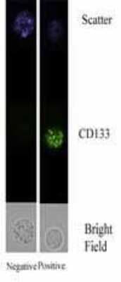
Chromatin Immunoprecipitation: CD133 Antibody - BSA Free [NB120-16518] - [NB 120-16518] - Staining CD133 in Bone Marrow Mononuclear Cells by Immunofluorescence.
![Immunohistochemistry-Paraffin: CD133 Antibody - BSA Free [NB120-16518]](https://resources.rndsystems.com/images/products/CD133-Antibody---BSA-Free-Immunohistochemistry-Paraffin-NB120-16518-img0002.jpg "Immunohistochemistry-Paraffin: CD133 Antibody - BSA Free [NB120-16518]")
Immunohistochemistry-Paraffin: CD133 Antibody - BSA Free [NB120-16518]
Immunohistochemistry-Paraffin: CD133 Antibody - BSA Free [NB120-16518] - Staining of CD133 in human hepatocarcinoma (HC) tissue. NB120-16518 was peroxidase-conjugated to the secondary antibody, followed by AEC staining.
Western Blot: CD133 Antibody - BSA Free [NB120-16518] -
TRPM7 is expressed in GBM and positively correlates with the activation of Notch1, and the upregulation of TRPM7 activates Notch1 signaling. (A) The U87MG cells were transfected with (A) wild-type human TRPM7 (M7-wt); (B) two alpha -kinase-inactive mutants, “ alpha -kinase-dead" point mutation (K1648R, or M7-KR) and alpha -kinase deleted mutant (M7-DK) along with controls followed by assaying protein expression of TRPM7, Notch1, Notch2, Notch3, and Notch4 by Western blot. (B) HEK-293 cells were transfected with a pcDNA4/TO plasmid that allowed tetracycline-inducible protein expression of TRPM7-wt tagged with HA. Then, protein expression of exogenous, endogenous TRPM7, Notch1, Notch2, Notch3, Notch4, and Notch target genes Hey2 and Survivin were determined by Western blot. Image collected and cropped by CiteAb from the following open publication (https://pubmed.ncbi.nlm.nih.gov/33381038), licensed under a CC-BY license. Not internally tested by Novus Biologicals.
Immunocytochemistry/ Immunofluorescence: CD133 Antibody - BSA Free [NB120-16518] -
Immunostaining for CD133 in normal ovarian and tumor tissue sections: [A] Mouse monoclonal anti-CD133 antibody was localized in both OSE (A, B) and ovarian cortex (C, D) by immunohistochemistry. Region between dotted boxes in A, C is magnified in B, D respectively. Polar staining of CD133 is obvious in OSE layer especially in NO, BL and HG ovaries. BL ovaries exhibit multi-layered OSE. Cortex comprised of CD133+ cells arranged in clusters with elongated/spindle shaped morphology in NO and BN ovaries. BL ovarian cortex harbours single spherical cell clusters distributed throughout. HG comprised more of large CD133+ cells in OSE and few clusters in the cortex per field focussed. Insets include magnified images of cells from different fields. Scale bar = 100 μm (A, C) and 25 μm (B, D) respectively. [B] Immunofluorescence staining of CD133 in OSE layer (A, B) as well as cortex (C) reveals specific CD133+ cells with relatively higher cell numbers in BL and HG. Area within dotted lines in BN OSE (A) are magnified in (B) while elliptical/spindle shaped CD133+ cells in cortex from various fields were represented in the composite image in (C) of BN and HG. Large CD133+ cells in cortex were also observed. White scale bar = 50 μm; blue scale bar = 10 μm. Secondary antibody employed was conjugated with Alexa fluor 568 and tissue sections were counterstained with nucleus specific dye DAPI Image collected and cropped by CiteAb from the following open publication (https://pubmed.ncbi.nlm.nih.gov/30121075), licensed under a CC-BY license. Not internally tested by Novus Biologicals.Applications for CD133 Antibody - BSA Free
Application
Recommended Usage
ELISA
1:1000
Flow Cytometry
1:10-1:1000
Immunocytochemistry/ Immunofluorescence
1:100
Immunohistochemistry
1:100-1:250
Immunohistochemistry-Paraffin
1:100-1:250
Western Blot
1:500-1:1000
Application Notes
Detects a band of approximately 120 kDa (predicted molecular weight: 97 kDa). IHC: Citrate buffer antigen retreival required. ICC/IF: Fix with 3% paraformaldehyde for 20 min at RT. Optimal dilutions/concentrations should be determined by the end user. Use in IHC-Frozen reported in scientific literature (PMID: 29352176).
Flow Cytometry Panel Builder
Bio-Techne Knows Flow Cytometry
Save time and reduce costly mistakes by quickly finding compatible reagents using the Panel Builder Tool.
Advanced Features
- Spectra Viewer - Custom analysis of spectra from multiple fluorochromes
- Spillover Popups - Visualize the spectra of individual fluorochromes
- Antigen Density Selector - Match fluorochrome brightness with antigen density

Formulation, Preparation, and Storage
Purification
Immunogen affinity purified
Formulation
PBS
Format
BSA Free
Preservative
0.02% Sodium Azide
Concentration
1.0 mg/ml
Shipping
The product is shipped with polar packs. Upon receipt, store it immediately at the temperature recommended below.
Stability & Storage
Store at 4C short term. Aliquot and store at -20C long term. Avoid freeze-thaw cycles.
Background: CD133
Alternate Names
AC133, CD133, PROM1, Prominin 1, PROML1
Gene Symbol
PROM1
Additional CD133 Products
Product Documents for CD133 Antibody - BSA Free
Certificate of Analysis
To download a Certificate of Analysis, please enter a lot or batch number in the search box below.
Product Specific Notices for CD133 Antibody - BSA Free
This product is for research use only and is not approved for use in humans or in clinical diagnosis. Primary Antibodies are guaranteed for 1 year from date of receipt.
Citations for CD133 Antibody - BSA Free
 Powered by Bioz
Customer Reviews for CD133 Antibody - BSA Free
There are currently no reviews for this product. Be the first to review CD133 Antibody - BSA Free and earn rewards!
Have you used CD133 Antibody - BSA Free?
Submit a review and receive an Amazon gift card!
$25/€18/£15/$25CAN/¥2500 Yen for a review with an image
$10/€7/£6/$10CAN/¥1110 Yen for a review without an image
Submit a review
Protocols
Find general support by application which include: protocols, troubleshooting, illustrated assays, videos and webinars.
- 7-Amino Actinomycin D (7-AAD) Cell Viability Flow Cytometry Protocol
- Antigen Retrieval Protocol (PIER)
- Antigen Retrieval for Frozen Sections Protocol
- Appropriate Fixation of IHC/ICC Samples
- Cellular Response to Hypoxia Protocols
- ChIP Protocol Video
- Chromatin Immunoprecipitation (ChIP) Protocol
- Chromatin Immunoprecipitation Protocol
- Chromogenic IHC Staining of Formalin-Fixed Paraffin-Embedded (FFPE) Tissue Protocol
- Chromogenic Immunohistochemistry Staining of Frozen Tissue
- ClariTSA™ Fluorophore Kits
- Detection & Visualization of Antibody Binding
- ELISA Sample Preparation & Collection Guide
- ELISA Troubleshooting Guide
- Extracellular Membrane Flow Cytometry Protocol
- Flow Cytometry Protocol for Cell Surface Markers
- Flow Cytometry Protocol for Staining Membrane Associated Proteins
- Flow Cytometry Staining Protocols
- Flow Cytometry Troubleshooting Guide
- Fluorescent IHC Staining of Frozen Tissue Protocol
- Graphic Protocol for Heat-induced Epitope Retrieval
- Graphic Protocol for the Preparation and Fluorescent IHC Staining of Frozen Tissue Sections
- Graphic Protocol for the Preparation and Fluorescent IHC Staining of Paraffin-embedded Tissue Sections
- Graphic Protocol for the Preparation of Gelatin-coated Slides for Histological Tissue Sections
- How to Run an R&D Systems DuoSet ELISA
- How to Run an R&D Systems Quantikine ELISA
- How to Run an R&D Systems Quantikine™ QuicKit™ ELISA
- ICC Cell Smear Protocol for Suspension Cells
- ICC Immunocytochemistry Protocol Videos
- ICC for Adherent Cells
- IHC Sample Preparation (Frozen sections vs Paraffin)
- Immunocytochemistry (ICC) Protocol
- Immunocytochemistry Troubleshooting
- Immunofluorescence of Organoids Embedded in Cultrex Basement Membrane Extract
- Immunofluorescent IHC Staining of Formalin-Fixed Paraffin-Embedded (FFPE) Tissue Protocol
- Immunohistochemistry (IHC) and Immunocytochemistry (ICC) Protocols
- Immunohistochemistry Frozen Troubleshooting
- Immunohistochemistry Paraffin Troubleshooting
- Intracellular Flow Cytometry Protocol Using Alcohol (Methanol)
- Intracellular Flow Cytometry Protocol Using Detergents
- Intracellular Nuclear Staining Flow Cytometry Protocol Using Detergents
- Intracellular Staining Flow Cytometry Protocol Using Alcohol Permeabilization
- Intracellular Staining Flow Cytometry Protocol Using Detergents to Permeabilize Cells
- Preparing Samples for IHC/ICC Experiments
- Preventing Non-Specific Staining (Non-Specific Binding)
- Primary Antibody Selection & Optimization
- Propidium Iodide Cell Viability Flow Cytometry Protocol
- Protocol for Heat-Induced Epitope Retrieval (HIER)
- Protocol for Liperfluo
- Protocol for Making a 4% Formaldehyde Solution in PBS
- Protocol for VisUCyte™ HRP Polymer Detection Reagent
- Protocol for the Characterization of Human Th22 Cells
- Protocol for the Characterization of Human Th9 Cells
- Protocol for the Fluorescent ICC Staining of Cell Smears - Graphic
- Protocol for the Fluorescent ICC Staining of Cultured Cells on Coverslips - Graphic
- Protocol for the Preparation & Fixation of Cells on Coverslips
- Protocol for the Preparation and Chromogenic IHC Staining of Frozen Tissue Sections
- Protocol for the Preparation and Chromogenic IHC Staining of Frozen Tissue Sections - Graphic
- Protocol for the Preparation and Chromogenic IHC Staining of Paraffin-embedded Tissue Sections
- Protocol for the Preparation and Chromogenic IHC Staining of Paraffin-embedded Tissue Sections - Graphic
- Protocol for the Preparation and Fluorescent ICC Staining of Cells on Coverslips
- Protocol for the Preparation and Fluorescent ICC Staining of Non-adherent Cells
- Protocol for the Preparation and Fluorescent ICC Staining of Stem Cells on Coverslips
- Protocol for the Preparation and Fluorescent IHC Staining of Frozen Tissue Sections
- Protocol for the Preparation and Fluorescent IHC Staining of Paraffin-embedded Tissue Sections
- Protocol for the Preparation of Gelatin-coated Slides for Histological Tissue Sections
- Protocol for the Preparation of a Cell Smear for Non-adherent Cell ICC - Graphic
- Protocol: Annexin V and PI Staining by Flow Cytometry
- Protocol: Annexin V and PI Staining for Apoptosis by Flow Cytometry
- Quantikine HS ELISA Kit Assay Principle, Alkaline Phosphatase
- Quantikine HS ELISA Kit Principle, Streptavidin-HRP Polymer
- R&D Systems Quality Control Western Blot Protocol
- Sandwich ELISA (Colorimetric) – Biotin/Streptavidin Detection Protocol
- Sandwich ELISA (Colorimetric) – Direct Detection Protocol
- TUNEL and Active Caspase-3 Detection by IHC/ICC Protocol
- The Importance of IHC/ICC Controls
- Troubleshooting Guide: ELISA
- Troubleshooting Guide: Fluorokine Flow Cytometry Kits
- Troubleshooting Guide: Immunohistochemistry
- Troubleshooting Guide: Western Blot Figures
- Western Blot Conditions
- Western Blot Protocol
- Western Blot Protocol for Cell Lysates
- Western Blot Troubleshooting
- Western Blot Troubleshooting Guide
- View all Protocols, Troubleshooting, Illustrated assays and Webinars

