CD31/PECAM-1 Antibody (TLD-3A12) - BSA Free
Novus Biologicals | Catalog # NB100-64796
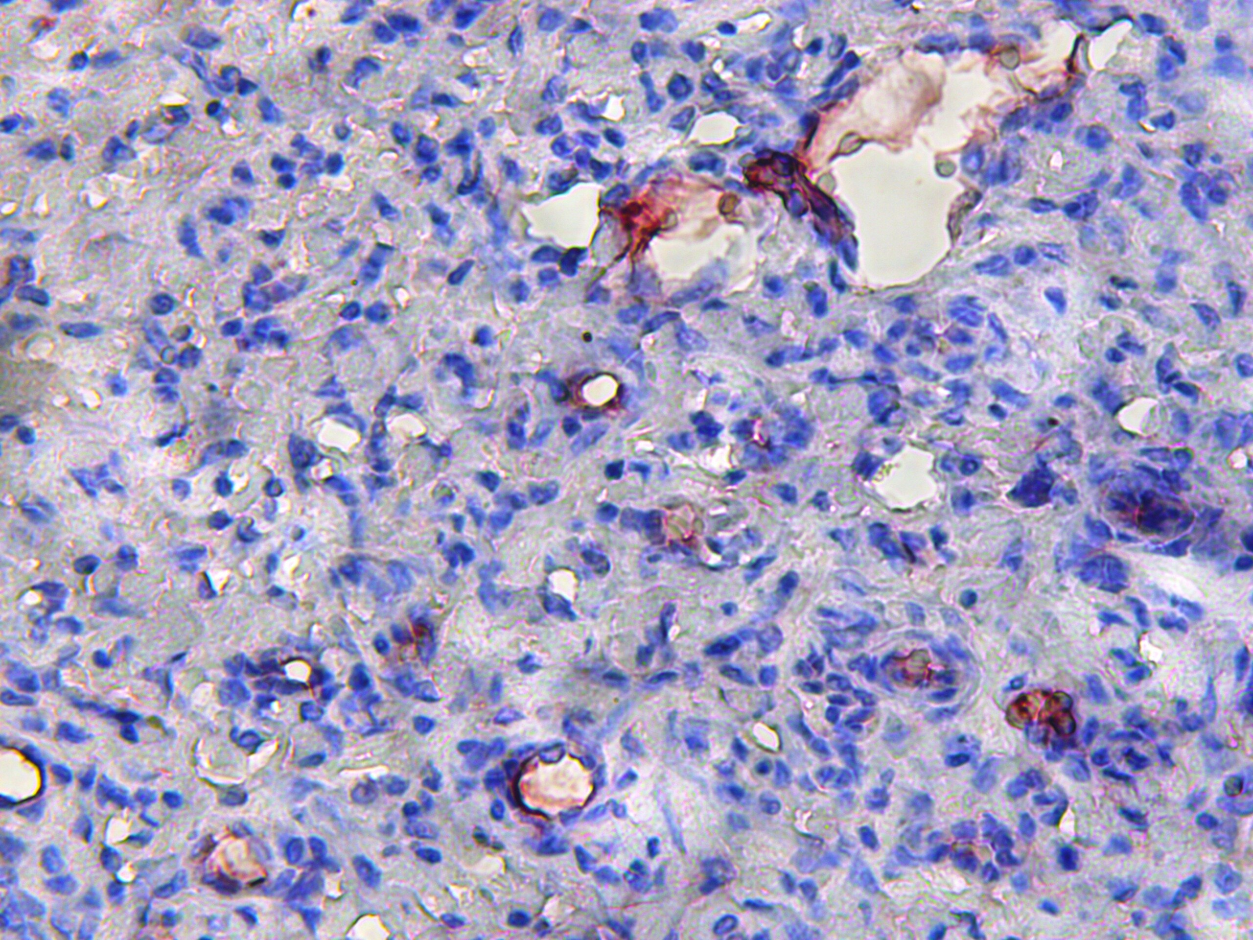
![Immunohistochemistry-Frozen: CD31/PECAM-1 Antibody (TLD-3A12) - BSA Free [NB100-64796]](https://resources.rndsystems.com/images/products/CD31-PECAM-1-Antibody-TLD-3A12-Immunohistochemistry-Frozen-NB100-64796-img0012.jpg "Immunohistochemistry-Frozen: CD31/PECAM-1 Antibody (TLD-3A12) - BSA Free [NB100-64796]")
Key Product Details
Species Reactivity
Validated:
Rat
Cited:
Human, Mouse, Rat, Rabbit
Applications
Validated:
Immunohistochemistry, Immunohistochemistry-Paraffin, Immunohistochemistry-Frozen, Western Blot, ELISA, Flow Cytometry, Immunocytochemistry/ Immunofluorescence
Cited:
Immunohistochemistry, Immunohistochemistry-Paraffin, Immunohistochemistry-Frozen, Flow Cytometry, Immunocytochemistry/ Immunofluorescence, IF/IHC
Label
Unconjugated
Antibody Source
Monoclonal Mouse IgG1 Clone # TLD-3A12
Format
BSA Free
Loading...
Product Specifications
Immunogen
Activated, Lewis rat derived microglial cells
Reactivity Notes
Predicted cross-reactivities: Rhesus Monkey, Porcine
Localization
Membrane; single pass type I membrane protein.
Clonality
Monoclonal
Host
Mouse
Isotype
IgG1
Theoretical MW
82.5 kDa.
Disclaimer note: The observed molecular weight of the protein may vary from the listed predicted molecular weight due to post translational modifications, post translation cleavages, relative charges, and other experimental factors.
Disclaimer note: The observed molecular weight of the protein may vary from the listed predicted molecular weight due to post translational modifications, post translation cleavages, relative charges, and other experimental factors.
Scientific Data Images for CD31/PECAM-1 Antibody (TLD-3A12) - BSA Free
Immunohistochemistry-Frozen: CD31/PECAM-1 Antibody (TLD-3A12) - BSA Free [NB100-64796]
Immunohistochemistry-Frozen: CD31/PECAM-1 Antibody (TLD-3A12) [NB100-64796] - Analysis using the FITC conjugate of CD31/PECAM-1 Antibody (TLD-3A12) (NB100-64796). Staining of rat brain cryosection with Mouse.![Flow Cytometry: CD31/PECAM-1 Antibody (TLD-3A12) - BSA Free [NB100-64796]](https://resources.rndsystems.com/images/products/CD31-PECAM-1-Antibody-TLD-3A12-Flow-Cytometry-NB100-64796-img0007.jpg "Flow Cytometry: CD31/PECAM-1 Antibody (TLD-3A12) - BSA Free [NB100-64796]")
Flow Cytometry: CD31/PECAM-1 Antibody (TLD-3A12) - BSA Free [NB100-64796]
Flow Cytometry: CD31/PECAM-1 Antibody (TLD-3A12) [NB100-64796] - Analysis using the Alexa Fluor (R) 488 conjugate of CD31/PECAM-1 Antibody (TLD-3A12) (NB100-64796). Staining of PBMC's (Monocyte gate at 1 x 10^6 cells/ml) with CD31 antibody (orange) stained at a dilution of 1:500. Shown with mIgG (AF488) isotype control (blue).![Immunohistochemistry-Paraffin: CD31/PECAM-1 Antibody (TLD-3A12) - BSA Free [NB100-64796]](https://resources.rndsystems.com/images/products/CD31-PECAM-1-Antibody-TLD-3A12-Immunohistochemistry-Paraffin-NB100-64796-img0005.jpg "Immunohistochemistry-Paraffin: CD31/PECAM-1 Antibody (TLD-3A12) - BSA Free [NB100-64796]")
Immunohistochemistry-Paraffin: CD31/PECAM-1 Antibody (TLD-3A12) - BSA Free [NB100-64796]
Immunohistochemistry-Paraffin: CD31/PECAM-1 Antibody (TLD-3A12) [NB100-64796] - Analysis of CD31 in 4th day cutaneous wound section of diabetic rat using CD31/PECAM-1 Antibody (TLD-3A12). Image from verified customer review.![Immunohistochemistry-Frozen: CD31/PECAM-1 Antibody (TLD-3A12) - BSA Free [NB100-64796]](https://resources.rndsystems.com/images/products/CD31-PECAM-1-Antibody-TLD-3A12-Immunohistochemistry-Frozen-NB100-64796-img0013.jpg "Immunohistochemistry-Frozen: CD31/PECAM-1 Antibody (TLD-3A12) - BSA Free [NB100-64796]")
Immunohistochemistry-Frozen: CD31/PECAM-1 Antibody (TLD-3A12) - BSA Free [NB100-64796]
Immunohistochemistry-Frozen: CD31/PECAM-1 Antibody (TLD-3A12) [NB100-64796] - Analysis using the FITC conjugate of CD31/PECAM-1 Antibody (TLD-3A12) (NB100-64796). Staining of rat spleen cryosection.![Immunohistochemistry-Paraffin: CD31/PECAM-1 Antibody (TLD-3A12) - BSA Free [NB100-64796]](https://resources.rndsystems.com/images/products/CD31-PECAM-1-Antibody-TLD-3A12-Immunohistochemistry-Paraffin-NB100-64796-img0014.jpg "Immunohistochemistry-Paraffin: CD31/PECAM-1 Antibody (TLD-3A12) - BSA Free [NB100-64796]")
Immunohistochemistry-Paraffin: CD31/PECAM-1 Antibody (TLD-3A12) - BSA Free [NB100-64796]
CD31-PECAM-1-Antibody-TLD-3A12-Immunohistochemistry-Paraffin-NB100-64796-img0014.jpg - BSA Free [NB100-64796] -")
Immunohistochemistry: CD31/PECAM-1 Antibody (TLD-3A12) - BSA Free [NB100-64796] -
(a) HE staining for wound tissues of different groups of rats (magnification ×40). (b) IHC analysis for the expression of CD31 of wound tissues of different groups of rats (magnification ×200). n = 6, ∗∗P < 0.01.Applications for CD31/PECAM-1 Antibody (TLD-3A12) - BSA Free
Application
Recommended Usage
ELISA
1:100-1:2000
Flow Cytometry
1:10-1:100
Immunocytochemistry/ Immunofluorescence
1:10-1:500
Immunohistochemistry
1:10-1:500
Immunohistochemistry-Frozen
1:10-1:100
Immunohistochemistry-Paraffin
1:10-1:500
Reviewed Applications
Read 1 review rated 5 using NB100-64796 in the following applications:
Flow Cytometry Panel Builder
Bio-Techne Knows Flow Cytometry
Save time and reduce costly mistakes by quickly finding compatible reagents using the Panel Builder Tool.
Advanced Features
- Spectra Viewer - Custom analysis of spectra from multiple fluorochromes
- Spillover Popups - Visualize the spectra of individual fluorochromes
- Antigen Density Selector - Match fluorochrome brightness with antigen density

Formulation, Preparation, and Storage
Purification
Protein A purified
Formulation
PBS
Format
BSA Free
Preservative
0.09% Sodium Azide
Concentration
1.0 mg/ml
Shipping
The product is shipped with polar packs. Upon receipt, store it immediately at the temperature recommended below.
Stability & Storage
Store at 4C short term. Aliquot and store at -20C long term. Avoid freeze-thaw cycles.
Background: CD31/PECAM-1
PECAM's intracellular cytoplasmic domain consists of a sequence of 118 amino acids and contains serine and tyrosine (also referred to as immunoreceptor tyrosine-based inhibitory motifs-ITIMs) residues, which may be phosphorylated upon cellular stimulation (3). ITIMs are phosphorylated by Src-family kinases and non-Src family kinases (e.g., Csk), leading to a conformational change which supports interactions with Src homology 2 (SH2) domain containing proteins such as protein-tyrosine phosphatase, SHP-2 (1,2). Formation of SHP-2/PECAM-1 complexes induces endothelial cell migration through the dephosphorylation of focal adhesion kinase and regulation of RhoA activity (1). Signaling downstream of ITIM tyrosine phosphorylations also plays a role in PECAM's anti-apoptotic activity, a function which is independent of its interaction with SHP-2. In platelets and leukocytes, phosphorylation of PECAM's cytosolic domain is inhibitory, preventing their activation.
References
1. Lertkiatmongkol, P., Liao, D., Mei, H., Hu, Y., & Newman, P. J. (2016). Endothelial functions of PECAM-1 (CD31). Current Opinion in Hematology. https://doi.org/10.1097/MOH.0000000000000239.Endothelial
2. Privratsky, J. R., & Newman, P. J. (2014). PECAM-1: Regulator of endothelial junctional integrity. Cell and Tissue Research. https://doi.org/10.1007/s00441-013-1779-3
3. Newman, P. J., & Newman, D. K. (2003). Signal transduction pathways mediated by PECAM-1: New roles for an old molecule in platelet and vascular cell biology. Arteriosclerosis, Thrombosis, and Vascular Biology. https://doi.org/10.1161/01.ATV.0000071347.69358.D9
Long Name
Platelet Endothelial Cell Adhesion Molecule 1
Alternate Names
CD31, EndoCAM, PECA1, PECAM-1, PECAM1, TLD-3A12 CD31, TLD-3A12 Clone
Gene Symbol
PECAM1
UniProt
Additional CD31/PECAM-1 Products
Product Documents for CD31/PECAM-1 Antibody (TLD-3A12) - BSA Free
Certificate of Analysis
To download a Certificate of Analysis, please enter a lot or batch number in the search box below.
Product Specific Notices for CD31/PECAM-1 Antibody (TLD-3A12) - BSA Free
This product is for research use only and is not approved for use in humans or in clinical diagnosis. Primary Antibodies are guaranteed for 1 year from date of receipt.
Related Research Areas
Citations for CD31/PECAM-1 Antibody (TLD-3A12) - BSA Free
 Powered by Bioz
Customer Reviews for CD31/PECAM-1 Antibody (TLD-3A12) - BSA Free (1)
5 out of 5
1 Customer Rating
Have you used CD31/PECAM-1 Antibody (TLD-3A12) - BSA Free?
Submit a review and receive an Amazon gift card!
$25/€18/£15/$25CAN/¥2500 Yen for a review with an image
$10/€7/£6/$10CAN/¥1110 Yen for a review without an image
Submit a review
Customer Images
Showing
1
-
1 of
1 review
Showing All
Filter By:
-
Application: ImmunocytochemistrySample Tested:Species: RatVerified Customer | Posted 05/29/2015Immunohistochemistry for CD31 in 14th day cutaneous wound section of diabetic rat.

There are no reviews that match your criteria.
Protocols
Find general support by application which include: protocols, troubleshooting, illustrated assays, videos and webinars.
- 7-Amino Actinomycin D (7-AAD) Cell Viability Flow Cytometry Protocol
- Antigen Retrieval Protocol (PIER)
- Antigen Retrieval for Frozen Sections Protocol
- Appropriate Fixation of IHC/ICC Samples
- Cellular Response to Hypoxia Protocols
- Chromogenic IHC Staining of Formalin-Fixed Paraffin-Embedded (FFPE) Tissue Protocol
- Chromogenic Immunohistochemistry Staining of Frozen Tissue
- ClariTSA™ Fluorophore Kits
- Detection & Visualization of Antibody Binding
- ELISA Sample Preparation & Collection Guide
- ELISA Troubleshooting Guide
- Extracellular Membrane Flow Cytometry Protocol
- Flow Cytometry Protocol for Cell Surface Markers
- Flow Cytometry Protocol for Staining Membrane Associated Proteins
- Flow Cytometry Staining Protocols
- Flow Cytometry Troubleshooting Guide
- Fluorescent IHC Staining of Frozen Tissue Protocol
- Graphic Protocol for Heat-induced Epitope Retrieval
- Graphic Protocol for the Preparation and Fluorescent IHC Staining of Frozen Tissue Sections
- Graphic Protocol for the Preparation and Fluorescent IHC Staining of Paraffin-embedded Tissue Sections
- Graphic Protocol for the Preparation of Gelatin-coated Slides for Histological Tissue Sections
- How to Run an R&D Systems DuoSet ELISA
- How to Run an R&D Systems Quantikine ELISA
- How to Run an R&D Systems Quantikine™ QuicKit™ ELISA
- ICC Cell Smear Protocol for Suspension Cells
- ICC Immunocytochemistry Protocol Videos
- ICC for Adherent Cells
- IHC Sample Preparation (Frozen sections vs Paraffin)
- Immunocytochemistry (ICC) Protocol
- Immunocytochemistry Troubleshooting
- Immunofluorescence of Organoids Embedded in Cultrex Basement Membrane Extract
- Immunofluorescent IHC Staining of Formalin-Fixed Paraffin-Embedded (FFPE) Tissue Protocol
- Immunohistochemistry (IHC) and Immunocytochemistry (ICC) Protocols
- Immunohistochemistry Frozen Troubleshooting
- Immunohistochemistry Paraffin Troubleshooting
- Intracellular Flow Cytometry Protocol Using Alcohol (Methanol)
- Intracellular Flow Cytometry Protocol Using Detergents
- Intracellular Nuclear Staining Flow Cytometry Protocol Using Detergents
- Intracellular Staining Flow Cytometry Protocol Using Alcohol Permeabilization
- Intracellular Staining Flow Cytometry Protocol Using Detergents to Permeabilize Cells
- Preparing Samples for IHC/ICC Experiments
- Preventing Non-Specific Staining (Non-Specific Binding)
- Primary Antibody Selection & Optimization
- Propidium Iodide Cell Viability Flow Cytometry Protocol
- Protocol for Heat-Induced Epitope Retrieval (HIER)
- Protocol for Liperfluo
- Protocol for Making a 4% Formaldehyde Solution in PBS
- Protocol for VisUCyte™ HRP Polymer Detection Reagent
- Protocol for the Characterization of Human Th22 Cells
- Protocol for the Characterization of Human Th9 Cells
- Protocol for the Fluorescent ICC Staining of Cell Smears - Graphic
- Protocol for the Fluorescent ICC Staining of Cultured Cells on Coverslips - Graphic
- Protocol for the Preparation & Fixation of Cells on Coverslips
- Protocol for the Preparation and Chromogenic IHC Staining of Frozen Tissue Sections
- Protocol for the Preparation and Chromogenic IHC Staining of Frozen Tissue Sections - Graphic
- Protocol for the Preparation and Chromogenic IHC Staining of Paraffin-embedded Tissue Sections
- Protocol for the Preparation and Chromogenic IHC Staining of Paraffin-embedded Tissue Sections - Graphic
- Protocol for the Preparation and Fluorescent ICC Staining of Cells on Coverslips
- Protocol for the Preparation and Fluorescent ICC Staining of Non-adherent Cells
- Protocol for the Preparation and Fluorescent ICC Staining of Stem Cells on Coverslips
- Protocol for the Preparation and Fluorescent IHC Staining of Frozen Tissue Sections
- Protocol for the Preparation and Fluorescent IHC Staining of Paraffin-embedded Tissue Sections
- Protocol for the Preparation of Gelatin-coated Slides for Histological Tissue Sections
- Protocol for the Preparation of a Cell Smear for Non-adherent Cell ICC - Graphic
- Protocol: Annexin V and PI Staining by Flow Cytometry
- Protocol: Annexin V and PI Staining for Apoptosis by Flow Cytometry
- Quantikine HS ELISA Kit Assay Principle, Alkaline Phosphatase
- Quantikine HS ELISA Kit Principle, Streptavidin-HRP Polymer
- R&D Systems Quality Control Western Blot Protocol
- Sandwich ELISA (Colorimetric) – Biotin/Streptavidin Detection Protocol
- Sandwich ELISA (Colorimetric) – Direct Detection Protocol
- TUNEL and Active Caspase-3 Detection by IHC/ICC Protocol
- The Importance of IHC/ICC Controls
- Troubleshooting Guide: ELISA
- Troubleshooting Guide: Fluorokine Flow Cytometry Kits
- Troubleshooting Guide: Immunohistochemistry
- Troubleshooting Guide: Western Blot Figures
- Western Blot Conditions
- Western Blot Protocol
- Western Blot Protocol for Cell Lysates
- Western Blot Troubleshooting
- Western Blot Troubleshooting Guide
- View all Protocols, Troubleshooting, Illustrated assays and Webinars
FAQs for CD31/PECAM-1 Antibody (TLD-3A12) - BSA Free
Showing
1
-
5 of
5 FAQs
Showing All
-
Q: Can you give me more information about this antibody?
A: Our mouse monoclonal antibody CD31/PECAM-1 Antibody (TLD-3A12) NB100-64796 was raised against activated Lewis rat derived microglial cells. Since we do not routinely epitope map our antibodies however, I am unable to tell you exactly where on the protein it binds.
-
Q: I potentially am interested in CD31/PECAM1 Antibody (TLD-3A12). I am woundering if this AB is suitable to use for immunohistochemistry with PFA-fixed section. Is it suitable?
A: Yes, this antibody is suitable for IHC with PFA
-
Q: What is the concentration NB100-64796?
A: The volume of the antibody provided is 250ul. The concentration of the antibody is 1.0mg/ml.
-
Q: What is the size of the target protein CD31/PECAM for this antibody?
A: In western blot, we see bands between 115-160kDa for this particular target. This weight varies from the theoretical molecular weight of 76kDa due to glycosylation of the protein.
-
Q: Would this antibody apply to tissue that fixed after the formalin?
A: Formalin fixation should be acceptable for use with this antibody. We incubate freshly dissected tissue in 10% formalin for 4-24 hours, although the incubation time is dependent on tissue type and size. We do not recommend fixation for longer than 24 hours since this can cause epitope masking.
-
Q: Can you give me more information about this antibody?
A: Our mouse monoclonal antibody CD31/PECAM-1 Antibody (TLD-3A12) NB100-64796 was raised against activated Lewis rat derived microglial cells. Since we do not routinely epitope map our antibodies however, I am unable to tell you exactly where on the protein it binds.
-
Q: I potentially am interested in CD31/PECAM1 Antibody (TLD-3A12). I am woundering if this AB is suitable to use for immunohistochemistry with PFA-fixed section. Is it suitable?
A: Yes, this antibody is suitable for IHC with PFA
-
Q: What is the concentration NB100-64796?
A: The volume of the antibody provided is 250ul. The concentration of the antibody is 1.0mg/ml.
-
Q: What is the size of the target protein CD31/PECAM for this antibody?
A: In western blot, we see bands between 115-160kDa for this particular target. This weight varies from the theoretical molecular weight of 76kDa due to glycosylation of the protein.
-
Q: Would this antibody apply to tissue that fixed after the formalin?
A: Formalin fixation should be acceptable for use with this antibody. We incubate freshly dissected tissue in 10% formalin for 4-24 hours, although the incubation time is dependent on tissue type and size. We do not recommend fixation for longer than 24 hours since this can cause epitope masking.
Loading...