CD31/PECAM-1 Antibody - BSA Free
Novus Biologicals | Catalog # NBP1-71663
![Immunocytochemistry/ Immunofluorescence: CD31/PECAM-1 Antibody - BSA Free [NBP1-71663]](https://resources.rndsystems.com/images/products/CD31-PECAM-1-Antibody-Immunocytochemistry-Immunofluorescence-NBP1-71663-img0009.jpg "Immunocytochemistry/ Immunofluorescence: CD31/PECAM-1 Antibody - BSA Free [NBP1-71663]")
Key Product Details
Validated by
Biological Validation
Species Reactivity
Validated:
Human, Mouse
Cited:
Human, Mouse
Applications
Validated:
Immunohistochemistry, Immunohistochemistry-Paraffin, Immunohistochemistry-Frozen, Western Blot, Flow Cytometry, Immunocytochemistry/ Immunofluorescence, Simple Western
Cited:
Immunohistochemistry, Immunohistochemistry-Paraffin, Immunohistochemistry-Frozen, Western Blot, Immunocytochemistry/ Immunofluorescence, In vivo assay, IF/IHC
Label
Unconjugated
Antibody Source
Polyclonal Rabbit IgG
Format
BSA Free
Loading...
Product Specifications
Immunogen
This CD31/PECAM-1 Antibody was developed against a genomic peptide made to an internal region of human CD31/PECAM1 (within residues 100-300) [Swiss-Prot P16284].
Localization
Membrane; Single-pass type I membrane protein. Cell junction. Note: Localizes to the lateral border recycling compartment (LBRC) and recycles from the LBRC to the junction in resting endothelial cells.
Clonality
Polyclonal
Host
Rabbit
Isotype
IgG
Theoretical MW
82.5 kDa.
Disclaimer note: The observed molecular weight of the protein may vary from the listed predicted molecular weight due to post translational modifications, post translation cleavages, relative charges, and other experimental factors.
Disclaimer note: The observed molecular weight of the protein may vary from the listed predicted molecular weight due to post translational modifications, post translation cleavages, relative charges, and other experimental factors.
Scientific Data Images for CD31/PECAM-1 Antibody - BSA Free
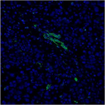
Immunocytochemistry/ Immunofluorescence: CD31/PECAM-1 Antibody - BSA Free [NBP1-71663]
Immunocytochemistry/Immunofluorescence: CD31/PECAM-1 Antibody [NBP1-71663] - Staining of mouse glioma tissue using CD31/PECAM-1 Antibody (NBP1-71663) at a dilution of 1:2500. Image provided by Dr. Eric Woolf of Dignity Health.![Simple Western: CD31/PECAM-1 AntibodyBSA Free [NBP1-71663]](https://resources.rndsystems.com/images/products/CD31-PECAM-1-Antibody-Simple-Western-NBP1-71663-img0010.jpg "Simple Western: CD31/PECAM-1 AntibodyBSA Free [NBP1-71663]")
Simple Western: CD31/PECAM-1 AntibodyBSA Free [NBP1-71663]
Simple Western: CD31/PECAM-1 Antibody [NBP1-71663] - Image shows a specific band for CD31/PECAM 1 in 0.5 mg/mL of HUVEC lysate. This experiment was performed under reducing conditions using the 12-230 kDa separation system.![Immunohistochemistry: CD31/PECAM-1 Antibody - BSA Free [NBP1-71663]](https://resources.rndsystems.com/images/products/CD31-PECAM-1-Antibody-Immunohistochemistry-NBP1-71663-img0012.jpg "Immunohistochemistry: CD31/PECAM-1 Antibody - BSA Free [NBP1-71663]")
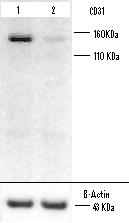
![Western Blot: CD31/PECAM-1 AntibodyBSA Free [NBP1-71663]](https://resources.rndsystems.com/images/products/CD31-PECAM-1-Antibody-Western-Blot-NBP1-71663-img0013.jpg "Western Blot: CD31/PECAM-1 AntibodyBSA Free [NBP1-71663]")
Western Blot: CD31/PECAM-1 AntibodyBSA Free [NBP1-71663]
CD31-PECAM-1-Antibody-Western-Blot-NBP1-71663-img0013.jpg![Immunocytochemistry/ Immunofluorescence: CD31/PECAM-1 Antibody - BSA Free [NBP1-71663]](https://resources.rndsystems.com/images/products/CD31-PECAM-1-Antibody-Immunocytochemistry-Immunofluorescence-NBP1-71663-img0011.jpg "Immunocytochemistry/ Immunofluorescence: CD31/PECAM-1 Antibody - BSA Free [NBP1-71663]")
Immunocytochemistry/ Immunofluorescence: CD31/PECAM-1 Antibody - BSA Free [NBP1-71663]
Immunocytochemistry/Immunofluorescence: CD31/PECAM-1 Antibody [NBP1-71663] - HUVEC cells were fixed for 10 minutes using 10% formalin and then permeabilized for 5 minutes using 1X TBS + 0.5% Triton X-100. The cells were incubated with CD31/PECAM-1 Antibody at 5 ug/mL overnight at 4C and detected with an anti-rabbit Dylight 488 (Green) at a 1:500 dilution. Alpha tubulin (DM1A) NB100-690 was used as a co-stain at a 1:1000 dilution and detected with an anti-mouse Dylight 550 (Red) at a 1:500 dilution. Nuclei were counterstained with DAPI (Blue). Cells were imaged using a 40X objective.![Flow Cytometry: CD31/PECAM-1 Antibody - BSA Free [NBP1-71663]](https://resources.rndsystems.com/images/products/CD31-PECAM-1-Antibody-Flow-Cytometry-NBP1-71663-img0005.jpg "Flow Cytometry: CD31/PECAM-1 Antibody - BSA Free [NBP1-71663]")
Flow Cytometry: CD31/PECAM-1 Antibody - BSA Free [NBP1-71663]
Flow Cytometry: CD31/PECAM-1 Antibody [NBP1-71663] - CD31/PECAM-1 Antibody was tested in WeHi cells with Dylight 488 (green) alongside a matched isotype control (black).![Western Blot: CD31/PECAM-1 AntibodyBSA Free [NBP1-71663]](https://resources.rndsystems.com/images/products/CD31-PECAM-1-Antibody-Western-Blot-NBP1-71663-img0008.jpg "Western Blot: CD31/PECAM-1 AntibodyBSA Free [NBP1-71663]")
Western Blot: CD31/PECAM-1 AntibodyBSA Free [NBP1-71663]
Western Blot: CD31/PECAM-1 Antibody [NBP1-71663] - Detection of CD31 in HUVEC cell lysates.Applications for CD31/PECAM-1 Antibody - BSA Free
Application
Recommended Usage
Flow Cytometry
1:1000
Immunocytochemistry/ Immunofluorescence
1:10-1:500
Immunohistochemistry
1:50
Immunohistochemistry-Frozen
reported in scientific literature (PMID 35879332)
Immunohistochemistry-Paraffin
1:50
Simple Western
1:1000
Western Blot
1:10000
Application Notes
In Western Blot a band is seen ~150 kDa. Prior to immunostaining paraffin tissues, antigen retrieval with sodium citrate buffer (pH 6.0) is recommended. Customer feedback has been negative on cryosections.
In Simple Western only 10 - 15 uL of the recommended dilution is used per data point.
See Simple Western Antibody Database for Simple Western validation: Tested in HUVEC lysate 0.5 mg/mL, separated by Size, antibody dilution of 1:1000, apparent MW was 166 kDa. Separated by Size-Wes, Sally Sue/Peggy Sue.
Reviewed Applications
Read 2 reviews rated 5 using NBP1-71663 in the following applications:
Flow Cytometry Panel Builder
Bio-Techne Knows Flow Cytometry
Save time and reduce costly mistakes by quickly finding compatible reagents using the Panel Builder Tool.
Advanced Features
- Spectra Viewer - Custom analysis of spectra from multiple fluorochromes
- Spillover Popups - Visualize the spectra of individual fluorochromes
- Antigen Density Selector - Match fluorochrome brightness with antigen density

Formulation, Preparation, and Storage
Purification
Immunogen affinity purified
Formulation
PBS, 30% Glycerol
Format
BSA Free
Preservative
0.02% Sodium Azide
Concentration
1.0 mg/ml
Shipping
The product is shipped with polar packs. Upon receipt, store it immediately at the temperature recommended below.
Stability & Storage
Store at 4C short term. Aliquot and store at -20C long term. Avoid freeze-thaw cycles.
Background: CD31/PECAM-1
PECAM's intracellular cytoplasmic domain consists of a sequence of 118 amino acids and contains serine and tyrosine (also referred to as immunoreceptor tyrosine-based inhibitory motifs-ITIMs) residues, which may be phosphorylated upon cellular stimulation (3). ITIMs are phosphorylated by Src-family kinases and non-Src family kinases (e.g., Csk), leading to a conformational change which supports interactions with Src homology 2 (SH2) domain containing proteins such as protein-tyrosine phosphatase, SHP-2 (1,2). Formation of SHP-2/PECAM-1 complexes induces endothelial cell migration through the dephosphorylation of focal adhesion kinase and regulation of RhoA activity (1). Signaling downstream of ITIM tyrosine phosphorylations also plays a role in PECAM's anti-apoptotic activity, a function which is independent of its interaction with SHP-2. In platelets and leukocytes, phosphorylation of PECAM's cytosolic domain is inhibitory, preventing their activation.
References
1. Lertkiatmongkol, P., Liao, D., Mei, H., Hu, Y., & Newman, P. J. (2016). Endothelial functions of PECAM-1 (CD31). Current Opinion in Hematology. https://doi.org/10.1097/MOH.0000000000000239.Endothelial
2. Privratsky, J. R., & Newman, P. J. (2014). PECAM-1: Regulator of endothelial junctional integrity. Cell and Tissue Research. https://doi.org/10.1007/s00441-013-1779-3
3. Newman, P. J., & Newman, D. K. (2003). Signal transduction pathways mediated by PECAM-1: New roles for an old molecule in platelet and vascular cell biology. Arteriosclerosis, Thrombosis, and Vascular Biology. https://doi.org/10.1161/01.ATV.0000071347.69358.D9
Long Name
Platelet Endothelial Cell Adhesion Molecule 1
Alternate Names
CD31, EndoCAM, PECA1, PECAM-1, PECAM1
Gene Symbol
PECAM1
Additional CD31/PECAM-1 Products
Product Documents for CD31/PECAM-1 Antibody - BSA Free
Certificate of Analysis
To download a Certificate of Analysis, please enter a lot or batch number in the search box below.
Product Specific Notices for CD31/PECAM-1 Antibody - BSA Free
Manufactured by Genomic Antibody Technology™. GAT FAQs
This product is for research use only and is not approved for use in humans or in clinical diagnosis. Primary Antibodies are guaranteed for 1 year from date of receipt.
Related Research Areas
Citations for CD31/PECAM-1 Antibody - BSA Free
 Powered by Bioz
Customer Reviews for CD31/PECAM-1 Antibody - BSA Free (2)
5 out of 5
2 Customer Ratings
Have you used CD31/PECAM-1 Antibody - BSA Free?
Submit a review and receive an Amazon gift card!
$25/€18/£15/$25CAN/¥2500 Yen for a review with an image
$10/€7/£6/$10CAN/¥1110 Yen for a review without an image
Submit a review
Customer Images
Showing
1
-
2 of
2 reviews
Showing All
Filter By:
-
Application: Western BlotSample Tested: Mouse glioma tissue and mouse normal brain tissue.Species: MouseVerified Customer | Posted 09/05/2013

-
Application: ImmunofluorescenceSample Tested: Mouse glioma tissueSpecies: MouseVerified Customer | Posted 09/05/2013
There are no reviews that match your criteria.
Protocols
View specific protocols for CD31/PECAM-1 Antibody - BSA Free (NBP1-71663):
Immunohistochemistry-Paraffin Embedded Sections
Antigen Unmasking:
Bring slides to a boil in 10 mM sodium citrate buffer (pH 6.0) then maintain at a sub-boiling temperature for 10 minutes. Cool slides on bench-top for 30 minutes.
Staining:
1. Wash sections in deionized water three times for 5 minutes each.
2. Wash sections in wash buffer for 5 minutes.
3. Block each section with 100-400 ul blocking solution for 1 hour at room temperature.
4. Remove blocking solution and add 100-400 ul diluted primary antibody. Incubate overnight at 4C.
5. Remove antibody solution and wash sections in wash buffer three times for 5 minutes each.
6. Add 100-400 ul biotinylated diluted secondary antibody. Incubate 30 minutes at room temperature.
7. Remove secondary antibody solution and wash sections three times with wash buffer for 5 minutes each.
8. Add 100-400 ul Streptavidin-HRP reagent to each section and incubate for 30 minutes at room temperature.
9. Wash sections three times in wash buffer for 5 minutes each.
10. Add 100-400 ul DAB substrate to each section and monitor staining closely.
11. As soon as the sections develop, immerse slides in deionized water.
12. Counterstain sections in hematoxylin.
13. Wash sections in deionized water two times for 5 minutes each.
14. Dehydrate sections.
15. Mount coverslips.
Western Blot Protocol
1. Perform SDS-PAGE on samples to be analyzed, loading 40 ug of total protein per lane.
2. Transfer proteins to membrane according to the instructions provided by the manufacturer of the membrane and transfer apparatus.
3. Stain according to standard Ponceau S procedure (or similar product) to assess transfer success, and mark molecular weight standards where appropriate.
4. Rinse the blot.
5. Block the membrane using standard blocking buffer for at least 1 hour.
6. Wash the membrane in wash buffer three times for 10 minutes each.
7. Dilute primary antibody in blocking buffer and incubate 1 hour at room temperature.
8. Wash the membrane in wash buffer three times for 10 minutes each.
9. Apply the diluted HRP conjugated secondary antibody in blocking buffer (as per manufacturers instructions) and incubate 1 hour at room temperature.
10. Wash the blot in wash buffer three times for 10 minutes each (this step can be repeated as required to reduce background).
11. Apply the detection reagent of choice in accordance with the manufacturers instructions.
*Note: Tween-20 can be added to the blocking or antibody dilution buffer at a final concentration of 0.05-0.2%.
Find general support by application which include: protocols, troubleshooting, illustrated assays, videos and webinars.
- 7-Amino Actinomycin D (7-AAD) Cell Viability Flow Cytometry Protocol
- Antigen Retrieval Protocol (PIER)
- Antigen Retrieval for Frozen Sections Protocol
- Appropriate Fixation of IHC/ICC Samples
- Cellular Response to Hypoxia Protocols
- Chromogenic IHC Staining of Formalin-Fixed Paraffin-Embedded (FFPE) Tissue Protocol
- Chromogenic Immunohistochemistry Staining of Frozen Tissue
- ClariTSA™ Fluorophore Kits
- Detection & Visualization of Antibody Binding
- Extracellular Membrane Flow Cytometry Protocol
- Flow Cytometry Protocol for Cell Surface Markers
- Flow Cytometry Protocol for Staining Membrane Associated Proteins
- Flow Cytometry Staining Protocols
- Flow Cytometry Troubleshooting Guide
- Fluorescent IHC Staining of Frozen Tissue Protocol
- Graphic Protocol for Heat-induced Epitope Retrieval
- Graphic Protocol for the Preparation and Fluorescent IHC Staining of Frozen Tissue Sections
- Graphic Protocol for the Preparation and Fluorescent IHC Staining of Paraffin-embedded Tissue Sections
- Graphic Protocol for the Preparation of Gelatin-coated Slides for Histological Tissue Sections
- ICC Cell Smear Protocol for Suspension Cells
- ICC Immunocytochemistry Protocol Videos
- ICC for Adherent Cells
- IHC Sample Preparation (Frozen sections vs Paraffin)
- Immunocytochemistry (ICC) Protocol
- Immunocytochemistry Troubleshooting
- Immunofluorescence of Organoids Embedded in Cultrex Basement Membrane Extract
- Immunofluorescent IHC Staining of Formalin-Fixed Paraffin-Embedded (FFPE) Tissue Protocol
- Immunohistochemistry (IHC) and Immunocytochemistry (ICC) Protocols
- Immunohistochemistry Frozen Troubleshooting
- Immunohistochemistry Paraffin Troubleshooting
- Intracellular Flow Cytometry Protocol Using Alcohol (Methanol)
- Intracellular Flow Cytometry Protocol Using Detergents
- Intracellular Nuclear Staining Flow Cytometry Protocol Using Detergents
- Intracellular Staining Flow Cytometry Protocol Using Alcohol Permeabilization
- Intracellular Staining Flow Cytometry Protocol Using Detergents to Permeabilize Cells
- Preparing Samples for IHC/ICC Experiments
- Preventing Non-Specific Staining (Non-Specific Binding)
- Primary Antibody Selection & Optimization
- Propidium Iodide Cell Viability Flow Cytometry Protocol
- Protocol for Heat-Induced Epitope Retrieval (HIER)
- Protocol for Liperfluo
- Protocol for Making a 4% Formaldehyde Solution in PBS
- Protocol for VisUCyte™ HRP Polymer Detection Reagent
- Protocol for the Characterization of Human Th22 Cells
- Protocol for the Characterization of Human Th9 Cells
- Protocol for the Fluorescent ICC Staining of Cell Smears - Graphic
- Protocol for the Fluorescent ICC Staining of Cultured Cells on Coverslips - Graphic
- Protocol for the Preparation & Fixation of Cells on Coverslips
- Protocol for the Preparation and Chromogenic IHC Staining of Frozen Tissue Sections
- Protocol for the Preparation and Chromogenic IHC Staining of Frozen Tissue Sections - Graphic
- Protocol for the Preparation and Chromogenic IHC Staining of Paraffin-embedded Tissue Sections
- Protocol for the Preparation and Chromogenic IHC Staining of Paraffin-embedded Tissue Sections - Graphic
- Protocol for the Preparation and Fluorescent ICC Staining of Cells on Coverslips
- Protocol for the Preparation and Fluorescent ICC Staining of Non-adherent Cells
- Protocol for the Preparation and Fluorescent ICC Staining of Stem Cells on Coverslips
- Protocol for the Preparation and Fluorescent IHC Staining of Frozen Tissue Sections
- Protocol for the Preparation and Fluorescent IHC Staining of Paraffin-embedded Tissue Sections
- Protocol for the Preparation of Gelatin-coated Slides for Histological Tissue Sections
- Protocol for the Preparation of a Cell Smear for Non-adherent Cell ICC - Graphic
- Protocol: Annexin V and PI Staining by Flow Cytometry
- Protocol: Annexin V and PI Staining for Apoptosis by Flow Cytometry
- R&D Systems Quality Control Western Blot Protocol
- TUNEL and Active Caspase-3 Detection by IHC/ICC Protocol
- The Importance of IHC/ICC Controls
- Troubleshooting Guide: Fluorokine Flow Cytometry Kits
- Troubleshooting Guide: Immunohistochemistry
- Troubleshooting Guide: Western Blot Figures
- Western Blot Conditions
- Western Blot Protocol
- Western Blot Protocol for Cell Lysates
- Western Blot Troubleshooting
- Western Blot Troubleshooting Guide
- View all Protocols, Troubleshooting, Illustrated assays and Webinars
FAQs for CD31/PECAM-1 Antibody - BSA Free
Showing
1
-
2 of
2 FAQs
Showing All
-
Q: I am looking for references concerning the use of NBP2-15797, NBP1-47913 and NB100-2284 in IHC on PFA- fixed section.
A: Among your mentioned catalog numbers (NBP2-15797, NBP1-47913 and NB100-2284), we have published references for NB100-2284 only. Hylander et al. 2013 (Journal of Translational Medicine 2013, 11:110) have cited the IHC-P use of NB100-2284 and this paper has also explained a nice detailed IHC-P protocol that you may follow if you want to. NBP1-71663 is our bestselling catalog number among all CD31/PECAM1 antibodies and it has been cited in two publications, both explaining its use in IHC-P assays (Pratheeshkumar et al. 2012 PLoS One. 2012;7(10):e47516; and Pratheeshkumar et al. 2012 PLoS One. 2012;7(12):e52279)
-
Q: I'm interested in purchasing primary CD31/PECAM1 antibody for immunohistochemistry staining on formalin fixed paraffin embedded mouse samples. You have two antibodies that would work for paraffin sections, NBP1-71663 and NB100-92205, and both are rabbit polyclonal antibody. Which one would you recommend I purchase?
A: I would recommend NBP1-71663 as the image is better than NB100-92205.
-
Q: I am looking for references concerning the use of NBP2-15797, NBP1-47913 and NB100-2284 in IHC on PFA- fixed section.
A: Among your mentioned catalog numbers (NBP2-15797, NBP1-47913 and NB100-2284), we have published references for NB100-2284 only. Hylander et al. 2013 (Journal of Translational Medicine 2013, 11:110) have cited the IHC-P use of NB100-2284 and this paper has also explained a nice detailed IHC-P protocol that you may follow if you want to. NBP1-71663 is our bestselling catalog number among all CD31/PECAM1 antibodies and it has been cited in two publications, both explaining its use in IHC-P assays (Pratheeshkumar et al. 2012 PLoS One. 2012;7(10):e47516; and Pratheeshkumar et al. 2012 PLoS One. 2012;7(12):e52279)
-
Q: I'm interested in purchasing primary CD31/PECAM1 antibody for immunohistochemistry staining on formalin fixed paraffin embedded mouse samples. You have two antibodies that would work for paraffin sections, NBP1-71663 and NB100-92205, and both are rabbit polyclonal antibody. Which one would you recommend I purchase?
A: I would recommend NBP1-71663 as the image is better than NB100-92205.
Loading...