Key Product Details
Species Reactivity
Human
Applications
Immunohistochemistry, Immunohistochemistry-Paraffin, Western Blot, Flow Cytometry, Immunocytochemistry/ Immunofluorescence
Label
Unconjugated
Antibody Source
Monoclonal Mouse IgG1 kappa Clone # SPM503
Loading...
Product Specifications
Immunogen
Myeloblastic KG1 cells
Localization
Cell surface
Marker
T-cell Marker
Specificity
It recognizes a cell surface glycoprotein of 95/115/135kDa (depending upon the extent of glycosylation), identified as CD43. 70-90% of T-cell lymphomas and from 22-37% of B-cell lymphomas express CD43. No reactivity has been observed with reactive B-cells. So, a B-lineage population that co-expresses CD43 is highly likely to be a malignant lymphoma, especially a low-grade lymphoma, rather than a reactive B-cell population. When CD43 antibody is used in combination with anti-CD20, effective immunophenotyping of the lymphomas in formalin-fixed tissues can be obtained. Co-staining of a lymphoid infiltrate with anti-CD20 and anti-CD43 argues against a reactive process and favors a diagnosis of lymphoma.
Clonality
Monoclonal
Host
Mouse
Isotype
IgG1 kappa
Description
200ug/ml of antibody purified from Bioreactor Concentrate by Protein A or G. Prepared in 10 mM PBS with 0.05% BSA & 0.05% azide. Also available WITHOUT BSA & azide at 1.0 mg/ml. (NBP2-34775)
Antibody with azide - store at 2 to 8C. Antibody without azide - store at -20 to -80 C.
Antibody with azide - store at 2 to 8C. Antibody without azide - store at -20 to -80 C.
Scientific Data Images for CD43/Sialophorin Antibody (SPM503)
![Western Blot: CD43/Sialophorin Antibody (SPM503) [NBP2-32822]](https://resources.rndsystems.com/images/products/CD43-Sialophorin-Antibody-SPM503-Western-Blot-NBP2-32822-img0004.jpg "Western Blot: CD43/Sialophorin Antibody (SPM503) [NBP2-32822]")
Western Blot: CD43/Sialophorin Antibody (SPM503) [NBP2-32822]
Western Blot: CD43/Sialophorin Antibody (SPM503) [NBP2-32822] - Western Blot Analysis of K562 cell lysate using CD43/Sialophorin Antibody (SPM503).![Immunocytochemistry/ Immunofluorescence: CD43/Sialophorin Antibody (SPM503) [NBP2-32822]](https://resources.rndsystems.com/images/products/CD43-Sialophorin-Antibody-SPM503-Immunocytochemistry-Immunofluorescence-NBP2-32822-img0003.jpg "Immunocytochemistry/ Immunofluorescence: CD43/Sialophorin Antibody (SPM503) [NBP2-32822]")
Immunocytochemistry/ Immunofluorescence: CD43/Sialophorin Antibody (SPM503) [NBP2-32822]
Immunocytochemistry/Immunofluorescence: CD43/Sialophorin Antibody (SPM503) [NBP2-32822] - Immunofluorescence Analysis of K562 cells labeling CD43 with CD43/Sialophorin Antibody (SPM503) followed by Goat anti-Mouse IgG-CF488 (Green). The nuclear counterstain is NucSpot Live 650.![Immunohistochemistry-Paraffin: CD43/Sialophorin Antibody (SPM503) [NBP2-32822]](https://resources.rndsystems.com/images/products/CD43-Sialophorin-Antibody-SPM503-Immunohistochemistry-Paraffin-NBP2-32822-img0006.jpg "Immunohistochemistry-Paraffin: CD43/Sialophorin Antibody (SPM503) [NBP2-32822]")
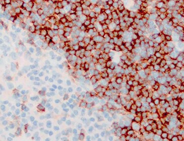
Immunohistochemistry-Paraffin: CD43/Sialophorin Antibody (SPM503) [NBP2-32822]
Immunohistochemistry-Paraffin: CD43/Sialophorin Antibody (SPM503) [NBP2-32822] - Formalin-fixed, paraffin-embedded human tonsil stained with CD43/Sialophorin antibody (SPM503). Primary antibody dilution: 1.0ug/mL Incubation: 1.0ug/mL for 30 minutes at room temperature. Image from verified customer review.![Flow Cytometry: CD43/Sialophorin Antibody (SPM503) [NBP2-32822]](https://resources.rndsystems.com/images/products/CD43-Sialophorin-Antibody-SPM503-Flow-Cytometry-NBP2-32822-img0001.jpg "Flow Cytometry: CD43/Sialophorin Antibody (SPM503) [NBP2-32822]")
Flow Cytometry: CD43/Sialophorin Antibody (SPM503) [NBP2-32822]
Flow Cytometry: CD43/Sialophorin Antibody (SPM503) [NBP2-32822] - CD43 expression by lymphocyte gated population of PBMC: PBMC stained either with CD43 MAb (SPM503) or isotype control.![Immunohistochemistry-Paraffin: CD43/Sialophorin Antibody (SPM503) [NBP2-32822]](https://resources.rndsystems.com/images/products/CD43-Sialophorin-Antibody-SPM503-Immunohistochemistry-Paraffin-NBP2-32822-img0005.jpg "Immunohistochemistry-Paraffin: CD43/Sialophorin Antibody (SPM503) [NBP2-32822]")
Immunohistochemistry-Paraffin: CD43/Sialophorin Antibody (SPM503) [NBP2-32822]
Immunohistochemistry-Paraffin: CD43/Sialophorin Antibody (SPM503) [NBP2-32822] - Formalin-fixed, paraffin-embedded human tonsil stained with CD43/Sialophorin Mouse Monoclonal Antibody (SPM503).Applications for CD43/Sialophorin Antibody (SPM503)
Application
Recommended Usage
Flow Cytometry
1-2 ug/million cells
Immunocytochemistry/ Immunofluorescence
1-2 ug/ml
Immunohistochemistry-Paraffin
1-2 ug/ml
Western Blot
1-2 ug/ml
Application Notes
Immunohistochemistry (Formalin-fixed): 1-2ug/ml for 30 minutes at RT. Staining of formalin-fixed tissues requires heating tissue sections in 10mM Tris with 1mM EDTA, pH 9.0, for 45 min at 95C followed by cooling at RT for 20 minutes.
Optimal dilution for a specific application should be determined.
Optimal dilution for a specific application should be determined.
Reviewed Applications
Read 1 review rated 5 using NBP2-32822 in the following applications:
Flow Cytometry Panel Builder
Bio-Techne Knows Flow Cytometry
Save time and reduce costly mistakes by quickly finding compatible reagents using the Panel Builder Tool.
Advanced Features
- Spectra Viewer - Custom analysis of spectra from multiple fluorochromes
- Spillover Popups - Visualize the spectra of individual fluorochromes
- Antigen Density Selector - Match fluorochrome brightness with antigen density

Formulation, Preparation, and Storage
Purification
Protein A or G purified
Formulation
10 mM PBS with 0.05% BSA
Preservative
0.05% Sodium Azide
Concentration
0.2 mg/ml
Shipping
The product is shipped with polar packs. Upon receipt, store it immediately at the temperature recommended below.
Stability & Storage
Store at 4C.
Background: CD43
Long Name
Cluster of Differentiation 43
Alternate Names
CD43, GPL115, Leukosialin, LSN, Ly-48, Sialophorin, SPN
Gene Symbol
SPN
UniProt
Additional CD43 Products
Product Documents for CD43/Sialophorin Antibody (SPM503)
Certificate of Analysis
To download a Certificate of Analysis, please enter a lot or batch number in the search box below.
Product Specific Notices for CD43/Sialophorin Antibody (SPM503)
This product is for research use only and is not approved for use in humans or in clinical diagnosis. Primary Antibodies are guaranteed for 1 year from date of receipt.
Customer Reviews for CD43/Sialophorin Antibody (SPM503) (1)
5 out of 5
1 Customer Rating
Have you used CD43/Sialophorin Antibody (SPM503)?
Submit a review and receive an Amazon gift card!
$25/€18/£15/$25CAN/¥2500 Yen for a review with an image
$10/€7/£6/$10CAN/¥1110 Yen for a review without an image
Submit a review
Customer Images
Showing
1
-
1 of
1 review
Showing All
Filter By:
-
Application: Immunohistochemistry-ParaffinSample Tested: Tonsil tissueSpecies: HumanVerified Customer | Posted 03/22/2022Tonsil stainingDilution: 1.0ug/ml Incubation: 1ug/ml for 30 minutes at RT

There are no reviews that match your criteria.
Protocols
Find general support by application which include: protocols, troubleshooting, illustrated assays, videos and webinars.
- 7-Amino Actinomycin D (7-AAD) Cell Viability Flow Cytometry Protocol
- Antigen Retrieval Protocol (PIER)
- Antigen Retrieval for Frozen Sections Protocol
- Appropriate Fixation of IHC/ICC Samples
- Cellular Response to Hypoxia Protocols
- Chromogenic IHC Staining of Formalin-Fixed Paraffin-Embedded (FFPE) Tissue Protocol
- Chromogenic Immunohistochemistry Staining of Frozen Tissue
- ClariTSA™ Fluorophore Kits
- Detection & Visualization of Antibody Binding
- Extracellular Membrane Flow Cytometry Protocol
- Flow Cytometry Protocol for Cell Surface Markers
- Flow Cytometry Protocol for Staining Membrane Associated Proteins
- Flow Cytometry Staining Protocols
- Flow Cytometry Troubleshooting Guide
- Fluorescent IHC Staining of Frozen Tissue Protocol
- Graphic Protocol for Heat-induced Epitope Retrieval
- Graphic Protocol for the Preparation and Fluorescent IHC Staining of Frozen Tissue Sections
- Graphic Protocol for the Preparation and Fluorescent IHC Staining of Paraffin-embedded Tissue Sections
- Graphic Protocol for the Preparation of Gelatin-coated Slides for Histological Tissue Sections
- ICC Cell Smear Protocol for Suspension Cells
- ICC Immunocytochemistry Protocol Videos
- ICC for Adherent Cells
- IHC Sample Preparation (Frozen sections vs Paraffin)
- Immunocytochemistry (ICC) Protocol
- Immunocytochemistry Troubleshooting
- Immunofluorescence of Organoids Embedded in Cultrex Basement Membrane Extract
- Immunofluorescent IHC Staining of Formalin-Fixed Paraffin-Embedded (FFPE) Tissue Protocol
- Immunohistochemistry (IHC) and Immunocytochemistry (ICC) Protocols
- Immunohistochemistry Frozen Troubleshooting
- Immunohistochemistry Paraffin Troubleshooting
- Intracellular Flow Cytometry Protocol Using Alcohol (Methanol)
- Intracellular Flow Cytometry Protocol Using Detergents
- Intracellular Nuclear Staining Flow Cytometry Protocol Using Detergents
- Intracellular Staining Flow Cytometry Protocol Using Alcohol Permeabilization
- Intracellular Staining Flow Cytometry Protocol Using Detergents to Permeabilize Cells
- Preparing Samples for IHC/ICC Experiments
- Preventing Non-Specific Staining (Non-Specific Binding)
- Primary Antibody Selection & Optimization
- Propidium Iodide Cell Viability Flow Cytometry Protocol
- Protocol for Heat-Induced Epitope Retrieval (HIER)
- Protocol for Liperfluo
- Protocol for Making a 4% Formaldehyde Solution in PBS
- Protocol for VisUCyte™ HRP Polymer Detection Reagent
- Protocol for the Characterization of Human Th22 Cells
- Protocol for the Characterization of Human Th9 Cells
- Protocol for the Fluorescent ICC Staining of Cell Smears - Graphic
- Protocol for the Fluorescent ICC Staining of Cultured Cells on Coverslips - Graphic
- Protocol for the Preparation & Fixation of Cells on Coverslips
- Protocol for the Preparation and Chromogenic IHC Staining of Frozen Tissue Sections
- Protocol for the Preparation and Chromogenic IHC Staining of Frozen Tissue Sections - Graphic
- Protocol for the Preparation and Chromogenic IHC Staining of Paraffin-embedded Tissue Sections
- Protocol for the Preparation and Chromogenic IHC Staining of Paraffin-embedded Tissue Sections - Graphic
- Protocol for the Preparation and Fluorescent ICC Staining of Cells on Coverslips
- Protocol for the Preparation and Fluorescent ICC Staining of Non-adherent Cells
- Protocol for the Preparation and Fluorescent ICC Staining of Stem Cells on Coverslips
- Protocol for the Preparation and Fluorescent IHC Staining of Frozen Tissue Sections
- Protocol for the Preparation and Fluorescent IHC Staining of Paraffin-embedded Tissue Sections
- Protocol for the Preparation of Gelatin-coated Slides for Histological Tissue Sections
- Protocol for the Preparation of a Cell Smear for Non-adherent Cell ICC - Graphic
- Protocol: Annexin V and PI Staining by Flow Cytometry
- Protocol: Annexin V and PI Staining for Apoptosis by Flow Cytometry
- R&D Systems Quality Control Western Blot Protocol
- TUNEL and Active Caspase-3 Detection by IHC/ICC Protocol
- The Importance of IHC/ICC Controls
- Troubleshooting Guide: Fluorokine Flow Cytometry Kits
- Troubleshooting Guide: Immunohistochemistry
- Troubleshooting Guide: Western Blot Figures
- Western Blot Conditions
- Western Blot Protocol
- Western Blot Protocol for Cell Lysates
- Western Blot Troubleshooting
- Western Blot Troubleshooting Guide
- View all Protocols, Troubleshooting, Illustrated assays and Webinars
Loading...
Associated Pathways

