![Immunocytochemistry/ Immunofluorescence: CFTR Antibody (CF3) [NB300-511]](https://resources.rndsystems.com/images/products/CFTR-Antibody-CF3-Immunocytochemistry-Immunofluorescence-NB300-511-img0006.jpg "Immunocytochemistry/ Immunofluorescence: CFTR Antibody (CF3) [NB300-511]")
Loading...
Key Product Details
Species Reactivity
Validated:
Human, Mouse, Rat, Chicken, Golden Syrian Hamster
Cited:
Human, Mouse, Rat, Avian - Chicken, Golden Syrian Hamster
Applications
Validated:
Immunohistochemistry, Immunohistochemistry-Paraffin, Western Blot, Block/Neutralize, Flow Cytometry, Immunocytochemistry/ Immunofluorescence, Immunoprecipitation
Cited:
Western Blot, Flow Cytometry, Immunocytochemistry/ Immunofluorescence, IF/IHC
Label
Unconjugated
Antibody Source
Monoclonal Mouse IgM Clone # CF3
Loading...
Product Specifications
Immunogen
Synthetic Peptide: G(103) R I I A S Y D P D N K E E R(117)
Reactivity Notes
Rat reactivity reported in scientific literature (see product references). Chicken reactivity reported in scientific literature (PMID: 12505864). Please note that this antibody is reactive to Mouse and derived from the same host, Mouse. Additional Mouse on Mouse blocking steps may be required for IHC and ICC experiments. Please contact Technical Support for more information. Use in Golden Syrian Hamster reported in scientific literature (PMID:22534621).
Localization
Membrane; multi-pass membrane protein.
Specificity
CFTR (CF3)
Clonality
Monoclonal
Host
Mouse
Isotype
IgM
Theoretical MW
168 kDa.
Disclaimer note: The observed molecular weight of the protein may vary from the listed predicted molecular weight due to post translational modifications, post translation cleavages, relative charges, and other experimental factors.
Disclaimer note: The observed molecular weight of the protein may vary from the listed predicted molecular weight due to post translational modifications, post translation cleavages, relative charges, and other experimental factors.
Scientific Data Images for CFTR Antibody (CF3)
Immunocytochemistry/ Immunofluorescence: CFTR Antibody (CF3) [NB300-511]
Immunocytochemistry/Immunofluorescence: CFTR Antibody (CF3) [NB300-511] - CFTR staining (green), F-Actin staining with Phalloidin (red) and nuclei with DAPI (blue) is shown. Cells were grown on chamber slides and fixed with formaldehyde prior to staining. Cells were probed without (control) or with or an antibody recognizing CFTR at a dilution of 1:100-1:200 over night at 4C, washed with PBS and incubated with a DyLight 488 conjugated antibody.![Immunohistochemistry-Paraffin: CFTR Antibody (CF3) [NB300-511]](https://resources.rndsystems.com/images/products/CFTR-Antibody-CF3-Immunohistochemistry-Paraffin-NB300-511-img0003.jpg "Immunohistochemistry-Paraffin: CFTR Antibody (CF3) [NB300-511]")
Immunohistochemistry-Paraffin: CFTR Antibody (CF3) [NB300-511]
Immunohistochemistry-Paraffin: CFTR Antibody (CF3) [NB300-511] - Normal biopsies of deparaffinized Human tonsil tissue.![Flow Cytometry: CFTR Antibody (CF3) [NB300-511]](https://resources.rndsystems.com/images/products/CFTR-Antibody-CF3-Flow-Cytometry-NB300-511-img0008.jpg "Flow Cytometry: CFTR Antibody (CF3) [NB300-511]")
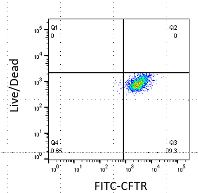
Flow Cytometry: CFTR Antibody (CF3) [NB300-511]
Flow Cytometry: CFTR Antibody (CF3) [NB300-511] - Human airway epithelial cells stained with CFTR (CF3) coupled with goat anti-mouse FITC conjugated secondary antibody. Flow cytometry image submitted by a verified customer review.![Immunocytochemistry/ Immunofluorescence: CFTR Antibody (CF3) [NB300-511]](https://resources.rndsystems.com/images/products/CFTR-Antibody-CF3-Immunocytochemistry-Immunofluorescence-NB300-511-img0007.jpg "Immunocytochemistry/ Immunofluorescence: CFTR Antibody (CF3) [NB300-511]")
Immunocytochemistry/ Immunofluorescence: CFTR Antibody (CF3) [NB300-511]
Immunocytochemistry/Immunofluorescence: CFTR Antibody (CF3) [NB300-511] - Staining of human pancreas cryosection (200x). Alpha cells are strongly labeled. ICC/IF image submitted by a verified customer review.![Immunocytochemistry/ Immunofluorescence: CFTR Antibody (CF3) [NB300-511]](https://resources.rndsystems.com/images/products/CFTR-Antibody-CF3-Immunocytochemistry-Immunofluorescence-NB300-511-img0004.jpg "Immunocytochemistry/ Immunofluorescence: CFTR Antibody (CF3) [NB300-511]")
Immunocytochemistry/ Immunofluorescence: CFTR Antibody (CF3) [NB300-511]
Immunocytochemistry/Immunofluorescence: CFTR Antibody (CF3) [NB300-511] - CFTR staining (green), F-Actin staining with Phalloidin (red) and nuclei with DAPI (blue) is shown. Cells were grown on chamber slides and fixed with formaldehyde prior to staining. Cells were probed without (control) or with or an antibody recognizing CFTR at a dilution of 1:100-1:200 over night at 4C, washed with PBS and incubated with a DyLight 488 conjugated antibody.![Immunocytochemistry/ Immunofluorescence: CFTR Antibody (CF3) [NB300-511]](https://resources.rndsystems.com/images/products/CFTR-Antibody-CF3-Immunocytochemistry-Immunofluorescence-NB300-511-img0005.jpg "Immunocytochemistry/ Immunofluorescence: CFTR Antibody (CF3) [NB300-511]")
Immunocytochemistry/ Immunofluorescence: CFTR Antibody (CF3) [NB300-511]
Immunocytochemistry/Immunofluorescence: CFTR Antibody (CF3) [NB300-511] - CFTR staining (green), F-Actin staining with Phalloidin (red) and nuclei with DAPI (blue) is shown. Cells were grown on chamber slides and fixed with formaldehyde prior to staining. Cells were probed without (control) or with or an antibody recognizing CFTR at a dilution of 1:100-1:200 over night at 4C, washed with PBS and incubated with a DyLight 488 conjugated antibody.![Immunohistochemistry-Paraffin: CFTR Antibody (CF3) [NB300-511]](https://resources.rndsystems.com/images/products/CFTR-Antibody-CF3-Immunohistochemistry-Paraffin-NB300-511-img0001.jpg "Immunohistochemistry-Paraffin: CFTR Antibody (CF3) [NB300-511]")
Immunohistochemistry-Paraffin: CFTR Antibody (CF3) [NB300-511]
Immunohistochemistry-Paraffin: CFTR Antibody (CF3) [NB300-511] - Cancer biopsies of deparaffinized Human colon carcinoma tissue.![Immunohistochemistry-Paraffin: CFTR Antibody (CF3) [NB300-511]](https://resources.rndsystems.com/images/products/CFTR-Antibody-CF3-Immunohistochemistry-Paraffin-NB300-511-img0002.jpg "Immunohistochemistry-Paraffin: CFTR Antibody (CF3) [NB300-511]")
Immunohistochemistry-Paraffin: CFTR Antibody (CF3) [NB300-511]
Immunohistochemistry-Paraffin: CFTR Antibody (CF3) [NB300-511] - Normal biopsies of deparaffinized Human pancreas tissue.Applications for CFTR Antibody (CF3)
Application
Recommended Usage
Flow Cytometry
1:100
Immunocytochemistry/ Immunofluorescence
1:500
Immunohistochemistry
1:200
Immunohistochemistry-Paraffin
1:200
Immunoprecipitation
1:10 - 1:500
Western Blot
1:500
Application Notes
Flow usage reported in literature and validated by a verified customer review. ICC usage reported in literature. IHC usage was reported in scientific literature. Blocking Assay was reported in the scientific literature (PMID: 9590693). WB: Detects an approx. 170 kDa protein. Also detects one or more immunologically related proteins in murine cell line Heb7a that does not contain CFTR mRNA. IF: Staining of CFTR in mouse epithelial cells results in cell surface staining, consistent with localization at the plasma membrane. Inhibit the epithelial uptake of S. typhi in some mouse cell lines.
Reviewed Applications
Read 2 reviews rated 4.5 using NB300-511 in the following applications:
Flow Cytometry Panel Builder
Bio-Techne Knows Flow Cytometry
Save time and reduce costly mistakes by quickly finding compatible reagents using the Panel Builder Tool.
Advanced Features
- Spectra Viewer - Custom analysis of spectra from multiple fluorochromes
- Spillover Popups - Visualize the spectra of individual fluorochromes
- Antigen Density Selector - Match fluorochrome brightness with antigen density

Formulation, Preparation, and Storage
Purification
Unpurified
Formulation
Ascites
Preservative
0.05% Sodium Azide
Concentration
This product is unpurified. The exact concentration of antibody is not quantifiable.
Shipping
The product is shipped with polar packs. Upon receipt, store it immediately at the temperature recommended below.
Stability & Storage
Store at -20C. Avoid freeze-thaw cycles.
Background: CFTR
Long Name
Cystic Fibrosis Transmembrane Conductance Regulator
Alternate Names
ABC35, ABCC7CF, ATP-binding cassette sub-family C member 7, ATP-binding cassette transporter sub-family C member 7, cAMP-dependent chloride channel, CFTR/MRP, Channel conductance-controlling ATPase, cystic fibrosis transmembrane conductance regulator, cystic fibrosis transmembrane conductance regulator (ATP-binding cassettesub-family C, member 7), cystic fibrosis transmembrane conductance regulator, ATP-binding cassette(sub-family C, member 7), dJ760C5.1, EC 3.6.3, MRP7EC 3.6.3.49, TNR-CFTR
Gene Symbol
CFTR
UniProt
Additional CFTR Products
Product Documents for CFTR Antibody (CF3)
Certificate of Analysis
To download a Certificate of Analysis, please enter a lot or batch number in the search box below.
Product Specific Notices for CFTR Antibody (CF3)
This product is for research use only and is not approved for use in humans or in clinical diagnosis. Primary Antibodies are guaranteed for 1 year from date of receipt.
Related Research Areas
Citations for CFTR Antibody (CF3)
 Powered by Bioz
Customer Reviews for CFTR Antibody (CF3) (2)
4.5 out of 5
2 Customer Ratings
Have you used CFTR Antibody (CF3)?
Submit a review and receive an Amazon gift card!
$25/€18/£15/$25CAN/¥2500 Yen for a review with an image
$10/€7/£6/$10CAN/¥1110 Yen for a review without an image
Submit a review
Customer Images
-(01-ml)_NB300-511_7431.jpg)
Showing
1
-
2 of
2 reviews
Showing All
Filter By:
-
Application: Flow CytometrySample Tested: human airway epithelial cellsSpecies: HumanVerified Customer | Posted 09/30/2020Human airway epithelial cells stained with CFTR (CF3) coupled with goat anti-mouse FITC conjugated 2nd ab.

-
Application: ImmunofluorescenceVerified Customer | Posted 05/09/2014NB300-511 on human pancreas cryosection (200x). Alpha cells are strongly labeled.
There are no reviews that match your criteria.
Protocols
Find general support by application which include: protocols, troubleshooting, illustrated assays, videos and webinars.
- 7-Amino Actinomycin D (7-AAD) Cell Viability Flow Cytometry Protocol
- Antigen Retrieval Protocol (PIER)
- Antigen Retrieval for Frozen Sections Protocol
- Appropriate Fixation of IHC/ICC Samples
- Cellular Response to Hypoxia Protocols
- Chromogenic IHC Staining of Formalin-Fixed Paraffin-Embedded (FFPE) Tissue Protocol
- Chromogenic Immunohistochemistry Staining of Frozen Tissue
- ClariTSA™ Fluorophore Kits
- Detection & Visualization of Antibody Binding
- Extracellular Membrane Flow Cytometry Protocol
- Flow Cytometry Protocol for Cell Surface Markers
- Flow Cytometry Protocol for Staining Membrane Associated Proteins
- Flow Cytometry Staining Protocols
- Flow Cytometry Troubleshooting Guide
- Fluorescent IHC Staining of Frozen Tissue Protocol
- Graphic Protocol for Heat-induced Epitope Retrieval
- Graphic Protocol for the Preparation and Fluorescent IHC Staining of Frozen Tissue Sections
- Graphic Protocol for the Preparation and Fluorescent IHC Staining of Paraffin-embedded Tissue Sections
- Graphic Protocol for the Preparation of Gelatin-coated Slides for Histological Tissue Sections
- ICC Cell Smear Protocol for Suspension Cells
- ICC Immunocytochemistry Protocol Videos
- ICC for Adherent Cells
- IHC Sample Preparation (Frozen sections vs Paraffin)
- Immunocytochemistry (ICC) Protocol
- Immunocytochemistry Troubleshooting
- Immunofluorescence of Organoids Embedded in Cultrex Basement Membrane Extract
- Immunofluorescent IHC Staining of Formalin-Fixed Paraffin-Embedded (FFPE) Tissue Protocol
- Immunohistochemistry (IHC) and Immunocytochemistry (ICC) Protocols
- Immunohistochemistry Frozen Troubleshooting
- Immunohistochemistry Paraffin Troubleshooting
- Immunoprecipitation Protocol
- Intracellular Flow Cytometry Protocol Using Alcohol (Methanol)
- Intracellular Flow Cytometry Protocol Using Detergents
- Intracellular Nuclear Staining Flow Cytometry Protocol Using Detergents
- Intracellular Staining Flow Cytometry Protocol Using Alcohol Permeabilization
- Intracellular Staining Flow Cytometry Protocol Using Detergents to Permeabilize Cells
- Preparing Samples for IHC/ICC Experiments
- Preventing Non-Specific Staining (Non-Specific Binding)
- Primary Antibody Selection & Optimization
- Propidium Iodide Cell Viability Flow Cytometry Protocol
- Protocol for Heat-Induced Epitope Retrieval (HIER)
- Protocol for Liperfluo
- Protocol for Making a 4% Formaldehyde Solution in PBS
- Protocol for VisUCyte™ HRP Polymer Detection Reagent
- Protocol for the Characterization of Human Th22 Cells
- Protocol for the Characterization of Human Th9 Cells
- Protocol for the Fluorescent ICC Staining of Cell Smears - Graphic
- Protocol for the Fluorescent ICC Staining of Cultured Cells on Coverslips - Graphic
- Protocol for the Preparation & Fixation of Cells on Coverslips
- Protocol for the Preparation and Chromogenic IHC Staining of Frozen Tissue Sections
- Protocol for the Preparation and Chromogenic IHC Staining of Frozen Tissue Sections - Graphic
- Protocol for the Preparation and Chromogenic IHC Staining of Paraffin-embedded Tissue Sections
- Protocol for the Preparation and Chromogenic IHC Staining of Paraffin-embedded Tissue Sections - Graphic
- Protocol for the Preparation and Fluorescent ICC Staining of Cells on Coverslips
- Protocol for the Preparation and Fluorescent ICC Staining of Non-adherent Cells
- Protocol for the Preparation and Fluorescent ICC Staining of Stem Cells on Coverslips
- Protocol for the Preparation and Fluorescent IHC Staining of Frozen Tissue Sections
- Protocol for the Preparation and Fluorescent IHC Staining of Paraffin-embedded Tissue Sections
- Protocol for the Preparation of Gelatin-coated Slides for Histological Tissue Sections
- Protocol for the Preparation of a Cell Smear for Non-adherent Cell ICC - Graphic
- Protocol: Annexin V and PI Staining by Flow Cytometry
- Protocol: Annexin V and PI Staining for Apoptosis by Flow Cytometry
- R&D Systems Quality Control Western Blot Protocol
- TUNEL and Active Caspase-3 Detection by IHC/ICC Protocol
- The Importance of IHC/ICC Controls
- Troubleshooting Guide: Fluorokine Flow Cytometry Kits
- Troubleshooting Guide: Immunohistochemistry
- Troubleshooting Guide: Western Blot Figures
- Western Blot Conditions
- Western Blot Protocol
- Western Blot Protocol for Cell Lysates
- Western Blot Troubleshooting
- Western Blot Troubleshooting Guide
- View all Protocols, Troubleshooting, Illustrated assays and Webinars
Loading...
Associated Pathways
