Collagen I alpha 1 Antibody - Azide and BSA Free
Novus Biologicals | Catalog # NBP1-30054
![Western Blot: Collagen I alpha 1 AntibodyAzide and BSA Free [NBP1-30054]](https://resources.rndsystems.com/images/products/Collagen-I-alpha-1-Antibody---Azide-and-BSA-Free-Western-Blot-NBP1-30054-img0005.jpg "Western Blot: Collagen I alpha 1 AntibodyAzide and BSA Free [NBP1-30054]")
Loading...
Key Product Details
Validated by
Biological Validation
Species Reactivity
Validated:
Human, Mouse, Rat, Amphibian, Avian, Mammal, Sheep
Cited:
Human, Mouse, Rat, Avian, Canine, Ovine
Predicted:
Bovine (100%), Canine (100%), Chicken (100%), Equine (100%), Feline (100%), Finch (100%), Goat (100%), Guinea Pig (100%), Hamster (100%), Primate (100%), Rabbit (100%), Vole (100%), Xenopus (100%). Backed by our 100% Guarantee.
Applications
Validated:
Immunohistochemistry, Immunohistochemistry-Paraffin, Immunohistochemistry-Frozen, Western Blot, Immunocytochemistry/ Immunofluorescence, Simple Western
Cited:
Immunohistochemistry, Immunohistochemistry-Paraffin, Immunohistochemistry-Frozen, Western Blot, Flow Cytometry, Immunocytochemistry/ Immunofluorescence, Simple Western, IF/IHC
Label
Unconjugated
Antibody Source
Polyclonal Rabbit IgG
Format
Azide and BSA Free
Loading...
Product Specifications
Immunogen
This Collagen I alpha 1 antibody was raised against synthetic peptide corresponding to amino acid residues within the C-terminal telo peptide portion (aa1193-1218) of the human COL1A1, conjugated to KLH. Accession # P02452
Reactivity Notes
Based on the homology of the immunogen, this antibody is expected to recognize the collagen I alpha1 polypeptide in all mammals, birds, and amphibians.
Specificity
This Collagen I alpha 1 antibody is specific for the ~ 140 kDa telopeptide portion of the collagen I alpha 1 polypeptide. The antibody works well for immunohistochemistry on paraformaldehyde-fixed sections with a simple antigen-retrieval protocol (incubate slides for 20 minutes at 90 degrees C in 10 mM sodium citrate (pH 6.0)/ 0.1 % Tween-20). Note that in paraffin sections of formaldehydefixed fibrotic mouse lung tissue, the antibody recognizes mature collagen I that has formed fibrils in the extracellular matrix.
Clonality
Polyclonal
Host
Rabbit
Isotype
IgG
Theoretical MW
140 kDa.
Disclaimer note: The observed molecular weight of the protein may vary from the listed predicted molecular weight due to post translational modifications, post translation cleavages, relative charges, and other experimental factors.
Disclaimer note: The observed molecular weight of the protein may vary from the listed predicted molecular weight due to post translational modifications, post translation cleavages, relative charges, and other experimental factors.
Description
Recommended that the undiluted antibody be aliquoted into smaller working volumes (10-30 uL/vial depending on usage).
Scientific Data Images for Collagen I alpha 1 Antibody - Azide and BSA Free
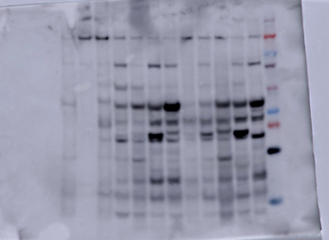
Western Blot: Collagen I alpha 1 AntibodyAzide and BSA Free [NBP1-30054]
Western Blot: Collagen I alpha 1 Antibody - Azide and BSA Free [NBP1-30054] - Collagen I Antibody [NBP1-30054] - Rat lung lysate showing specific immunolabeling of the collagen protein with an observed molecular weight ~140.![Immunohistochemistry: Collagen I alpha 1 Antibody - Azide and BSA Free [NBP1-30054]](https://resources.rndsystems.com/images/products/Collagen-I-alpha-1-Antibody---Azide-and-BSA-Free-Immunohistochemistry-NBP1-30054-img0006.jpg "Immunohistochemistry: Collagen I alpha 1 Antibody - Azide and BSA Free [NBP1-30054]")
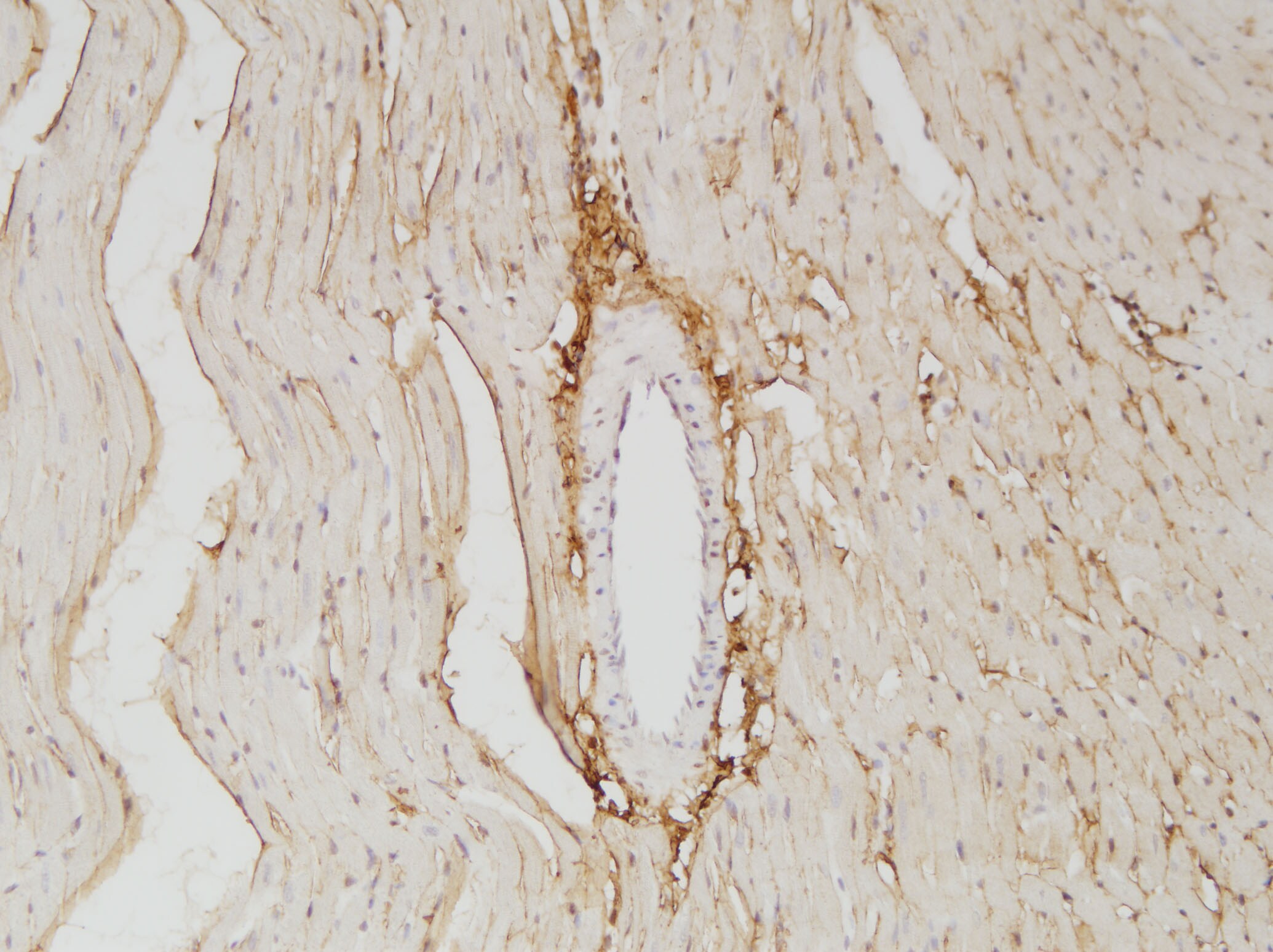
![Immunohistochemistry: Collagen I alpha 1 Antibody - Azide and BSA Free [NBP1-30054]](https://resources.rndsystems.com/images/products/Collagen-I-alpha-1-Antibody---Azide-and-BSA-Free-Immunohistochemistry-NBP1-30054-img0004.jpg "Immunohistochemistry: Collagen I alpha 1 Antibody - Azide and BSA Free [NBP1-30054]")
Immunohistochemistry: Collagen I alpha 1 Antibody - Azide and BSA Free [NBP1-30054]
Immunohistochemistry: Collagen I alpha 1 Antibody - Azide and BSA Free [NBP1-30054] - Collagen I Antibody [NBP1-30054] - Formaldehyde-fixed fibrotic mouse lung tissue. The antibody recognizes mature collagen I (red) that has formed fibrils in the extracellular matrix.![Simple Western: Collagen I alpha 1 AntibodyAzide and BSA Free [NBP1-30054]](https://resources.rndsystems.com/images/products/Collagen-I-alpha-1-Antibody---Azide-and-BSA-Free-Simple-Western-NBP1-30054-img0003.jpg "Simple Western: Collagen I alpha 1 AntibodyAzide and BSA Free [NBP1-30054]")
Simple Western: Collagen I alpha 1 AntibodyAzide and BSA Free [NBP1-30054]
Simple Western: Collagen I alpha 1 Antibody - Azide and BSA Free [NBP1-30054] - Simple Western lane view shows a specific band for Collagen I Alpha 1 in 0.5 mg/ml of Human Lung (left) and Human Kidney (right) lysate. This experiment was performed under reducing conditions using the 66-440 kDa separation system. * Non-specific interaction with the 230 kDa Simple Western standard may be seen with this antibody
Western Blot: Collagen I alpha 1 Antibody - Azide and BSA Free [NBP1-30054] -
Western Blot: Collagen I alpha 1 Antibody - Azide and BSA Free [NBP1-30054] - TNC deficiency reduced kidney fibrosis in animal models.Deletion of TNC (TNC−/−) significantly attenuated the induction of collagen I assessed by IF following UUO by approximate 30% at day 7 & 10 compared with their wild-type littermates (A, n = 4 for each time point, two-way ANOVA p < 0.05). Consistently, the expression of fibrosis markers, such as collagen I alpha, fibronectin & plasminogen activator inhibitor-1 (PAI-1), were significantly lower in TNC−/− mice at UUO day 7 & 10 (B, n = 4 for each time point, two-way ANOVA, p < 0.05). Western blot showed that the proteins of collagen I alpha & alpha -SMA were also significantly reduced in TNC−/− mice at UUO day 7 (C, n = 7, p < 0.05). Image collected & cropped by CiteAb from the following publication (https://pubmed.ncbi.nlm.nih.gov/36522320), licensed under a CC-BY license. Not internally tested by Novus Biologicals.
Immunocytochemistry/ Immunofluorescence: Collagen I alpha 1 Antibody - Azide and BSA Free [NBP1-30054] -
Pathological staining analysis of 28-day animal experiment induced by BAPN. (A), representative images from elastic van Gieson (EVG) staining, along with statistical analysis of elastin degradation. (B), representative images from hematoxylin and eosin (HE) staining are presented, accompanied by statistical analysis of aortic wall thickness. The scale bar for both panels (A) and (B) is 200 μm. (C), immunofluorescence staining of Col1a1, accompanied by quantitative analysis of the Col1a1 positive area in the vascular wall, scale bar 50 μm Image collected and cropped by CiteAb from the following open publication (https://nutritionandmetabolism.biomedcentral.com/articles/10.1186/s1298…), licensed under a CC-BY license. Not internally tested by Novus Biologicals.
Immunocytochemistry/ Immunofluorescence: Collagen I alpha 1 Antibody - Azide and BSA Free [NBP1-30054] -
Pathological staining analysis of 28-day animal experiment induced by BAPN. (A), representative images from elastic van Gieson (EVG) staining, along with statistical analysis of elastin degradation. (B), representative images from hematoxylin and eosin (HE) staining are presented, accompanied by statistical analysis of aortic wall thickness. The scale bar for both panels (A) and (B) is 200 μm. (C), immunofluorescence staining of Col1a1, accompanied by quantitative analysis of the Col1a1 positive area in the vascular wall, scale bar 50 μm Image collected and cropped by CiteAb from the following open publication (https://nutritionandmetabolism.biomedcentral.com/articles/10.1186/s1298…), licensed under a CC-BY license. Not internally tested by Novus Biologicals.Applications for Collagen I alpha 1 Antibody - Azide and BSA Free
Application
Recommended Usage
Immunohistochemistry
1:10-1:500
Immunohistochemistry-Frozen
1:10-1:500
Immunohistochemistry-Paraffin
1:100
Simple Western
1:100
Western Blot
1:1000
Application Notes
Use in Immunohistochemistry-Frozen sections was reported in the scientific literature (PMID: 21939397), IHC reactivity reported in scientific literature (PMID: 26045736). Use in Immunocytochemistry/immunofluorescence reported in scientific literature (PMID: 26651081).
In Simple Western only 10 - 15 uL of the recommended dilution is used per data point.
See Simple Western Antibody Database for Simple Western validation: Tested in Human Lung and Human Kidney lysate 0.5 mg/mL, separated by Size, antibody dilution of 1:100. Separated by Size-Wes, Sally Sue/Peggy Sue.
Reviewed Applications
Read 2 reviews rated 4 using NBP1-30054 in the following applications:
Formulation, Preparation, and Storage
Purification
Antigen Affinity-purified
Formulation
PBS
Format
Azide and BSA Free
Preservative
No Preservative
Concentration
Please see the vial label for concentration. If unlisted please contact technical services.
Shipping
The product is shipped with polar packs. Upon receipt, store it immediately at the temperature recommended below.
Stability & Storage
Store at -20C. Avoid freeze-thaw cycles.
Background: Collagen I alpha 1
Type I collagen is a fibril-forming collagen found in most connective tissues and is abundant in bone, cornea, dermis and tendon tissue. Collagens are fibrous, extracellular matrix proteins with high tensile strength and are the major components of connective tissue. Several collagens play a role in cell adhesion, responsible for maintaining normal tissue architecture and function. All collagens contain a triple helix domain and frequently show lateral self-association in order to form complex connective tissues. Post-Golgi LH3 trafficking is essential for collagen homeostasis and for the development and function of multiple organs and tissues (1).
The COL1A1 gene encodes the pro-alpha1 chains of type I collagen protein, whose triple helix is comprised of two alpha1 chains and one alpha2 chain. Mutations in the encoding COL1A1 gene are associated with brittle bone disease (Osteogenesis Imperfecta), cortical hyperostosis (Caffey disease) and disorders that affect the connective tissues (Ehlers-Danlos syndrome) (2). Studies have found that HIF-1 transcription regulation of collagen prolyl hydroxylases regulates collagen deposition, promoting cancer cell alignment along collagen fibers, which enhances invasion and metastasis to lymph nodes and lung tissue by breast cancer cells (3).
References
1. Banushi, B., Forneris, F., Straatman-Iwanowska, A., Strange, A., Lyne, A. M., Rogerson, C.,... Gissen, P. (2016). Regulation of post-Golgi LH3 trafficking is essential for collagen homeostasis. Nat Commun, 7, 12111. doi:10.1038/ncomms12111
2. Lu, Y., Zhang, S., Wang, Y., Ren, X., & Han, J. (2019). Molecular mechanisms and clinical manifestations of rare genetic disorders associated with type I collagen. Intractable Rare Dis Res, 8(2), 98-107. doi:10.5582/irdr.2019.01064
3. Gilkes, D. M., Chaturvedi, P., Bajpai, S., Wong, C. C., Wei, H., Pitcairn, S.,... Semenza, G. L. (2013). Collagen prolyl hydroxylases are essential for breast cancer metastasis. Cancer Res, 73(11), 3285-3296. doi:10.1158/0008-5472.Can-12-3963
Alternate Names
COL1A1, OI4
Gene Symbol
COL1A1
UniProt
Additional Collagen I alpha 1 Products
Product Documents for Collagen I alpha 1 Antibody - Azide and BSA Free
Certificate of Analysis
To download a Certificate of Analysis, please enter a lot or batch number in the search box below.
Product Specific Notices for Collagen I alpha 1 Antibody - Azide and BSA Free
This product is for research use only and is not approved for use in humans or in clinical diagnosis. Primary Antibodies are guaranteed for 1 year from date of receipt.
Related Research Areas
Citations for Collagen I alpha 1 Antibody - Azide and BSA Free
 Powered by Bioz
Customer Reviews for Collagen I alpha 1 Antibody - Azide and BSA Free (2)
4 out of 5
2 Customer Ratings
Have you used Collagen I alpha 1 Antibody - Azide and BSA Free?
Submit a review and receive an Amazon gift card!
$25/€18/£15/$25CAN/¥2500 Yen for a review with an image
$10/€7/£6/$10CAN/¥1110 Yen for a review without an image
Submit a review
Customer Images
Showing
1
-
2 of
2 reviews
Showing All
Filter By:
-
Application: Western BlotSample Tested: Mouse PancreasSpecies: MouseVerified Customer | Posted 10/02/2020Caerulein dosed mouse pancreas

-
Application: Immunohistochemistry-ParaffinSample Tested: Adult heartSpecies: RatVerified Customer | Posted 12/20/2017collagen I for sd rat‘s heart
There are no reviews that match your criteria.
Protocols
Find general support by application which include: protocols, troubleshooting, illustrated assays, videos and webinars.
- Antigen Retrieval Protocol (PIER)
- Antigen Retrieval for Frozen Sections Protocol
- Appropriate Fixation of IHC/ICC Samples
- Cellular Response to Hypoxia Protocols
- Chromogenic IHC Staining of Formalin-Fixed Paraffin-Embedded (FFPE) Tissue Protocol
- Chromogenic Immunohistochemistry Staining of Frozen Tissue
- ClariTSA™ Fluorophore Kits
- Detection & Visualization of Antibody Binding
- Fluorescent IHC Staining of Frozen Tissue Protocol
- Graphic Protocol for Heat-induced Epitope Retrieval
- Graphic Protocol for the Preparation and Fluorescent IHC Staining of Frozen Tissue Sections
- Graphic Protocol for the Preparation and Fluorescent IHC Staining of Paraffin-embedded Tissue Sections
- Graphic Protocol for the Preparation of Gelatin-coated Slides for Histological Tissue Sections
- ICC Cell Smear Protocol for Suspension Cells
- ICC Immunocytochemistry Protocol Videos
- ICC for Adherent Cells
- IHC Sample Preparation (Frozen sections vs Paraffin)
- Immunocytochemistry (ICC) Protocol
- Immunocytochemistry Troubleshooting
- Immunofluorescence of Organoids Embedded in Cultrex Basement Membrane Extract
- Immunofluorescent IHC Staining of Formalin-Fixed Paraffin-Embedded (FFPE) Tissue Protocol
- Immunohistochemistry (IHC) and Immunocytochemistry (ICC) Protocols
- Immunohistochemistry Frozen Troubleshooting
- Immunohistochemistry Paraffin Troubleshooting
- Preparing Samples for IHC/ICC Experiments
- Preventing Non-Specific Staining (Non-Specific Binding)
- Primary Antibody Selection & Optimization
- Protocol for Heat-Induced Epitope Retrieval (HIER)
- Protocol for Making a 4% Formaldehyde Solution in PBS
- Protocol for VisUCyte™ HRP Polymer Detection Reagent
- Protocol for the Fluorescent ICC Staining of Cell Smears - Graphic
- Protocol for the Fluorescent ICC Staining of Cultured Cells on Coverslips - Graphic
- Protocol for the Preparation & Fixation of Cells on Coverslips
- Protocol for the Preparation and Chromogenic IHC Staining of Frozen Tissue Sections
- Protocol for the Preparation and Chromogenic IHC Staining of Frozen Tissue Sections - Graphic
- Protocol for the Preparation and Chromogenic IHC Staining of Paraffin-embedded Tissue Sections
- Protocol for the Preparation and Chromogenic IHC Staining of Paraffin-embedded Tissue Sections - Graphic
- Protocol for the Preparation and Fluorescent ICC Staining of Cells on Coverslips
- Protocol for the Preparation and Fluorescent ICC Staining of Non-adherent Cells
- Protocol for the Preparation and Fluorescent ICC Staining of Stem Cells on Coverslips
- Protocol for the Preparation and Fluorescent IHC Staining of Frozen Tissue Sections
- Protocol for the Preparation and Fluorescent IHC Staining of Paraffin-embedded Tissue Sections
- Protocol for the Preparation of Gelatin-coated Slides for Histological Tissue Sections
- Protocol for the Preparation of a Cell Smear for Non-adherent Cell ICC - Graphic
- R&D Systems Quality Control Western Blot Protocol
- TUNEL and Active Caspase-3 Detection by IHC/ICC Protocol
- The Importance of IHC/ICC Controls
- Troubleshooting Guide: Immunohistochemistry
- Troubleshooting Guide: Western Blot Figures
- Western Blot Conditions
- Western Blot Protocol
- Western Blot Protocol for Cell Lysates
- Western Blot Troubleshooting
- Western Blot Troubleshooting Guide
- View all Protocols, Troubleshooting, Illustrated assays and Webinars
FAQs for Collagen I alpha 1 Antibody - Azide and BSA Free
Showing
1
-
5 of
7 FAQs
Showing All
-
Q: Do you have any references on IHC-P performed on sheep tissue?
A: Unfortunately we do not have any references of IHC-P performed on sheep tissue. However, I can confirm that this has been tested and will be backed by our 100% guarantee. Therefore if the antibody fails to stain sheep tissue that expresses the protein in an IHC-P assay, Novus would be happy to issue a refund or replacement.
-
Q: How long does delivery take for your collagen 1 alpha 1 antibody?
A: Our most popular collagen I alpha 1 antibody (NB600-408) is usually in stock and ships priority overnight for next business day delivery in the U.S.
-
Q: I am looking for anti collagen 1 antibody, I see your product calls it collagen I alpha 1 is there a difference?
A: Collagen I alpha 1 is the most common protein for the collagen 1 protein. There are other subunits as well (such as collagen 1 alpha 2…etc.).
-
Q: I intend buying anti-collagen I antibody for IF and WB. Kindly let me know its isotype
A: NBP1-30054 is a rabbit IgG polyclonal antibody.
-
Q: The predicted molecular weight says 150kDA but the band on the gel appears at 130kDA why is that?
A: Differences between the predicted and observed MW can be affected by a number of factors including PTMs, relative charges and experimental factors. These cumulative effect of these differences can be quite substantial with larger molecules such as collagen I alpha 1.
-
Q: We are looking for an anti-Collagen antibody for Flow Cytometry (FACS) analysis in Swine sample. Would you please provide some references if there is any available as well?
A:
We currently have one antibody for porcine Collagen validated in flow cytometry (catalog # NBP1-94202).
-
Q: What secondary antibody do you recommend for collagen I alpha 1 antibodies?
A: Because this collagen I alpha 1 antibody is raised in rabbit you would want to use any anti-rabbit IgG secondary antibody. NB7160 is a popular choice for use in WB.
-
Q: Do you have any references on IHC-P performed on sheep tissue?
A: Unfortunately we do not have any references of IHC-P performed on sheep tissue. However, I can confirm that this has been tested and will be backed by our 100% guarantee. Therefore if the antibody fails to stain sheep tissue that expresses the protein in an IHC-P assay, Novus would be happy to issue a refund or replacement.
-
Q: How long does delivery take for your collagen 1 alpha 1 antibody?
A: Our most popular collagen I alpha 1 antibody (NB600-408) is usually in stock and ships priority overnight for next business day delivery in the U.S.
-
Q: I am looking for anti collagen 1 antibody, I see your product calls it collagen I alpha 1 is there a difference?
A: Collagen I alpha 1 is the most common protein for the collagen 1 protein. There are other subunits as well (such as collagen 1 alpha 2…etc.).
-
Q: I intend buying anti-collagen I antibody for IF and WB. Kindly let me know its isotype
A: NBP1-30054 is a rabbit IgG polyclonal antibody.
-
Q: The predicted molecular weight says 150kDA but the band on the gel appears at 130kDA why is that?
A: Differences between the predicted and observed MW can be affected by a number of factors including PTMs, relative charges and experimental factors. These cumulative effect of these differences can be quite substantial with larger molecules such as collagen I alpha 1.
-
Q: We are looking for an anti-Collagen antibody for Flow Cytometry (FACS) analysis in Swine sample. Would you please provide some references if there is any available as well?
A:
We currently have one antibody for porcine Collagen validated in flow cytometry (catalog # NBP1-94202).
-
Q: What secondary antibody do you recommend for collagen I alpha 1 antibodies?
A: Because this collagen I alpha 1 antibody is raised in rabbit you would want to use any anti-rabbit IgG secondary antibody. NB7160 is a popular choice for use in WB.
Loading...