Collagen III alpha 1/COL3A1 Antibody [Biotin]
Novus Biologicals | Catalog # NB120-6580
Key Product Details
Species Reactivity
Human, Mouse, Rat, Bovine, Feline, Sheep
Applications
Immunohistochemistry, Immunohistochemistry-Paraffin, Western Blot, ELISA, Simple Western, Immunoprecipitation
Label
Biotin
Antibody Source
Polyclonal Rabbit IgG
Loading...
Product Specifications
Immunogen
Collagen III alpha 1/COL3A1 from human and bovine placenta (Uniprot: P02461)
Reactivity Notes
This antibody reacts with most mammalian Collagen III alpha 1/COL3A1 and has expected cross-reactivity with Type I and negligible cross reactivity with Type II, IV, V or VI collagens.
Mouse reactivity reported in multiple pieces of scientific literature.
Rat reactivity reported in scientific literature (PMID: 23370982)
Feline reactivity reported in scientific literature (PMID: 33091431)
Mouse reactivity reported in multiple pieces of scientific literature.
Rat reactivity reported in scientific literature (PMID: 23370982)
Feline reactivity reported in scientific literature (PMID: 33091431)
Localization
Extracellular matrix
Specificity
Some class-specific anti-collagens may be specific for three-dimensional epitopes which may result in diminished reactivity with denatured collagen or formalin-fixed, paraffin embedded tissues. This antibody reacts with most mammalian Collagen III alpha 1/COL3A1 and has expected cross-reactivity with Type I and negligible cross reactivity with Type II, IV, V or VI collagens. Non-specific cross-reaction of anti-collagen antibodies with other human serum proteins or non-collagen extracellular matrix proteins has not been tested.
Clonality
Polyclonal
Host
Rabbit
Isotype
IgG
Description
Store vial at -20C or below. Avoid cycles of freezing and thawing. Centrifuge product if not completely clear after standing at room temperature. This product is stable for several weeks at 4C as an undiluted liquid. Dilute only prior to immediate use.
This product has been prepared by immunoaffinity chromatography using immobilized antigens followed by extensive cross-adsorption against other collagens, human serum proteins and non-collagen extracellular matrix proteins to remove any unwanted specificities
This product has been prepared by immunoaffinity chromatography using immobilized antigens followed by extensive cross-adsorption against other collagens, human serum proteins and non-collagen extracellular matrix proteins to remove any unwanted specificities
Scientific Data Images for Collagen III alpha 1/COL3A1 Antibody [Biotin]
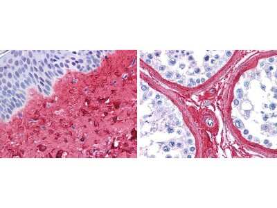
Immunohistochemistry: Collagen III alpha 1/COL3A1 Antibody [Biotin] [NB120-6580] - Strong staining in FFPE sections of human skin(left, dermis) with moderate to strong red staining and testis (right) where strong staining was observed within connective tissue between seminiferous tubules. The antibody showed strong extracellular staining within connective tissues across many organs with minimal background staining. Slides were steamed in 0.01 M sodium citrate buffer, pH 6.0 at 99-100C. 20 minutes for antigen retrieval.
Applications for Collagen III alpha 1/COL3A1 Antibody [Biotin]
Application
Recommended Usage
ELISA
1:10000 - 1:50000
Immunohistochemistry
1:200 - 1:1000
Immunohistochemistry-Paraffin
1:200 - 1:1000
Immunoprecipitation
1:100
Western Blot
1:1000 - 1:5000
Application Notes
Collagen antibodies have been used for indirect trapping ELISA for quantitation of antigen in serum using a standard curve, immunoprecipitation, native (non-denaturing, non-dissociating) PAGE, immunohistochemistry, and western blotting for highly sensitive qualitative analysis.
See Simple Western Antibody Database for Simple Western validation
See Simple Western Antibody Database for Simple Western validation
Formulation, Preparation, and Storage
Purification
Immunogen affinity purified
Reconstitution
Reconstitute with 100 ul deionized water (or equivalent)
Formulation
Lyophilized from 0.02 M Potassium Phosphate, 0.15 M Sodium Chloride, pH 7.2, 10 mg/mL Bovine Serum Albumin (BSA) - Immunoglobulin and Protease free
Preservative
0.01% Sodium Azide
Concentration
LYOPH mg/ml
Shipping
The product is shipped with polar packs. Upon receipt, store it immediately at the temperature recommended below.
Stability & Storage
Store lyophilized antibody at -20C prior to opening. Aliquot reconstituted liquid and and freeze at -20C or below for extended storage. Avoid cycles of freezing and thawing.
Calculators
Background: Collagen III alpha 1/COL3A1
Collagen III is a fibrillar collagen that constitutes 5-20% of all collagen in the body (1). It provides structural integrity and is found in many hallow organs and soft connective tissue including the vascular system, skin, lung, uterus, and intestine (1,2). Additionally, collagen III has be found to be associated with type I collagen in the same fibrils (1). Collagen III interacts with signaling integrins to carry out other key functions including cell adhesion, proliferation, and differentiation (1).
Mutations in the COL3A1 gene has been associated with a variety of human diseases, the most well-known being a group of connective tissue disorders termed Ehlers-Danlos Syndromes (1,2,4). Vascular Ehlers-Danlos Syndrome is a specific subtype that is considered the most severe and although the clinical manifestations vary, symptoms include thin skin and fragile blood vessels and can often result in both lung and heart complications (1,4). COL3A1 is also associated with glomerulopathies, or diseases of the glomeruli, which are characterized by an abundance of extracellular matrix (3). Collagenofibrotic glomerulopathy is one specific rare renal disease that is characterized by excessive levels of collagen III (3).
References
1. Kuivaniemi, H., & Tromp, G. (2019). Type III collagen (COL3A1): Gene and protein structure, tissue distribution, and associated diseases. Gene. https://doi.org/10.1016/j.gene.2019.05.003
2. Ricard-Blum S. (2011). The collagen family. Cold Spring Harbor perspectives in biology. https://doi.org/10.1101/cshperspect.a004978
3. Cohen A. H. (2012). Collagen Type III Glomerulopathies. Advances in chronic kidney disease. https://doi.org/10.1053/j.ackd.2012.02.017
4. Olson, S. L., Murray, M. L., & Skeik, N. (2019). A Novel Frameshift COL3A1 Variant in Vascular Ehlers-Danlos Syndrome. Annals of vascular surgery. https://doi.org/10.1016/j.avsg.2019.05.057
Additional Collagen III alpha 1/COL3A1 Products
Product Documents for Collagen III alpha 1/COL3A1 Antibody [Biotin]
Certificate of Analysis
To download a Certificate of Analysis, please enter a lot or batch number in the search box below.
Product Specific Notices for Collagen III alpha 1/COL3A1 Antibody [Biotin]
This product is for research use only and is not approved for use in humans or in clinical diagnosis. Primary Antibodies are guaranteed for 1 year from date of receipt.
Related Research Areas
Citations for Collagen III alpha 1/COL3A1 Antibody [Biotin]
 Powered by Bioz
Customer Reviews for Collagen III alpha 1/COL3A1 Antibody [Biotin]
There are currently no reviews for this product. Be the first to review Collagen III alpha 1/COL3A1 Antibody [Biotin] and earn rewards!
Have you used Collagen III alpha 1/COL3A1 Antibody [Biotin]?
Submit a review and receive an Amazon gift card!
$25/€18/£15/$25CAN/¥2500 Yen for a review with an image
$10/€7/£6/$10CAN/¥1110 Yen for a review without an image
Submit a review
Protocols
Find general support by application which include: protocols, troubleshooting, illustrated assays, videos and webinars.
- Antigen Retrieval Protocol (PIER)
- Antigen Retrieval for Frozen Sections Protocol
- Appropriate Fixation of IHC/ICC Samples
- Cellular Response to Hypoxia Protocols
- Chromogenic IHC Staining of Formalin-Fixed Paraffin-Embedded (FFPE) Tissue Protocol
- Chromogenic Immunohistochemistry Staining of Frozen Tissue
- ClariTSA™ Fluorophore Kits
- Detection & Visualization of Antibody Binding
- ELISA Sample Preparation & Collection Guide
- ELISA Troubleshooting Guide
- Fluorescent IHC Staining of Frozen Tissue Protocol
- Graphic Protocol for Heat-induced Epitope Retrieval
- Graphic Protocol for the Preparation and Fluorescent IHC Staining of Frozen Tissue Sections
- Graphic Protocol for the Preparation and Fluorescent IHC Staining of Paraffin-embedded Tissue Sections
- Graphic Protocol for the Preparation of Gelatin-coated Slides for Histological Tissue Sections
- How to Run an R&D Systems DuoSet ELISA
- How to Run an R&D Systems Quantikine ELISA
- How to Run an R&D Systems Quantikine™ QuicKit™ ELISA
- IHC Sample Preparation (Frozen sections vs Paraffin)
- Immunofluorescent IHC Staining of Formalin-Fixed Paraffin-Embedded (FFPE) Tissue Protocol
- Immunohistochemistry (IHC) and Immunocytochemistry (ICC) Protocols
- Immunohistochemistry Frozen Troubleshooting
- Immunohistochemistry Paraffin Troubleshooting
- Immunoprecipitation Protocol
- Preparing Samples for IHC/ICC Experiments
- Preventing Non-Specific Staining (Non-Specific Binding)
- Primary Antibody Selection & Optimization
- Protocol for Heat-Induced Epitope Retrieval (HIER)
- Protocol for Making a 4% Formaldehyde Solution in PBS
- Protocol for VisUCyte™ HRP Polymer Detection Reagent
- Protocol for the Preparation & Fixation of Cells on Coverslips
- Protocol for the Preparation and Chromogenic IHC Staining of Frozen Tissue Sections
- Protocol for the Preparation and Chromogenic IHC Staining of Frozen Tissue Sections - Graphic
- Protocol for the Preparation and Chromogenic IHC Staining of Paraffin-embedded Tissue Sections
- Protocol for the Preparation and Chromogenic IHC Staining of Paraffin-embedded Tissue Sections - Graphic
- Protocol for the Preparation and Fluorescent IHC Staining of Frozen Tissue Sections
- Protocol for the Preparation and Fluorescent IHC Staining of Paraffin-embedded Tissue Sections
- Protocol for the Preparation of Gelatin-coated Slides for Histological Tissue Sections
- Quantikine HS ELISA Kit Assay Principle, Alkaline Phosphatase
- Quantikine HS ELISA Kit Principle, Streptavidin-HRP Polymer
- R&D Systems Quality Control Western Blot Protocol
- Sandwich ELISA (Colorimetric) – Biotin/Streptavidin Detection Protocol
- Sandwich ELISA (Colorimetric) – Direct Detection Protocol
- TUNEL and Active Caspase-3 Detection by IHC/ICC Protocol
- The Importance of IHC/ICC Controls
- Troubleshooting Guide: ELISA
- Troubleshooting Guide: Immunohistochemistry
- Troubleshooting Guide: Western Blot Figures
- Western Blot Conditions
- Western Blot Protocol
- Western Blot Protocol for Cell Lysates
- Western Blot Troubleshooting
- Western Blot Troubleshooting Guide
- View all Protocols, Troubleshooting, Illustrated assays and Webinars
Loading...