Collagen III alpha 1/COL3A1 Antibody - BSA Free
Novus Biologicals | Catalog # NB600-594
![Immunohistochemistry: Collagen III alpha 1/COL3A1 Antibody [NB600-594]](https://resources.rndsystems.com/images/products/Collagen-III-alpha-1-COL3A1-Antibody-Immunohistochemistry-NB600-594-img0018.jpg "Immunohistochemistry: Collagen III alpha 1/COL3A1 Antibody [NB600-594]")
Key Product Details
Validated by
Biological Validation
Species Reactivity
Validated:
Human, Mouse, Rat, Bovine, Feline, Sheep
Cited:
Human, Mouse, Rat, Feline, Mammal, Ovine
Applications
Validated:
Immunohistochemistry, Immunohistochemistry-Paraffin, Western Blot, ELISA, Flow Cytometry, Immunocytochemistry/ Immunofluorescence, Simple Western, Immunoprecipitation
Cited:
Immunohistochemistry, Immunohistochemistry-Paraffin, Immunohistochemistry-Frozen, Western Blot, Flow Cytometry, Immunocytochemistry/ Immunofluorescence, IF/IHC
Label
Unconjugated
Antibody Source
Polyclonal Rabbit IgG
Format
BSA Free
Loading...
Product Specifications
Immunogen
Collagen III alpha 1/COL3A1 from human and bovine placenta (Uniprot: P02461)
Reactivity Notes
This antibody reacts with most mammalian Collagen III alpha 1/COL3A1 and has expected cross-reactivity with Type I and negligible cross reactivity with Type II, IV, V or VI collagens.
Mouse reactivity reported in multiple pieces of scientific literature.
Rat reactivity reported in scientific literature (PMID: 23370982)
Feline reactivity reported in scientific literature (PMID: 33091431)
Mouse reactivity reported in multiple pieces of scientific literature.
Rat reactivity reported in scientific literature (PMID: 23370982)
Feline reactivity reported in scientific literature (PMID: 33091431)
Localization
Extracellular matrix
Specificity
Some class-specific anti-collagens may be specific for three-dimensional epitopes which may result in diminished reactivity with denatured collagen or formalin-fixed, paraffin embedded tissues. This antibody reacts with most mammalian Collagen III alpha 1/COL3A1 and has expected cross-reactivity with Type I and negligible cross reactivity with Type II, IV, V or VI collagens. Non-specific cross-reaction of anti-collagen antibodies with other human serum proteins or non-collagen extracellular matrix proteins has not been tested.
Clonality
Polyclonal
Host
Rabbit
Isotype
IgG
Description
This antibody has been prepared by immunoaffinity chromatography using immobilized antigens followed by extensive cross-adsorption against other collagens, human serum proteins and non-collagen extracellular matrix proteins to remove any unwanted specificities. Some class-specific anti-collagens may be specific for three-dimensional epitopes which may result in diminished reactivity with denatured collagen or formalin-fixed, paraffin embedded tissues.
Store vial at 4C prior to opening. This product is stable at 4C as an undiluted liquid. Dilute only prior to immediate use. For extended storage, mix with an equal volume of glycerol, aliquot contents and freeze at -20C or below. Avoid cycles of freezing and thawing.
Store vial at 4C prior to opening. This product is stable at 4C as an undiluted liquid. Dilute only prior to immediate use. For extended storage, mix with an equal volume of glycerol, aliquot contents and freeze at -20C or below. Avoid cycles of freezing and thawing.
Scientific Data Images for Collagen III alpha 1/COL3A1 Antibody - BSA Free
Immunohistochemistry: Collagen III alpha 1/COL3A1 Antibody [NB600-594]
Immunohistochemistry: Collagen III alpha 1/COL3A1 Antibody [NB600-594] - Tissue: right lobe of the liver section. A:Central Vein (CV) fibrosis, B: Non-fibrotic CV, C: Perisinusodial fibrosis, D: Non-fibrotic area, E: Protat tract fibrosis, F: Septal fibrosis (arrow). Fixation: FFPE. Antigen retrieval: not required. Primary antibody: Anti-collagen type I at 1:500 for 4 degrees Celsius for 24hr. Secondary antibody: Peroxidase biotin-streptavidin rabbit secondary antibody at 1:10,000 for 45 min at RT. Localization: Anti-collagen type III is intra and extracellular. Staining: 3.3'-diaminobenzidine tetrahydrochloride was used as the chromogen. Nuclei were counterstained purple with hematoxylin.![Western Blot: Collagen III alpha 1/COL3A1 Antibody [NB600-594]](https://resources.rndsystems.com/images/products/Collagen-III-alpha-1-COL3A1-Antibody-Western-Blot-NB600-594-img0017.jpg "Western Blot: Collagen III alpha 1/COL3A1 Antibody [NB600-594]")
Western Blot: Collagen III alpha 1/COL3A1 Antibody [NB600-594]
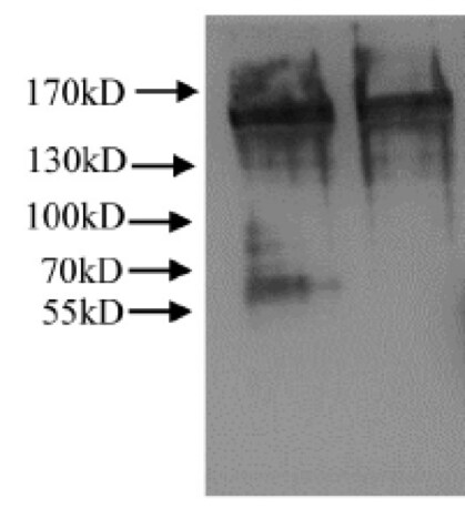
Western Blot: Collagen III alpha 1/COL3A1 Antibody [NB600-594] - Lane 1: Human Collagen III Load: 100 ng per lane Primary antibody: Collagen III Antibody at 1:1000 o/n at 4C Secondary antibody: DyLight 649 Goat anti-rabbit at 1:20,000 for 30 min at RT Block: incubated with blocking buffer for 30 min at RT Predicted/Observed size: 138 kDa/138 kDa.![Immunocytochemistry/ Immunofluorescence: Collagen III alpha 1/COL3A1 Antibody [NB600-594]](https://resources.rndsystems.com/images/products/Collagen-III-alpha-1-COL3A1-Antibody-Immunocytochemistry-Immunofluorescence-NB600-594-img0012.jpg "Immunocytochemistry/ Immunofluorescence: Collagen III alpha 1/COL3A1 Antibody [NB600-594]")
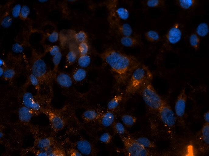
Immunocytochemistry/ Immunofluorescence: Collagen III alpha 1/COL3A1 Antibody [NB600-594]
Immunocytochemistry/Immunofluorescence: Collagen III alpha 1/COL3A1 Antibody [NB600-594] - Human primary ventricular cardiac fibroblasts were stained with anti-Collagen III antibody. Cells were cultured for 3 days in DMEM with 10% fetal calf serum. ICC/IF image submitted by a verified customer review.![Immunohistochemistry: Collagen III alpha 1/COL3A1 Antibody [NB600-594]](https://resources.rndsystems.com/images/products/Collagen-III-alpha-1-COL3A1-Antibody-Immunohistochemistry-NB600-594-img0016.jpg "Immunohistochemistry: Collagen III alpha 1/COL3A1 Antibody [NB600-594]")
Immunohistochemistry: Collagen III alpha 1/COL3A1 Antibody [NB600-594]
Immunohistochemistry: Collagen III alpha 1/COL3A1 Antibody [NB600-594] - Staining in FFPE sections of human skin(left, dermis) with moderate to strong red staining and testis (right) where strong staining was observed within connective tissue between seminiferous tubules. The antibody showed strong extracellular staining within connective tissues across many organs with minimal background staining. Slides were steamed in 0.01 M sodium citrate buffer, pH 6.0 at 99-100C. 20 minutes for antigen retrieval.![Western Blot: Collagen III alpha 1/COL3A1 Antibody [NB600-594]](https://resources.rndsystems.com/images/products/Collagen-III-alpha-1-COL3A1-Antibody-Western-Blot-NB600-594-img0023.jpg "Western Blot: Collagen III alpha 1/COL3A1 Antibody [NB600-594]")
![Immunohistochemistry: Collagen III alpha 1/COL3A1 Antibody [NB600-594]](https://resources.rndsystems.com/images/products/Collagen%20III%20alpha%201-COL3A1%20Antibody-Immunohistochemistry-NB600-594-img0024.jpg "Immunohistochemistry: Collagen III alpha 1/COL3A1 Antibody [NB600-594]")
Immunohistochemistry: Collagen III alpha 1/COL3A1 Antibody [NB600-594]
Collagen III alpha 1-COL3A1 Antibody-Immunohistochemistry-NB600-594-img0024.jpg![Western Blot: Collagen III alpha 1/COL3A1 Antibody [NB600-594]](https://resources.rndsystems.com/images/products/Collagen-III-alpha-1-COL3A1-Antibody-Western-Blot-NB600-594-img0019.jpg "Western Blot: Collagen III alpha 1/COL3A1 Antibody [NB600-594]")
Western Blot: Collagen III alpha 1/COL3A1 Antibody [NB600-594]
Collagen-III-alpha-1-COL3A1-Antibody-Western-Blot-NB600-594-img0019.jpg![Immunocytochemistry/ Immunofluorescence: Collagen III alpha 1/COL3A1 Antibody [NB600-594]](https://resources.rndsystems.com/images/products/Collagen-III-alpha-1-COL3A1-Antibody-Immunocytochemistry-Immunofluorescence-NB600-594-img0008.jpg "Immunocytochemistry/ Immunofluorescence: Collagen III alpha 1/COL3A1 Antibody [NB600-594]")
Immunocytochemistry/ Immunofluorescence: Collagen III alpha 1/COL3A1 Antibody [NB600-594]
Immunocytochemistry/Immunofluorescence: Collagen III alpha 1/COL3A1 Antibody [NB600-594] - Collagen III alpha 1 expression in HT-1080 cells. ICC/IF image submitted by a verified customer review.![Immunohistochemistry-Paraffin: Collagen III alpha 1/COL3A1 Antibody [NB600-594]](https://resources.rndsystems.com/images/products/Collagen-III-alpha-1-COL3A1-Antibody-Immunohistochemistry-Paraffin-NB600-594-img0013.jpg "Immunohistochemistry-Paraffin: Collagen III alpha 1/COL3A1 Antibody [NB600-594]")
Immunohistochemistry-Paraffin: Collagen III alpha 1/COL3A1 Antibody [NB600-594]
Immunohistochemistry-Paraffin: Collagen III alpha 1/COL3A1 Antibody [NB600-594] - Human lung tissue.![Immunohistochemistry-Paraffin: Collagen III alpha 1/COL3A1 Antibody [NB600-594]](https://resources.rndsystems.com/images/products/Collagen-III-alpha-1-COL3A1-Antibody-Immunohistochemistry-Paraffin-NB600-594-img0014.jpg "Immunohistochemistry-Paraffin: Collagen III alpha 1/COL3A1 Antibody [NB600-594]")
Immunohistochemistry-Paraffin: Collagen III alpha 1/COL3A1 Antibody [NB600-594]
Immunohistochemistry-Paraffin: Collagen III alpha 1/COL3A1 Antibody [NB600-594] - Human lung tissue![Immunohistochemistry: Collagen III alpha 1/COL3A1 Antibody [NB600-594]](https://resources.rndsystems.com/images/products/Collagen-III-alpha-1-COL3A1-Antibody-Immunohistochemistry-NB600-594-img0020.jpg "Immunohistochemistry: Collagen III alpha 1/COL3A1 Antibody [NB600-594]")
Immunohistochemistry: Collagen III alpha 1/COL3A1 Antibody [NB600-594]
Collagen-III-alpha-1-COL3A1-Antibody-Immunohistochemistry-NB600-594-img0020.jpg![Immunohistochemistry: Collagen III alpha 1/COL3A1 Antibody [NB600-594]](https://resources.rndsystems.com/images/products/Collagen-III-alpha-1-COL3A1-Antibody-Immunohistochemistry-NB600-594-img0021.jpg "Immunohistochemistry: Collagen III alpha 1/COL3A1 Antibody [NB600-594]")

Collagen III alpha 1/COL3A1 Antibody
anti collagen III antibody ( Lot 26016, 1:400, 45 min RT) showed strong staining in FFPE sections of human skin(left, dermis) with moderate to strong red staining and testis (right) where strong staining was observed within connective tissue between seminiferous tubules. The antibody showed strong extracellular staining within connective tissues across many organs with minimal background staining. Slides were steamed in 0.01 M sodium citrate buffer, pH 6.0 at 99-100C - 20 minutes for antigen retrieval. Images provided courtesy of LifeSpan Biosciences, Seattle, WA
Collagen III alpha 1/COL3A1 Antibody
Immunohistochemistry of Rabbit Anti-collagen type III antibody. Tissue: right lobe of the liver section. A:Central Vein (CV) fibrosis, B: Non-fibrotic CV, C: Perisinusodial fibrosis, D: Non-fibrotic area, E: Protat tract fibrosis, F: Septal fibrosis (arrow). Fixation: formalin fixed paraffin embedded. Antigen retrieval: not required. Primary antibody: Anti-collagen type III at 1:500 for 4C for 24hr. Secondary antibody: Peroxidase biotin-streptavidin rabbit secondary antibody at 1:10,000 for 45 min at RT. Localization: Anti-collagen type III is intra and extracellular. Staining: 3.3'-diaminobenzidine tetrahydrochloride was used as the chromogen. Nuclei were counterstained purple with hematoxylin.
Collagen III alpha 1/COL3A1 Antibody
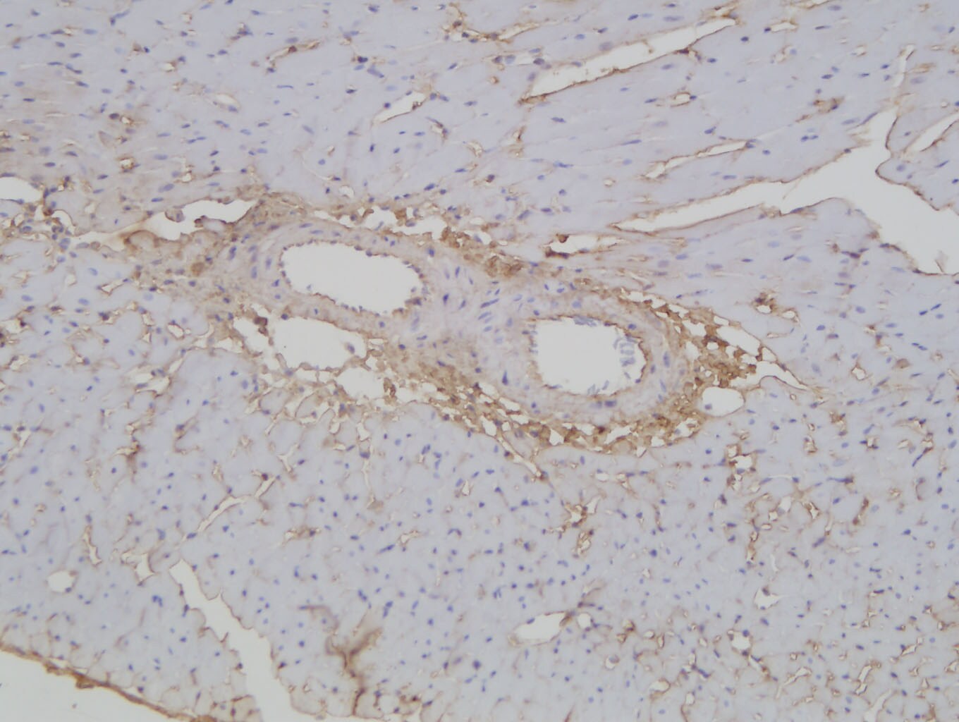
Immunohistochemistry of Rabbit Collagen III alpha 1/COL3A1 Antibody. Tissue: FFPE normal human spleen tissues (10X). Antigen Retrieval: 0.01 M sodium citrate buffer for 20 minutes. Primary Antibody: Anti-Collagen Type III at 5uL/mL for 45 mins at RT. Staining: Anti-Rabbit biotinylated secondary antibody for 30 min at RT. Alkaline phosphatase streptavidin for 30 min at RT. Alkaline phosphatase chromogen substrate for 30 min at RT. The stained slides were evaluated by a pathologist to confirm staining specificity.
Immunohistochemistry: Collagen III alpha 1/COL3A1 Antibody [NB600-594] -
FSP‐1–positive cells only partially responsible for collagen I & III production in irradiated bladders. Bladder strips from irradiated mice were costained for collagen I or for collagen III mRNA (pink) & for fibroblasts (FSP‐1, brown). U: urothelium; LP: lamina propria. Black arrow: FSP‐1 positive cells. Gray arrow: collagen only positive cells. Scale bar = 50 µm Image collected & cropped by CiteAb from the following publication (https://pubmed.ncbi.nlm.nih.gov/32109348), licensed under a CC-BY license. Not internally tested by Novus Biologicals.
Western Blot: Collagen III alpha 1/COL3A1 Antibody [NB600-594] -
Western Blot: Collagen III alpha 1/COL3A1 Antibody [NB600-594] - SCE attenuates myocardial fibrosis in heart tissue after MI. (a) RT-qPCR analysis for mRNA expression of collagen type I alpha 1 (Col1a1), collagen type III alpha 1 (Col3a1), actin, alpha 2 smooth muscle (Acta2), Mmp2, & Mmp9 in the border area (BA) & remote area (RA) 7 days, 14 days, & 21 days after MI. (b) Immunoblot analysis of protein expressions of alpha -SMA, collagen I, & collagen III in RA & BA in the heart tissues 7 days, 14 days, & 21 days after MI. Data are from three independent experiments ((a), mean ± SEM) or are representative of three independent experiments with similar results (b). ∗P < 0.05 & ∗∗P < 0.01, ANOVA with LSD t-test. Image collected & cropped by CiteAb from the following publication (https://pubmed.ncbi.nlm.nih.gov/31275414), licensed under a CC-BY license. Not internally tested by Novus Biologicals.
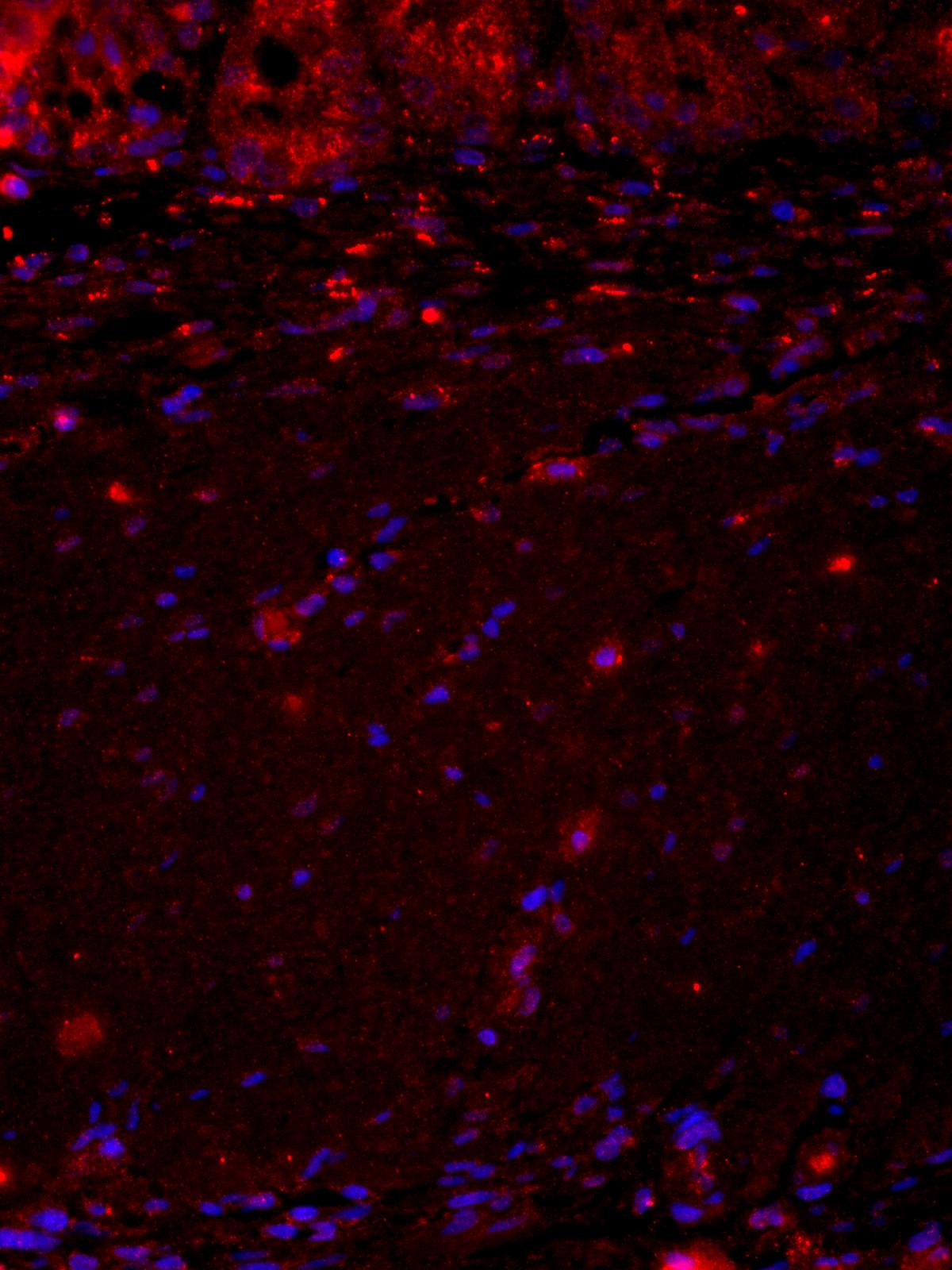
Immunocytochemistry/ Immunofluorescence: Collagen III alpha 1/COL3A1 Antibody [NB600-594] -
Immunocytochemistry/ Immunofluorescence: Collagen III alpha 1/COL3A1 Antibody [NB600-594] - Collagen I & collagen III accumulate in C57BL/6 mice in response to radiation. Bladder strips from irradiated & control mice were stained for collagen I or for collagen III & imaged using fluorescent microscopy. (a) Representative images (100 x 100 µm) of each mouse strain & treatment group are provided for collagen I & collagen III immunofluorescence. (b–c) Percentage of tissue‐positive for collagen I or collagen III staining. Collagen I & III density is significantly elevated in C57BL/6 mice, but not in the C3H or BALB/c strains. Results are mean ± SD of n = 3–6 mice. Dashed line: ANOVA; Full line: multiple t‐test. **p <.01, *** p < .001 Image collected & cropped by CiteAb from the following publication (https://pubmed.ncbi.nlm.nih.gov/32109348), licensed under a CC-BY license. Not internally tested by Novus Biologicals.
Immunohistochemistry: Collagen III alpha 1/COL3A1 Antibody [NB600-594] -
Immunohistochemistry: Collagen III alpha 1/COL3A1 Antibody [NB600-594] - Number of collagen I‐expressing cells is increased in irradiated C57BL/6 detrusor. Bladder strips from irradiated & control mice were stained for collagen I or for collagen III mRNA & counterstained with methyl green. Collagen I & III mRNA (pink) is apparent in both control & irradiated bladder sections in all three mouse strains. The number of collagen I & III‐producing cells in the detrusor is represented in the bar graphs. The number of collagen I‐expressing cells is significantly elevated in C57BL/6 mice after irradiation. Dashed line separates the urothelium (U) & lamina propria (LP) from the detrusor muscle (D). Results are mean ± SD of n = 3–4 mice. Scale bar = 100 µm. op ≤ .08, * p < .05 Image collected & cropped by CiteAb from the following publication (https://pubmed.ncbi.nlm.nih.gov/32109348), licensed under a CC-BY license. Not internally tested by Novus Biologicals.
Western Blot: Collagen III alpha 1/COL3A1 Antibody [NB600-594] -
Western Blot: Collagen III alpha 1/COL3A1 Antibody [NB600-594] - SCE attenuates fibrotic responses in CFs by inhibiting TGF-beta /Smad3 signaling activation. (a) RT-qPCR analysis of mRNA expression for genes of Col1a1, Col3a1, & Acta2 in cardiac fibroblasts (CFs) pretreated with different concentrations of SCE (5 μl/ml, 10 μl/ml, & 15 μl/ml) followed by TGF-beta stimulation (20 ng/ml) for 24 h. (b) RT-qPCR analysis of mRNA expression for genes of Col1a1, Col3a1, & Acta2 in CFs pretreated with SCE (10 μl/ml) followed by TGF-beta stimulation (20 ng/ml) for 12 h & 24 h. (c) RT-qPCR analysis of mRNA expression for genes of Smad2, Smad3, & Smad7 in CFs pretreated with SCE (10 μl/ml) followed by TGF-beta stimulation (20 ng/ml) for 12 h & 24 h. (d) Immunoblot analysis of expression of Smad2, Smad3, & Smad7 in CFs pretreated with SCE (10 μl/ml) followed by TGF-beta stimulation (20 ng/ml) for 24 h & 36 h. Data are from three independent experiments ((a)–(c), mean ± SEM) or are representative of three independent experiments with similar results (d). ∗P < 0.05 & ∗∗P < 0.01, Student's t-test. Image collected & cropped by CiteAb from the following publication (https://pubmed.ncbi.nlm.nih.gov/31275414), licensed under a CC-BY license. Not internally tested by Novus Biologicals.Applications for Collagen III alpha 1/COL3A1 Antibody - BSA Free
Application
Recommended Usage
ELISA
1:5000-1:50000
Immunocytochemistry/ Immunofluorescence
1:10 - 1:500
Immunohistochemistry
1:50-1:200
Immunohistochemistry-Paraffin
1:50 - 1:200
Immunoprecipitation
1:100
Western Blot
1:1000-1:10000
Application Notes
This product has been tested by dot Blot, western blot, and IHC and is useful for indirect trapping ELISA for quantitation of antigen in serum using a standard curve, immunoprecipitation, native (non-denaturing, non-dissociating) PAGE, immunohistochemistry, and western blotting for highly sensitive qualitative analysis.
See Simple Western Antibody Database for Simple Western validation: tested in skin; antibody dilution of 1:50; separated by size; detects a band at 139 kDa
See Simple Western Antibody Database for Simple Western validation: tested in skin; antibody dilution of 1:50; separated by size; detects a band at 139 kDa
Reviewed Applications
Read 7 reviews rated 4.7 using NB600-594 in the following applications:
Flow Cytometry Panel Builder
Bio-Techne Knows Flow Cytometry
Save time and reduce costly mistakes by quickly finding compatible reagents using the Panel Builder Tool.
Advanced Features
- Spectra Viewer - Custom analysis of spectra from multiple fluorochromes
- Spillover Popups - Visualize the spectra of individual fluorochromes
- Antigen Density Selector - Match fluorochrome brightness with antigen density

Formulation, Preparation, and Storage
Purification
Immunogen affinity purified
Formulation
0.02 M Potassium Phosphate, 0.15 M Sodium Chloride, pH 7.2
Format
BSA Free
Preservative
0.01% Sodium Azide
Concentration
Please see the vial label for concentration. If unlisted please contact technical services.
Shipping
The product is shipped with polar packs. Upon receipt, store it immediately at the temperature recommended below.
Stability & Storage
Store at 4C short term. For extended storage, add an equal volume of glycerol, aliquot and store at -20C or below. Avoid repeated freeze-thaw cycles.
Background: Collagen III alpha 1/COL3A1
Collagen III is a fibrillar collagen that constitutes 5-20% of all collagen in the body (1). It provides structural integrity and is found in many hallow organs and soft connective tissue including the vascular system, skin, lung, uterus, and intestine (1,2). Additionally, collagen III has be found to be associated with type I collagen in the same fibrils (1). Collagen III interacts with signaling integrins to carry out other key functions including cell adhesion, proliferation, and differentiation (1).
Mutations in the COL3A1 gene has been associated with a variety of human diseases, the most well-known being a group of connective tissue disorders termed Ehlers-Danlos Syndromes (1,2,4). Vascular Ehlers-Danlos Syndrome is a specific subtype that is considered the most severe and although the clinical manifestations vary, symptoms include thin skin and fragile blood vessels and can often result in both lung and heart complications (1,4). COL3A1 is also associated with glomerulopathies, or diseases of the glomeruli, which are characterized by an abundance of extracellular matrix (3). Collagenofibrotic glomerulopathy is one specific rare renal disease that is characterized by excessive levels of collagen III (3).
References
1. Kuivaniemi, H., & Tromp, G. (2019). Type III collagen (COL3A1): Gene and protein structure, tissue distribution, and associated diseases. Gene. https://doi.org/10.1016/j.gene.2019.05.003
2. Ricard-Blum S. (2011). The collagen family. Cold Spring Harbor perspectives in biology. https://doi.org/10.1101/cshperspect.a004978
3. Cohen A. H. (2012). Collagen Type III Glomerulopathies. Advances in chronic kidney disease. https://doi.org/10.1053/j.ackd.2012.02.017
4. Olson, S. L., Murray, M. L., & Skeik, N. (2019). A Novel Frameshift COL3A1 Variant in Vascular Ehlers-Danlos Syndrome. Annals of vascular surgery. https://doi.org/10.1016/j.avsg.2019.05.057
Additional Collagen III alpha 1/COL3A1 Products
Product Documents for Collagen III alpha 1/COL3A1 Antibody - BSA Free
Certificate of Analysis
To download a Certificate of Analysis, please enter a lot or batch number in the search box below.
Product Specific Notices for Collagen III alpha 1/COL3A1 Antibody - BSA Free
This product is for research use only and is not approved for use in humans or in clinical diagnosis. Primary Antibodies are guaranteed for 1 year from date of receipt.
Related Research Areas
Citations for Collagen III alpha 1/COL3A1 Antibody - BSA Free
 Powered by Bioz
Customer Reviews for Collagen III alpha 1/COL3A1 Antibody - BSA Free (7)
4.7 out of 5
7 Customer Ratings
Have you used Collagen III alpha 1/COL3A1 Antibody - BSA Free?
Submit a review and receive an Amazon gift card!
$25/€18/£15/$25CAN/¥2500 Yen for a review with an image
$10/€7/£6/$10CAN/¥1110 Yen for a review without an image
Submit a review
Customer Images
-(01-mg)_NB600-594_10821.jpg)
Showing
1
-
5 of
7 reviews
Showing All
Filter By:
-
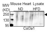
Application: Western BlotSample Tested: Heart lysatesSpecies: MouseVerified Customer | Posted 04/07/2021High fat diet-induced Col3a1 expression in mouse heart.Mouse heart lysate was prepared after 3 months of normal diet (ND) and high fat diet (HFD) consumption. Western blot was performed with anti-Col3a1 antibody.

-
Application: Western BlotSample Tested: whole cell lysateSpecies: HumanVerified Customer | Posted 06/07/2018
-
Application: Immunohistochemistry-FrozenSample Tested: Connective tissueSpecies: FelineVerified Customer | Posted 02/18/2018All images were captured using Leica SP8 conforcal microscope with X20 objective. Those primary antibodies were used at 1:200 dilution.
-
Application: Immunohistochemistry-ParaffinSample Tested: H9c2 whole cell lysateSpecies: RatVerified Customer | Posted 12/20/2017heart of SD rat;
-
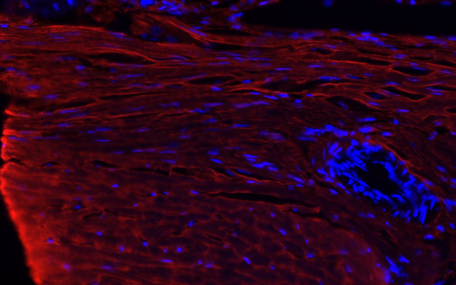
Application: Immunohistochemistry-ParaffinSample Tested: rat colonSpecies: RatVerified Customer | Posted 07/20/2016Rat colon collagen III (red) Hoechst (blue)
-
Application: ImmunofluorescenceSample Tested: HT-1080 (Fibrosarcoma)Species: HumanVerified Customer | Posted 04/02/2015Collagen III-positive Human Fibrosarcoma (HT-1080)
-
Application: ImmunofluorescenceSample Tested: human primary ventricular cardiac fibroblasts (from ScienCell)Species: HumanVerified Customer | Posted 10/04/2014NB600-594 in primary human cardiac fibroblasts
There are no reviews that match your criteria.
Protocols
Find general support by application which include: protocols, troubleshooting, illustrated assays, videos and webinars.
- 7-Amino Actinomycin D (7-AAD) Cell Viability Flow Cytometry Protocol
- Antigen Retrieval Protocol (PIER)
- Antigen Retrieval for Frozen Sections Protocol
- Appropriate Fixation of IHC/ICC Samples
- Cellular Response to Hypoxia Protocols
- Chromogenic IHC Staining of Formalin-Fixed Paraffin-Embedded (FFPE) Tissue Protocol
- Chromogenic Immunohistochemistry Staining of Frozen Tissue
- ClariTSA™ Fluorophore Kits
- Detection & Visualization of Antibody Binding
- ELISA Sample Preparation & Collection Guide
- ELISA Troubleshooting Guide
- Extracellular Membrane Flow Cytometry Protocol
- Flow Cytometry Protocol for Cell Surface Markers
- Flow Cytometry Protocol for Staining Membrane Associated Proteins
- Flow Cytometry Staining Protocols
- Flow Cytometry Troubleshooting Guide
- Fluorescent IHC Staining of Frozen Tissue Protocol
- Graphic Protocol for Heat-induced Epitope Retrieval
- Graphic Protocol for the Preparation and Fluorescent IHC Staining of Frozen Tissue Sections
- Graphic Protocol for the Preparation and Fluorescent IHC Staining of Paraffin-embedded Tissue Sections
- Graphic Protocol for the Preparation of Gelatin-coated Slides for Histological Tissue Sections
- How to Run an R&D Systems DuoSet ELISA
- How to Run an R&D Systems Quantikine ELISA
- How to Run an R&D Systems Quantikine™ QuicKit™ ELISA
- ICC Cell Smear Protocol for Suspension Cells
- ICC Immunocytochemistry Protocol Videos
- ICC for Adherent Cells
- IHC Sample Preparation (Frozen sections vs Paraffin)
- Immunocytochemistry (ICC) Protocol
- Immunocytochemistry Troubleshooting
- Immunofluorescence of Organoids Embedded in Cultrex Basement Membrane Extract
- Immunofluorescent IHC Staining of Formalin-Fixed Paraffin-Embedded (FFPE) Tissue Protocol
- Immunohistochemistry (IHC) and Immunocytochemistry (ICC) Protocols
- Immunohistochemistry Frozen Troubleshooting
- Immunohistochemistry Paraffin Troubleshooting
- Immunoprecipitation Protocol
- Intracellular Flow Cytometry Protocol Using Alcohol (Methanol)
- Intracellular Flow Cytometry Protocol Using Detergents
- Intracellular Nuclear Staining Flow Cytometry Protocol Using Detergents
- Intracellular Staining Flow Cytometry Protocol Using Alcohol Permeabilization
- Intracellular Staining Flow Cytometry Protocol Using Detergents to Permeabilize Cells
- Preparing Samples for IHC/ICC Experiments
- Preventing Non-Specific Staining (Non-Specific Binding)
- Primary Antibody Selection & Optimization
- Propidium Iodide Cell Viability Flow Cytometry Protocol
- Protocol for Heat-Induced Epitope Retrieval (HIER)
- Protocol for Liperfluo
- Protocol for Making a 4% Formaldehyde Solution in PBS
- Protocol for VisUCyte™ HRP Polymer Detection Reagent
- Protocol for the Characterization of Human Th22 Cells
- Protocol for the Characterization of Human Th9 Cells
- Protocol for the Fluorescent ICC Staining of Cell Smears - Graphic
- Protocol for the Fluorescent ICC Staining of Cultured Cells on Coverslips - Graphic
- Protocol for the Preparation & Fixation of Cells on Coverslips
- Protocol for the Preparation and Chromogenic IHC Staining of Frozen Tissue Sections
- Protocol for the Preparation and Chromogenic IHC Staining of Frozen Tissue Sections - Graphic
- Protocol for the Preparation and Chromogenic IHC Staining of Paraffin-embedded Tissue Sections
- Protocol for the Preparation and Chromogenic IHC Staining of Paraffin-embedded Tissue Sections - Graphic
- Protocol for the Preparation and Fluorescent ICC Staining of Cells on Coverslips
- Protocol for the Preparation and Fluorescent ICC Staining of Non-adherent Cells
- Protocol for the Preparation and Fluorescent ICC Staining of Stem Cells on Coverslips
- Protocol for the Preparation and Fluorescent IHC Staining of Frozen Tissue Sections
- Protocol for the Preparation and Fluorescent IHC Staining of Paraffin-embedded Tissue Sections
- Protocol for the Preparation of Gelatin-coated Slides for Histological Tissue Sections
- Protocol for the Preparation of a Cell Smear for Non-adherent Cell ICC - Graphic
- Protocol: Annexin V and PI Staining by Flow Cytometry
- Protocol: Annexin V and PI Staining for Apoptosis by Flow Cytometry
- Quantikine HS ELISA Kit Assay Principle, Alkaline Phosphatase
- Quantikine HS ELISA Kit Principle, Streptavidin-HRP Polymer
- R&D Systems Quality Control Western Blot Protocol
- Sandwich ELISA (Colorimetric) – Biotin/Streptavidin Detection Protocol
- Sandwich ELISA (Colorimetric) – Direct Detection Protocol
- TUNEL and Active Caspase-3 Detection by IHC/ICC Protocol
- The Importance of IHC/ICC Controls
- Troubleshooting Guide: ELISA
- Troubleshooting Guide: Fluorokine Flow Cytometry Kits
- Troubleshooting Guide: Immunohistochemistry
- Troubleshooting Guide: Western Blot Figures
- Western Blot Conditions
- Western Blot Protocol
- Western Blot Protocol for Cell Lysates
- Western Blot Troubleshooting
- Western Blot Troubleshooting Guide
- View all Protocols, Troubleshooting, Illustrated assays and Webinars
FAQs for Collagen III alpha 1/COL3A1 Antibody - BSA Free
Showing
1
-
3 of
3 FAQs
Showing All
-
Q: I am wondering if you could please confirm for me whether this product Collagen III antibody - catalogue # NB600-594 is able to be used under denaturing conditions with Western blots? Or whether it only binds to the collagen in its natural (non-denatured)
A: The Western blot image is not generated from native collagen - reducing conditions were used to obtain the image. The intact native molecule is too large to be run on standard 4-20% gels so the sample was run under reducing conditions. The image shows the sub-unit strand(s), not the intact collagen that consists of 3 subunit strands. These are not considered optimal or specific binding conditions for the anti-collagen antibodies, however the decision was made to run and post this on the website as an example.
-
Q: May I know if you carry the un-conjugated version of NB120-6580 (biotinylated anti-Collagen Type iII)?
A: I would recommend this catalog number for an unconjugated anti-Collagen Type III antibody: NB600-594.
-
Q: What is the dilution factor for IHC for starting? Because the range of dilution factor on the datasheet is so broad, I am confused which dilution factor is suitable for starting.
A: I can recommend the following dilution to start. Keep in mind that these will all vary greatly by sample type, so these are just suggestions. You may need to use more or less antibody. For catalog number NB600-594, the dilution range is 1:50-1:200.
-
Q: I am wondering if you could please confirm for me whether this product Collagen III antibody - catalogue # NB600-594 is able to be used under denaturing conditions with Western blots? Or whether it only binds to the collagen in its natural (non-denatured)
A: The Western blot image is not generated from native collagen - reducing conditions were used to obtain the image. The intact native molecule is too large to be run on standard 4-20% gels so the sample was run under reducing conditions. The image shows the sub-unit strand(s), not the intact collagen that consists of 3 subunit strands. These are not considered optimal or specific binding conditions for the anti-collagen antibodies, however the decision was made to run and post this on the website as an example.
-
Q: May I know if you carry the un-conjugated version of NB120-6580 (biotinylated anti-Collagen Type iII)?
A: I would recommend this catalog number for an unconjugated anti-Collagen Type III antibody: NB600-594.
-
Q: What is the dilution factor for IHC for starting? Because the range of dilution factor on the datasheet is so broad, I am confused which dilution factor is suitable for starting.
A: I can recommend the following dilution to start. Keep in mind that these will all vary greatly by sample type, so these are just suggestions. You may need to use more or less antibody. For catalog number NB600-594, the dilution range is 1:50-1:200.
Loading...