Collagens comprise a large family of insoluble extracellular glycoproteins that are essential components of connective tissues such as tendons, ligaments, cartilage, bone and skin. The mature polypeptides are secreted as coiled, left-handed helices that subsequently assemble into rope-like collagen fibers. Collagen I is a fibril-forming collagen that requires N- and C- terminal processing. Collagen IV is a network forming collagen whose C-terminus forms dimers and N-terminus forms tetramers.
Collagen IV Antibody - BSA Free
Novus Biologicals | Catalog # NB120-6586
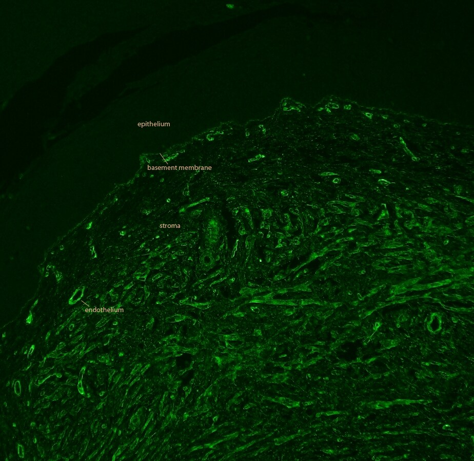
![Immunocytochemistry/ Immunofluorescence: Collagen IV Antibody [NB120-6586]](https://resources.rndsystems.com/images/products/Collagen-IV-Antibody-Immunocytochemistry-Immunofluorescence-NB120-6586-img0017.jpg "Immunocytochemistry/ Immunofluorescence: Collagen IV Antibody [NB120-6586]")
Key Product Details
Validated by
Knockout/Knockdown
Species Reactivity
Validated:
Human, Mouse, Rat, Bovine, Feline
Cited:
Human, Mouse, Rat, Feline
Applications
Validated:
Knockout Validated, Multiplex Immunofluorescence, Immunohistochemistry, Immunohistochemistry-Paraffin, Immunohistochemistry-Frozen, Immunohistochemistry Free-Floating, Western Blot, ELISA, Immunocytochemistry/ Immunofluorescence, Simple Western, Immunoprecipitation, Super-Resolution Microscopy
Cited:
Immunohistochemistry, Immunohistochemistry-Paraffin, Immunohistochemistry-Frozen, Immunohistochemistry Free-Floating, Western Blot, Immunocytochemistry/ Immunofluorescence, Simple Western, IF/IHC, Super-Resolution Microscopy
Label
Unconjugated
Antibody Source
Polyclonal Rabbit IgG
Format
BSA Free
Loading...
Product Specifications
Immunogen
Collagen IV from human and bovine placenta (Uniprot: P02462)
Reactivity Notes
This antibody reacts with most mammalian Collagen IV and has negligible cross-reactivity with Type I, II, III, V or VI collagens. Non-specific cross-reaction of anti-collagen antibodies with other human serum proteins or non-collagen extracellular matrix proteins is negligible.
Rat reactivity reported in scientific literature (PMID: 16774905)
Feline reactivity reported in scientific literature (PMID: 33091431).
Rat reactivity reported in scientific literature (PMID: 16774905)
Feline reactivity reported in scientific literature (PMID: 33091431).
Localization
Secreted and Cell surface (Potential).
Specificity
Some class-specific anti-collagens may be specific for three-dimensional epitopes which may result in diminished reactivity with denatured collagen or formalin-fixed, paraffin embedded tissues. This antibody reacts with most mammalian Collagen IV and has negligible cross-reactivity with Type I, II, III, V or VI collagens. Non-specific cross-reaction of anti-collagen antibodies with other human serum proteins or non-collagen extracellular matrix proteins is negligible.
Clonality
Polyclonal
Host
Rabbit
Isotype
IgG
Description
This antibody has been prepared by immunoaffinity chromatography using immobilized antigens followed by extensive cross-adsorption against other collagens, human serum proteins and non-collagen extracellular matrix proteins to remove any unwanted specificities. Some class-specific anti-collagens may be specific for three-dimensional epitopes which may result in diminished reactivity with denatured collagen or formalin-fixed, paraffin embedded tissues.
Store vial at 4C prior to opening. This product is stable at 4C as an undiluted liquid. Dilute only prior to immediate use. For extended storage, mix with an equal volume of glycerol, aliquot contents and freeze at -20C or below. Avoid cycles of freezing and thawing.
Store vial at 4C prior to opening. This product is stable at 4C as an undiluted liquid. Dilute only prior to immediate use. For extended storage, mix with an equal volume of glycerol, aliquot contents and freeze at -20C or below. Avoid cycles of freezing and thawing.
Scientific Data Images for Collagen IV Antibody - BSA Free
Immunocytochemistry/ Immunofluorescence: Collagen IV Antibody [NB120-6586]
Collagen-IV-Antibody-Immunocytochemistry-Immunofluorescence-NB120-6586-img0017.jpg![Immunohistochemistry-Paraffin: Collagen IV Antibody [NB120-6586]](https://resources.rndsystems.com/images/products/Collagen-IV-Antibody-Immunohistochemistry-Paraffin-NB120-6586-img0013.jpg "Immunohistochemistry-Paraffin: Collagen IV Antibody [NB120-6586]")
Immunohistochemistry-Paraffin: Collagen IV Antibody [NB120-6586]
Immunohistochemistry-Paraffin: Collagen IV Antibody [NB120-6586] - IHC-P image submitted by a verified customer review.![Immunohistochemistry-Paraffin: Collagen IV Antibody [NB120-6586]](https://resources.rndsystems.com/images/products/Collagen-IV-Antibody-Immunohistochemistry-Paraffin-NB120-6586-img0006.jpg "Immunohistochemistry-Paraffin: Collagen IV Antibody [NB120-6586]")
Immunohistochemistry-Paraffin: Collagen IV Antibody [NB120-6586]
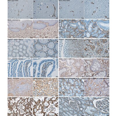
Immunohistochemistry-Paraffin: Collagen IV Antibody [NB120-6586] - Analysis of Collagen IV in various paraffin embedded human tissues. Image courtesy of product review submitted by Daniel Pirici.![Immunohistochemistry-Paraffin: Collagen IV Antibody [NB120-6586]](https://resources.rndsystems.com/images/products/Collagen-IV-Antibody-Immunohistochemistry-Paraffin-NB120-6586-img0008.jpg "Immunohistochemistry-Paraffin: Collagen IV Antibody [NB120-6586]")
Immunohistochemistry-Paraffin: Collagen IV Antibody [NB120-6586]
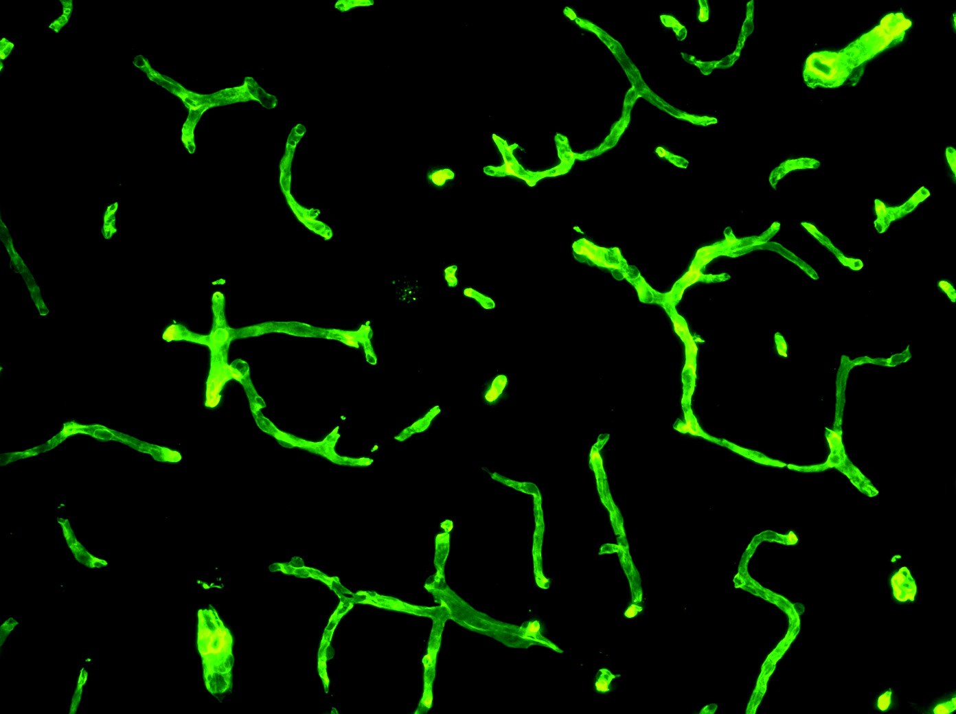
Immunohistochemistry-Paraffin: Collagen IV Antibody [NB120-6586] - Collagen IV antibody at 1:400, 45 min RT showed strong staining in FFPE sections of human kidney (left) with strong red staining observed in glomeruli and liver (right) with strong staining in sinusoids. Staining for both tissues was consistent with a basement membrane distribution. Slides were steamed in 0.01 M sodium citrate buffer, pH 6.0 at 100C for 20 minutes for antigen retrieval.![Immunohistochemistry-Paraffin: Collagen IV Antibody [NB120-6586]](https://resources.rndsystems.com/images/products/Collagen-IV-Antibody-Immunohistochemistry-Paraffin-NB120-6586-img0010.jpg "Immunohistochemistry-Paraffin: Collagen IV Antibody [NB120-6586]")
Immunohistochemistry-Paraffin: Collagen IV Antibody [NB120-6586]
Immunohistochemistry-Paraffin: Collagen IV Antibody [NB120-6586] - Analysis of Collagen IV in paraffin embedded mouse cervix tissue using anti-Collagen IV antibody. IHC-P image submitted by a verified customer review.![Immunohistochemistry-Paraffin: Collagen IV Antibody [NB120-6586]](https://resources.rndsystems.com/images/products/Collagen-IV-Antibody-Immunohistochemistry-Paraffin-NB120-6586-img0009.jpg "Immunohistochemistry-Paraffin: Collagen IV Antibody [NB120-6586]")
Immunohistochemistry-Paraffin: Collagen IV Antibody [NB120-6586]
Immunohistochemistry-Paraffin: Collagen IV Antibody [NB120-6586] - Analysis using the Biotin conjugate of NB120-6586. Staining of Collagen IV (red) and Insulin (green) in mouse pancreas. IHC-P image submitted by a verified customer review.![Immunohistochemistry-Frozen: Collagen IV Antibody [NB120-6586]](https://resources.rndsystems.com/images/products/Collagen-IV-Antibody-Immunohistochemistry-Frozen-NB120-6586-img0011.jpg "Immunohistochemistry-Frozen: Collagen IV Antibody [NB120-6586]")
Immunohistochemistry-Frozen: Collagen IV Antibody [NB120-6586]
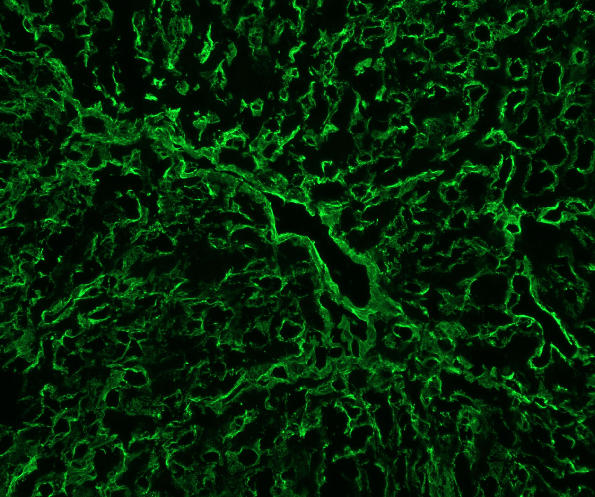
Immunohistochemistry-Frozen: Collagen IV Antibody [NB120-6586] - Mouse liver tissue. Collagen IV antibody at 1:200 dilution. Overnight incubation, no permeabilization. IHC-Fr image submitted by a verified customer review.![Immunohistochemistry-Frozen: Collagen IV Antibody [NB120-6586]](https://resources.rndsystems.com/images/products/Collagen-IV-Antibody-Immunohistochemistry-Frozen-NB120-6586-img0012.jpg "Immunohistochemistry-Frozen: Collagen IV Antibody [NB120-6586]")
Immunohistochemistry-Frozen: Collagen IV Antibody [NB120-6586]
Immunohistochemistry-Frozen: Collagen IV Antibody [NB120-6586] - Mouse brain tissue. Antibody at 1:500. IHC-Fr image submitted by a verified customer review.![Knockout Validated: Collagen IV Antibody [NB120-6586]](https://resources.rndsystems.com/images/products/Collagen-IV-Antibody-Knockout-Validated-NB120-6586-img0016.jpg "Immunocytochemistry/ Immunofluorescence: Collagen IV Antibody [NB120-6586]")
Immunocytochemistry/ Immunofluorescence: Collagen IV Antibody [NB120-6586]
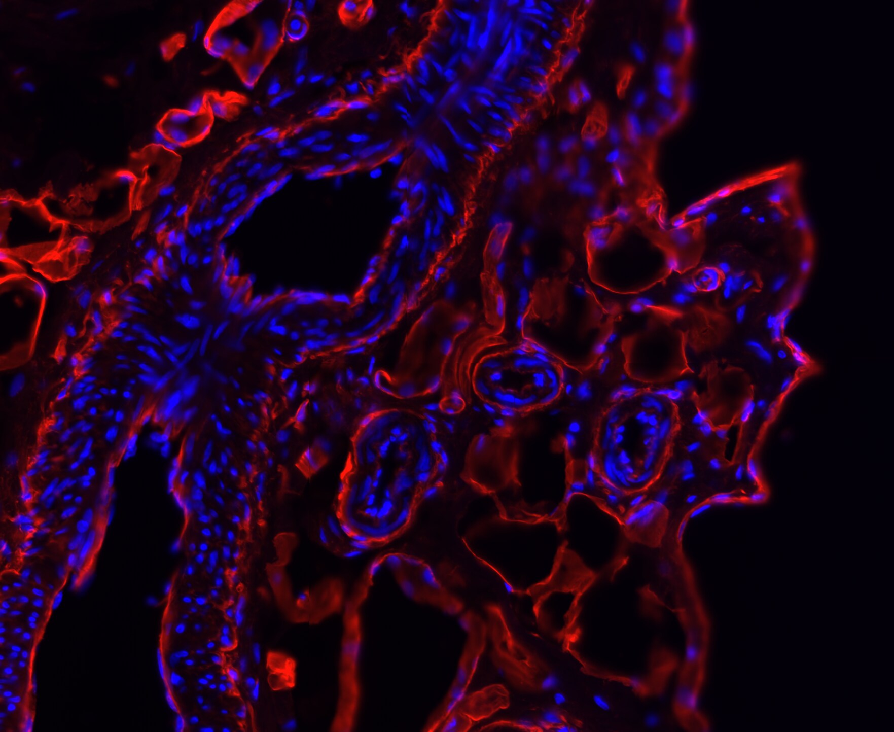
Immunocytochemistry/ Immunofluorescence: Collagen IV Antibody [NB120-6586] - Collagen IV was detected in immersion fixed THP-1 (positive) but not THP-1 KO (negative) cells using 10 ug/mL Rabbit Anti-Human/Bovine Polyclonal Antibody (Catalog # NB120-6586) for 3 hours at room temperature. Cells were stained with the NorthernLights(TM) 557-conjugated Donkey anti-Rabbit IgG Secondary Antibody [NL557] (red; Catalog # NL004) and counterstained with DAPI (blue).Applications for Collagen IV Antibody - BSA Free
Application
Recommended Usage
ELISA
1:5000-1:50000
Immunocytochemistry/ Immunofluorescence
1:10-1:500
Immunohistochemistry
1:50-1:200
Immunohistochemistry-Paraffin
1:50-1:200
Immunoprecipitation
1:100
Simple Western
1:50
Western Blot
1:1000-1:10000
Application Notes
This product has been tested by dot blot and IHC and is suitable for indirect trapping ELISA for quantitation of antigen in serum using a standard curve, immunoprecipitation, native (non-denaturing, non-dissociating) PAGE, immunohistochemistry, and western blotting for highly sensitive qualitative analysis.
Use in Immunohistochemistry Free-Floating reported in scientific literature (PMID: 32245949).
See Simple Western Antibody Database for Simple Western validation: Tested in Lungs, separated by Size, antibody dilution of 1:50
Use in Immunohistochemistry Free-Floating reported in scientific literature (PMID: 32245949).
See Simple Western Antibody Database for Simple Western validation: Tested in Lungs, separated by Size, antibody dilution of 1:50
Reviewed Applications
Read 7 reviews rated 4.9 using NB120-6586 in the following applications:
Formulation, Preparation, and Storage
Purification
Immunogen affinity purified
Formulation
0.02 M Potassium Phosphate, 0.15 M Sodium Chloride, pH 7.2
Format
BSA Free
Preservative
0.01% Sodium Azide
Concentration
Please see the vial label for concentration. If unlisted please contact technical services.
Shipping
The product is shipped with polar packs. Upon receipt, store it immediately at the temperature recommended below.
Stability & Storage
Store at 4C short term. For extended storage, add an equal volume of glycerol, aliquot and store at -20C or below. Avoid repeated freeze-thaw cycles.
Background: Collagen IV
Alternate Names
BSVD;BSVD1;COL4A1s;Collagen alpha-1(IV) chain;PADMAL;RATOR
Gene Symbol
COL4A1
UniProt
Additional Collagen IV Products
Product Documents for Collagen IV Antibody - BSA Free
Certificate of Analysis
To download a Certificate of Analysis, please enter a lot or batch number in the search box below.
Product Specific Notices for Collagen IV Antibody - BSA Free
This product is for research use only and is not approved for use in humans or in clinical diagnosis. Primary Antibodies are guaranteed for 1 year from date of receipt.
Related Research Areas
Citations for Collagen IV Antibody - BSA Free
 Powered by Bioz
Customer Reviews for Collagen IV Antibody - BSA Free (7)
4.9 out of 5
7 Customer Ratings
Have you used Collagen IV Antibody - BSA Free?
Submit a review and receive an Amazon gift card!
$25/€18/£15/$25CAN/¥2500 Yen for a review with an image
$10/€7/£6/$10CAN/¥1110 Yen for a review without an image
Submit a review
Customer Images
Showing
1
-
5 of
7 reviews
Showing All
Filter By:
-
Application: Immunohistochemistry-ParaffinSample Tested: bladeSpecies: HumanVerified Customer | Posted 10/09/2019

-
Application: Immunohistochemistry-FrozenSample Tested: Mouse brainSpecies: MouseVerified Customer | Posted 09/10/20191:500
-
Application: Immunohistochemistry-FrozenSample Tested: LiverSpecies: MouseVerified Customer | Posted 08/09/2019Dilution 1:200 Incubation: Overnight 4C No permeabilization
-
Application: Immunohistochemistry-FrozenSample Tested: Connective tissueSpecies: FelineVerified Customer | Posted 02/18/2018All images were captured using Leica SP8 conforcal microscope with X20 objective. Those primary antibodies were used at 1:200 dilution.
-
Application: Immunohistochemistry-ParaffinSample Tested: Mouse cervixSpecies: MouseVerified Customer | Posted 04/27/2016Mouse cervix
-
Application: Immunohistochemistry-ParaffinVerified Customer | Posted 09/15/2012
-
Application: Immunohistochemistry-ParaffinSample Tested: Human tissueSpecies: HumanVerified Customer | Posted 09/15/2012
There are no reviews that match your criteria.
Protocols
Find general support by application which include: protocols, troubleshooting, illustrated assays, videos and webinars.
- Antigen Retrieval Protocol (PIER)
- Antigen Retrieval for Frozen Sections Protocol
- Appropriate Fixation of IHC/ICC Samples
- Cellular Response to Hypoxia Protocols
- Chromogenic IHC Staining of Formalin-Fixed Paraffin-Embedded (FFPE) Tissue Protocol
- Chromogenic Immunohistochemistry Staining of Frozen Tissue
- ClariTSA™ Fluorophore Kits
- Detection & Visualization of Antibody Binding
- ELISA Sample Preparation & Collection Guide
- ELISA Troubleshooting Guide
- Fluorescent IHC Staining of Frozen Tissue Protocol
- Graphic Protocol for Heat-induced Epitope Retrieval
- Graphic Protocol for the Preparation and Fluorescent IHC Staining of Frozen Tissue Sections
- Graphic Protocol for the Preparation and Fluorescent IHC Staining of Paraffin-embedded Tissue Sections
- Graphic Protocol for the Preparation of Gelatin-coated Slides for Histological Tissue Sections
- How to Run an R&D Systems DuoSet ELISA
- How to Run an R&D Systems Quantikine ELISA
- How to Run an R&D Systems Quantikine™ QuicKit™ ELISA
- ICC Cell Smear Protocol for Suspension Cells
- ICC Immunocytochemistry Protocol Videos
- ICC for Adherent Cells
- IHC Sample Preparation (Frozen sections vs Paraffin)
- Immunocytochemistry (ICC) Protocol
- Immunocytochemistry Troubleshooting
- Immunofluorescence of Organoids Embedded in Cultrex Basement Membrane Extract
- Immunofluorescent IHC Staining of Formalin-Fixed Paraffin-Embedded (FFPE) Tissue Protocol
- Immunohistochemistry (IHC) and Immunocytochemistry (ICC) Protocols
- Immunohistochemistry Frozen Troubleshooting
- Immunohistochemistry Paraffin Troubleshooting
- Immunoprecipitation Protocol
- Preparing Samples for IHC/ICC Experiments
- Preventing Non-Specific Staining (Non-Specific Binding)
- Primary Antibody Selection & Optimization
- Protocol for Heat-Induced Epitope Retrieval (HIER)
- Protocol for Making a 4% Formaldehyde Solution in PBS
- Protocol for VisUCyte™ HRP Polymer Detection Reagent
- Protocol for the Fluorescent ICC Staining of Cell Smears - Graphic
- Protocol for the Fluorescent ICC Staining of Cultured Cells on Coverslips - Graphic
- Protocol for the Preparation & Fixation of Cells on Coverslips
- Protocol for the Preparation and Chromogenic IHC Staining of Frozen Tissue Sections
- Protocol for the Preparation and Chromogenic IHC Staining of Frozen Tissue Sections - Graphic
- Protocol for the Preparation and Chromogenic IHC Staining of Paraffin-embedded Tissue Sections
- Protocol for the Preparation and Chromogenic IHC Staining of Paraffin-embedded Tissue Sections - Graphic
- Protocol for the Preparation and Fluorescent ICC Staining of Cells on Coverslips
- Protocol for the Preparation and Fluorescent ICC Staining of Non-adherent Cells
- Protocol for the Preparation and Fluorescent ICC Staining of Stem Cells on Coverslips
- Protocol for the Preparation and Fluorescent IHC Staining of Frozen Tissue Sections
- Protocol for the Preparation and Fluorescent IHC Staining of Paraffin-embedded Tissue Sections
- Protocol for the Preparation of Gelatin-coated Slides for Histological Tissue Sections
- Protocol for the Preparation of a Cell Smear for Non-adherent Cell ICC - Graphic
- Quantikine HS ELISA Kit Assay Principle, Alkaline Phosphatase
- Quantikine HS ELISA Kit Principle, Streptavidin-HRP Polymer
- R&D Systems Quality Control Western Blot Protocol
- Sandwich ELISA (Colorimetric) – Biotin/Streptavidin Detection Protocol
- Sandwich ELISA (Colorimetric) – Direct Detection Protocol
- TUNEL and Active Caspase-3 Detection by IHC/ICC Protocol
- The Importance of IHC/ICC Controls
- Troubleshooting Guide: ELISA
- Troubleshooting Guide: Immunohistochemistry
- Troubleshooting Guide: Western Blot Figures
- Western Blot Conditions
- Western Blot Protocol
- Western Blot Protocol for Cell Lysates
- Western Blot Troubleshooting
- Western Blot Troubleshooting Guide
- View all Protocols, Troubleshooting, Illustrated assays and Webinars


