Collagen IV Antibody - Azide and BSA Free
Novus Biologicals | Catalog # NBP1-26549
![Immunohistochemistry-Frozen: Collagen IV Antibody [NBP1-26549]](https://resources.rndsystems.com/images/products/Collagen-IV-Antibody-Immunohistochemistry-Frozen-NBP1-26549-img0004.jpg "Immunohistochemistry-Frozen: Collagen IV Antibody [NBP1-26549]")
Key Product Details
Validated by
Biological Validation
Species Reactivity
Validated:
Human, Mouse, Rat, Monkey
Cited:
Human, Mouse, Rat, Primate - Macaca mulatta (Rhesus Macaque)
Applications
Validated:
Immunohistochemistry, Immunohistochemistry-Paraffin, Immunohistochemistry-Frozen, Western Blot, ELISA, Immunocytochemistry/ Immunofluorescence
Cited:
Immunohistochemistry, Immunohistochemistry-Paraffin, Immunohistochemistry-Frozen, Western Blot, Immunocytochemistry/ Immunofluorescence, IF/IHC
Label
Unconjugated
Antibody Source
Polyclonal Goat IgG
Format
Azide and BSA Free
Loading...
Product Specifications
Immunogen
Goats were hyperimmunized with human type IV collagen.
Reactivity Notes
Monkey reactivity reported in scientific literature (PMID: 27417518), Mouse reactivity reported in scientific literature (PMID: 32479919), Rat reactivity reported in scientific literature (PMID: 30362896).
Specificity
Reacts with conformational determinants on human type IV collagen as demonstrated by ELISA. May react with type IV collagen from other species. Exhibits <10% cross reactivity with collagen type I, II, III, V and VI. The antibody has not been tested for reactivity with other ECM proteins (e.g., laminin, fibronectin).
Clonality
Polyclonal
Host
Goat
Isotype
IgG
Scientific Data Images for Collagen IV Antibody - Azide and BSA Free
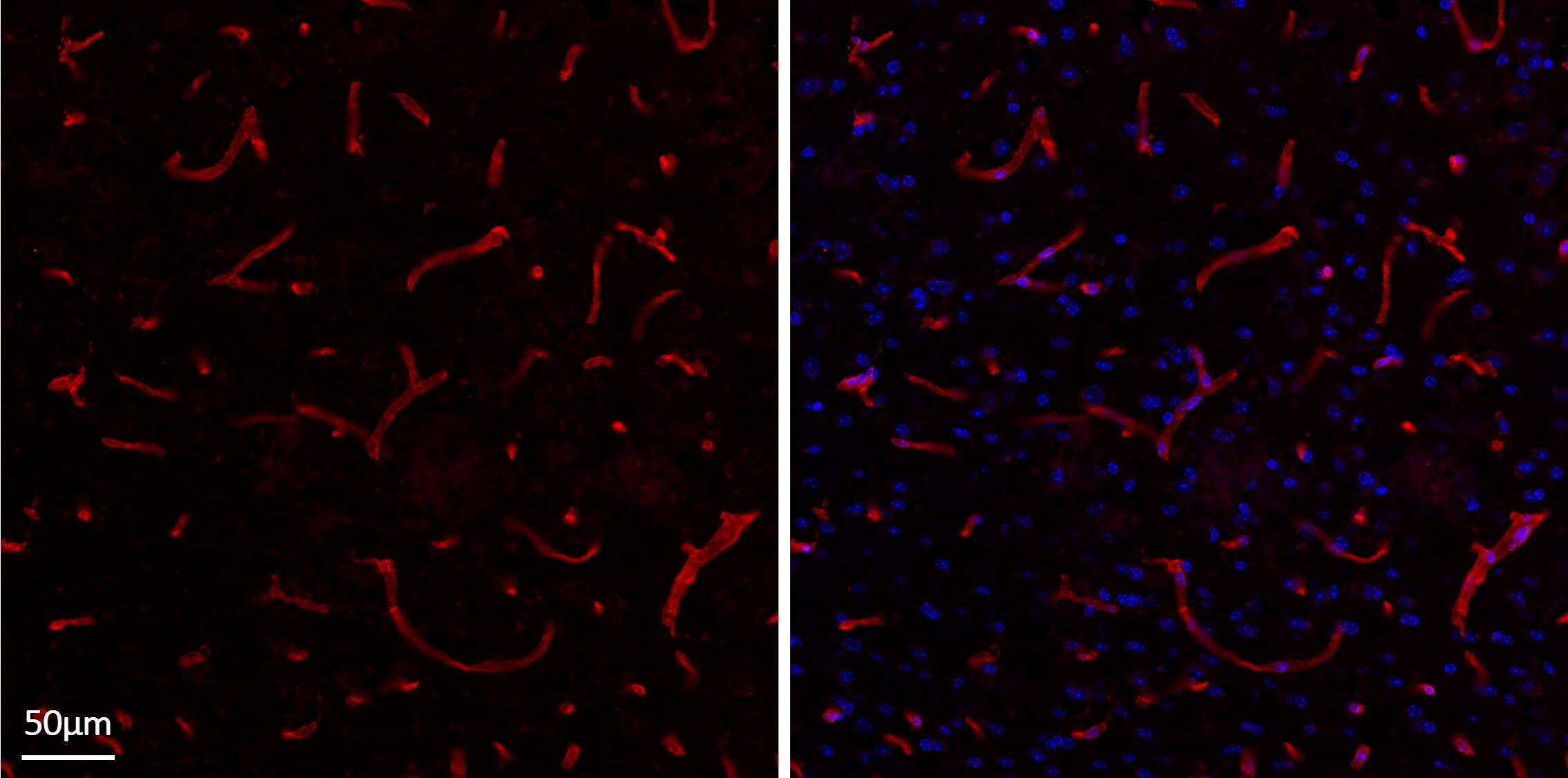
Immunohistochemistry-Frozen: Collagen IV Antibody [NBP1-26549]
Immunohistochemistry-Frozen: Collagen IV Antibody [NBP1-26549] - Left panel: NBP1-26549 signal, identifying Collagen IV in extracellular basement membrane of brain endothelial cells. Right panel: counterstained image with DAPI. Mouse brain tissue (12um thickness) was incubated overnight with NBP1-26549 (1:400) in PBS 1x at 4C. The day after, the slide was washed 3 x PBS 1x and then incubated with secondary anti-goat antibody (Alex Fluor 568-conjugated), 1:1000 for 2 hours at r.t. After counterstaining with DAPI, image was acquired with confocal microscopy. Left panel: NBP1-26549 signal, identifying Collagen IV in extracellular basement membrane of brain endothelial cells. Right panel: counterstained image with DAPI. Image from verified customer review.![Immunohistochemistry-Paraffin: Collagen IV Antibody [NBP1-26549]](https://resources.rndsystems.com/images/products/Collagen-IV-alpha-1-Antibody-Immunohistochemistry-Paraffin-NBP1-26549-img0001.jpg "Immunohistochemistry-Paraffin: Collagen IV Antibody [NBP1-26549]")
Immunohistochemistry-Paraffin: Collagen IV Antibody [NBP1-26549]
Immunohistochemistry-Paraffin: Collagen IV Antibody [NBP1-26549] - Human gastric cancer tissue was stained with Goat IgG-UNLB isotype control (left) and Goat Anti-Type IV Collagen-UNLB; (right) followed by Swine Anti-Goat IgG(H+L), Human/Rat/Mouse SP ads-HRP DAB, and hematoxylin.![Immunohistochemistry: Collagen IV Antibody [NBP1-26549]](https://resources.rndsystems.com/images/products/Collagen-IV-Antibody-Immunohistochemistry-NBP1-26549-img0002.jpg "Immunohistochemistry: Collagen IV Antibody [NBP1-26549]")
![Immunohistochemistry: Collagen IV Antibody [NBP1-26549]](https://resources.rndsystems.com/images/products/Collagen-IV-Antibody-Immunohistochemistry-NBP1-26549-img0003.jpg "Immunohistochemistry: Collagen IV Antibody [NBP1-26549]")

Immunohistochemistry: Collagen IV Antibody [NBP1-26549] -
Immunohistochemistry: Collagen IV Antibody [NBP1-26549] - Photographs showing H&E staining & immunostaining for Col-I, Col-III, Col-IV & Col-V in the rhesus macaque & baboon intramural tube. Images of open fallopian tube (a, c, e, g, i, k, m, o, q & s) were obtained from control animals. Occluded tube (b, d, f, h, j, l, n, p, r & t) were from animals treated with 5% PF. Arrows indicate fibrotic response. All images were captured at original magnification ×200. Insets (frame r & s) show negative control with an irrelevant antibody. Treatment with PF increased collagen immunoreactivity in the lamina propria of the intramural tube. Image collected & cropped by CiteAb from the following publication (https://linkinghub.elsevier.com/retrieve/pii/S0010782416301445), licensed under a CC-BY license. Not internally tested by Novus Biologicals.
Western Blot: Collagen IV Antibody - Azide and BSA Free [NBP1-26549] -
MiR-29a regulation of target gene expression in cochlea from miR-29a+/+ and miR-29a–/– mice. (A–G) With beta -actin as a control, COL4A1, COL4A2, COL4A3, COL4A4, COL4A5, LAMB2, and LAMC1 protein levels in cochlea were analyzed by Western blot. The independent t-test was used to compare data between two groups [COL4A1, t(4) = 9.920, P < 0.001; COL4A2, t(4) = 3.847, P = 0.018; COL4A3, t(4) = 3.389, P = 0.028; COL4A4, t(4) = 2.842, P = 0.047; COL4A5, t(4) = 3.782, P = 0.019; LAMB2, t(4) = 3.915, P = 0.017; LAMC1, t(4) = 3.088, P = 0.037]. (H) The mRNA expression levels of target genes as measured by qRT-PCR [independent t-test, Col4a1, t(4) = 6.050, P = 0.004; Col4a2, t(4) = 13.145, P < 0.001; Col4a3, t(4) = 3.735, P = 0.02; Col4a4, t(4) = 7.238, P = 0.002; Col4a5, t(4) = 13.141, P < 0.001; Lamb2, t(4) = 8.529, P = 0.001; Lamc1, t(4) = 3.126, P = 0.035]. All data are shown as the mean +/- SD, n = 3 biological replicates, *P < 0.05, **P < 0.01, and ***P < 0.001 between two groups. Image collected and cropped by CiteAb from the following open publication (https://pubmed.ncbi.nlm.nih.gov/37275774), licensed under a CC-BY license. Not internally tested by Novus Biologicals.
Western Blot: Collagen IV Antibody - Azide and BSA Free [NBP1-26549] -
Nimbidiol treatment normalized the expression of collagen IV, fibronectin and elastin in the diabetic kidney. Representative images from (A) Semi-quantitative RT-PCR analyses showing gene expression and (B) western blot analyses showing protein expression of Col IV, fibronectin and elastin in the kidney. The bar diagrams represent the fold change from WT + Saline. Data are mean +/- SD (n = 6/group). *p < 0.05 versus WT + Saline, WT + Nimbidiol and Akita + Nimbidiol, †p < 0.05 versus Akita + Saline. Image collected and cropped by CiteAb from the following open publication (https://pubmed.ncbi.nlm.nih.gov/36522378), licensed under a CC-BY license. Not internally tested by Novus Biologicals.
Immunocytochemistry/ Immunofluorescence: Collagen IV Antibody - Azide and BSA Free [NBP1-26549] -
Decreased angiogenesis and increased integrin/basement membrane signaling in TNF lungs.(A) Loss of angiogenesis signaling in TNF-Tg mice. MultiNicheNet was used to identify ligand-receptor pairs that were specific to either WT or TNF conditions. WT lungs demonstrated strong Tek, Flt1, Kdr, and Ednrb interactions with Angt1, Vegfa, Gpc3, and Edn3, which were absent TNF-Tg conditions. (B) Increased TGF-beta /integrin/basement membrane signaling in TNF-Tg lungs. TNF-Tg lungs demonstrated the presence of interactions between Itga2 (primarily in gCAP2 cells) and multiple collagen genes, Hspg2, and Tgfb1. (C) Decreased expression of Flt1 and Tek and increased expression of Col4a2 and Itga2 in TNF-Tg gCAP cells. Violin plots demonstrate expression across cell types. (D) Immunostaining confirms increased expression of Col4a1, Itga2, and Igfbp7 in TNF-Tg lungs. Original magnification × 200. (E) Circos plots indicating most differentially regulated ligand-receptor pairs ascertained by MultiNicheNet in WT lungs (left) and TNF-Tg (right) lungs; sender cells are listed on the bottom, while arrows point to receiver cells on the top. Image collected and cropped by CiteAb from the following open publication (https://pubmed.ncbi.nlm.nih.gov/40569693), licensed under a CC-BY license. Not internally tested by Novus Biologicals.
Immunocytochemistry/ Immunofluorescence: Collagen IV Antibody - Azide and BSA Free [NBP1-26549] -
Nimbidiol inhibited upregulation of collagen IV and fibronectin, and downregulation of elastin expression in the diabetic kidney. Representative immunofluorescence images of the renal cortex showing expression of (A) Col IV, (B) fibronectin and (C) elastin. The nuclear counterstaining was performed using DAPI (blue). The bar diagrams represent the fold change in the fluorescence intensity from WT + Saline for (D) Col IV (E) fibronectin and (F) elastin. Data are mean +/- SD (n = 6/group). *p < 0.05 versus WT + Saline, WT + Nimbidiol and Akita + Nimbidiol, †p < 0.05 versus Akita + Saline. Scale bar: 20 um; magnification × 60. Image collected and cropped by CiteAb from the following open publication (https://pubmed.ncbi.nlm.nih.gov/36522378), licensed under a CC-BY license. Not internally tested by Novus Biologicals.Applications for Collagen IV Antibody - Azide and BSA Free
Application
Recommended Usage
ELISA
1:1000-1:4000
Immunohistochemistry-Paraffin
1:10-1:500
Western Blot
1:100-1:2000
Application Notes
IHC, IHC-FR reported in (PMID: 32029859), ICC/IF reported in (PMID: 32479919), IHC-P reported in (PMID: 27417518).
Reviewed Applications
Read 1 review rated 5 using NBP1-26549 in the following applications:
Formulation, Preparation, and Storage
Purification
Immunogen affinity purified
Formulation
0.1M BBS (pH 8.2)
Format
Azide and BSA Free
Preservative
No Preservative
Concentration
0.4 mg/ml
Shipping
The product is shipped with polar packs. Upon receipt, store it immediately at the temperature recommended below.
Stability & Storage
Store at 4C. Do not freeze.
Background: Collagen IV
Alternate Names
BSVD;BSVD1;COL4A1s;Collagen alpha-1(IV) chain;PADMAL;RATOR
Gene Symbol
COL4A1
Additional Collagen IV Products
Product Documents for Collagen IV Antibody - Azide and BSA Free
Certificate of Analysis
To download a Certificate of Analysis, please enter a lot or batch number in the search box below.
Product Specific Notices for Collagen IV Antibody - Azide and BSA Free
This product is for research use only and is not approved for use in humans or in clinical diagnosis. Primary Antibodies are guaranteed for 1 year from date of receipt.
Related Research Areas
Citations for Collagen IV Antibody - Azide and BSA Free
 Powered by Bioz
Customer Reviews for Collagen IV Antibody - Azide and BSA Free (1)
5 out of 5
1 Customer Rating
Have you used Collagen IV Antibody - Azide and BSA Free?
Submit a review and receive an Amazon gift card!
$25/€18/£15/$25CAN/¥2500 Yen for a review with an image
$10/€7/£6/$10CAN/¥1110 Yen for a review without an image
Submit a review
Customer Images
Showing
1
-
1 of
1 review
Showing All
Filter By:
-
Application: Immunohistochemistry-FrozenSample Tested: Adult mouse brain tissueSpecies: MouseVerified Customer | Posted 05/24/2022Left panel: NBP1-26549 signal, identifying Collagen IV in extracellular basement membrane of brain endothelial cells. Right panel: counterstained image with DAPI.Mouse brain tissue (12um thickness) was incubated overnight with NBP1-26549 (1:400) in PBS 1x at 4C. The day after, the slide was washed 3 x PBS 1x and then incubated with secondary anti-goat antibody (Alex Fluor 568-conjugated), 1:1000 for 2 hours at r.t. After counterstaining with DAPI, image was acquired with confocal microscopy. Left panel: NBP1-26549 signal, identifying Collagen IV in extracellular basement membrane of brain endothelial cells. Right panel: counterstained image with DAPI.

There are no reviews that match your criteria.
Protocols
Find general support by application which include: protocols, troubleshooting, illustrated assays, videos and webinars.
- Antigen Retrieval Protocol (PIER)
- Antigen Retrieval for Frozen Sections Protocol
- Appropriate Fixation of IHC/ICC Samples
- Cellular Response to Hypoxia Protocols
- Chromogenic IHC Staining of Formalin-Fixed Paraffin-Embedded (FFPE) Tissue Protocol
- Chromogenic Immunohistochemistry Staining of Frozen Tissue
- ClariTSA™ Fluorophore Kits
- Detection & Visualization of Antibody Binding
- ELISA Sample Preparation & Collection Guide
- ELISA Troubleshooting Guide
- Fluorescent IHC Staining of Frozen Tissue Protocol
- Graphic Protocol for Heat-induced Epitope Retrieval
- Graphic Protocol for the Preparation and Fluorescent IHC Staining of Frozen Tissue Sections
- Graphic Protocol for the Preparation and Fluorescent IHC Staining of Paraffin-embedded Tissue Sections
- Graphic Protocol for the Preparation of Gelatin-coated Slides for Histological Tissue Sections
- How to Run an R&D Systems DuoSet ELISA
- How to Run an R&D Systems Quantikine ELISA
- How to Run an R&D Systems Quantikine™ QuicKit™ ELISA
- ICC Cell Smear Protocol for Suspension Cells
- ICC Immunocytochemistry Protocol Videos
- ICC for Adherent Cells
- IHC Sample Preparation (Frozen sections vs Paraffin)
- Immunocytochemistry (ICC) Protocol
- Immunocytochemistry Troubleshooting
- Immunofluorescence of Organoids Embedded in Cultrex Basement Membrane Extract
- Immunofluorescent IHC Staining of Formalin-Fixed Paraffin-Embedded (FFPE) Tissue Protocol
- Immunohistochemistry (IHC) and Immunocytochemistry (ICC) Protocols
- Immunohistochemistry Frozen Troubleshooting
- Immunohistochemistry Paraffin Troubleshooting
- Preparing Samples for IHC/ICC Experiments
- Preventing Non-Specific Staining (Non-Specific Binding)
- Primary Antibody Selection & Optimization
- Protocol for Heat-Induced Epitope Retrieval (HIER)
- Protocol for Making a 4% Formaldehyde Solution in PBS
- Protocol for VisUCyte™ HRP Polymer Detection Reagent
- Protocol for the Fluorescent ICC Staining of Cell Smears - Graphic
- Protocol for the Fluorescent ICC Staining of Cultured Cells on Coverslips - Graphic
- Protocol for the Preparation & Fixation of Cells on Coverslips
- Protocol for the Preparation and Chromogenic IHC Staining of Frozen Tissue Sections
- Protocol for the Preparation and Chromogenic IHC Staining of Frozen Tissue Sections - Graphic
- Protocol for the Preparation and Chromogenic IHC Staining of Paraffin-embedded Tissue Sections
- Protocol for the Preparation and Chromogenic IHC Staining of Paraffin-embedded Tissue Sections - Graphic
- Protocol for the Preparation and Fluorescent ICC Staining of Cells on Coverslips
- Protocol for the Preparation and Fluorescent ICC Staining of Non-adherent Cells
- Protocol for the Preparation and Fluorescent ICC Staining of Stem Cells on Coverslips
- Protocol for the Preparation and Fluorescent IHC Staining of Frozen Tissue Sections
- Protocol for the Preparation and Fluorescent IHC Staining of Paraffin-embedded Tissue Sections
- Protocol for the Preparation of Gelatin-coated Slides for Histological Tissue Sections
- Protocol for the Preparation of a Cell Smear for Non-adherent Cell ICC - Graphic
- Quantikine HS ELISA Kit Assay Principle, Alkaline Phosphatase
- Quantikine HS ELISA Kit Principle, Streptavidin-HRP Polymer
- R&D Systems Quality Control Western Blot Protocol
- Sandwich ELISA (Colorimetric) – Biotin/Streptavidin Detection Protocol
- Sandwich ELISA (Colorimetric) – Direct Detection Protocol
- TUNEL and Active Caspase-3 Detection by IHC/ICC Protocol
- The Importance of IHC/ICC Controls
- Troubleshooting Guide: ELISA
- Troubleshooting Guide: Immunohistochemistry
- Troubleshooting Guide: Western Blot Figures
- Western Blot Conditions
- Western Blot Protocol
- Western Blot Protocol for Cell Lysates
- Western Blot Troubleshooting
- Western Blot Troubleshooting Guide
- View all Protocols, Troubleshooting, Illustrated assays and Webinars


