![Western Blot: COX-2 Antibody [NB100-868]](https://resources.rndsystems.com/images/products/COX-2-Antibody-Western-Blot-NB100-868-img0007.jpg "Western Blot: COX-2 Antibody [NB100-868]")
Key Product Details
Species Reactivity
Validated:
Cited:
Applications
Validated:
Cited:
Label
Antibody Source
Product Specifications
Immunogen
Reactivity Notes
Localization
Clonality
Host
Isotype
Scientific Data Images for COX-2 Antibody
Western Blot: COX-2 Antibody [NB100-868]
Western Blot: COX-2 Antibody [NB100-868] - A549 (A) and Daudi (B) cell lysates (35 ug protein in RIPA buffer). Detected by chemiluminescence.![Immunocytochemistry/ Immunofluorescence: COX-2 Antibody [NB100-868]](https://resources.rndsystems.com/images/products/COX-2-Antibody-Immunocytochemistry-Immunofluorescence-NB100-868-img0006.jpg "Immunocytochemistry/ Immunofluorescence: COX-2 Antibody [NB100-868]")
Immunocytochemistry/ Immunofluorescence: COX-2 Antibody [NB100-868]
Immunocytochemistry/Immunofluorescence: COX-2 Antibody [NB100-868] - Analysis of paraformaldehyde fixed NIH3T3 cells, permeabilized with 0.15%Triton. Primary incubation 1hr (10ug/ml) followed by Alexa Fluor 488 secondary antibody (2ug/ml), showing cytoplasm and vesicle staining. The nuclear stain is DAPI (blue). Negative control: Unimmunized goat IgG (10ug/ml) followed by Alexa Fluor 488 secondary antibody (2ug/ml).![Immunohistochemistry-Paraffin: COX-2 Antibody [NB100-868]](https://resources.rndsystems.com/images/products/COX-2-Antibody-Immunohistochemistry-Paraffin-NB100-868-img0008.jpg "Immunohistochemistry-Paraffin: COX-2 Antibody [NB100-868]")
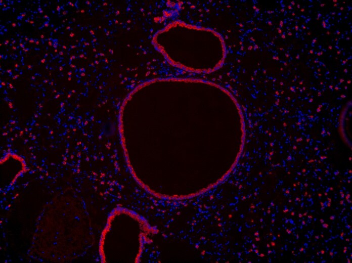
Immunohistochemistry-Paraffin: COX-2 Antibody [NB100-868]
Immunohistochemistry-Paraffin: COX-2 Antibody [NB100-868] - 10x image of mouse lung tissue expressing cox-2. Secondary antibody: Alexa Fluor 546 (red), counterstain: DAPI (blue). Goat anti-mouse cox-2 immunofluorescence staining of paraffin embedded fixed mouse lung tissue sections. Antigen retrieval in citrate buffer (95C, 20 min) followed by permeabilization with 0.2% Triton X-100 for 20 min. Sections were blocked in 5% donkey serum for 1 hr at RT, and incubated with NB100-868 1:100 dilution in 3% donkey serum overnight at 4 C. Incubated with secondary donkey anti-goat AF546 at 1:500 dilution 1 hr at RT. Image submitted by a verified customer review.![Flow Cytometry: COX-2 Antibody [NB100-868]](https://resources.rndsystems.com/images/products/COX-2-Antibody-Flow-Cytometry-NB100-868-img0004.jpg "Flow Cytometry: COX-2 Antibody [NB100-868]")
Flow Cytometry: COX-2 Antibody [NB100-868]
Flow Cytometry: COX-2 Antibody [NB100-868] - Paraformaldehyde fixed HeLa cells (blue line), permeabilized with 0.5% Triton. Primary incubation 1hr (10 ug/ml followed by Alexa Fluor 488 secondary antibody (2 ug/ml). IgG control: Unimmunized goat IgG (black line)![Immunocytochemistry/ Immunofluorescence: COX-2 Antibody [NB100-868]](https://resources.rndsystems.com/images/products/COX-2-Antibody-Immunocytochemistry-Immunofluorescence-NB100-868-img0005.jpg "Immunocytochemistry/ Immunofluorescence: COX-2 Antibody [NB100-868]")
Immunocytochemistry/ Immunofluorescence: COX-2 Antibody [NB100-868]
Immunocytochemistry/Immunofluorescence: COX-2 Antibody [NB100-868] - Paraformaldehyde fixed HepG2 cells, permeabilized with 0.15% Triton. Primary incubation 1hr (10 ug/ml followed by Alexa Fluor 488 secondary antibody (2 ug/ml, showing cytoplasmic/vesicle staining. The nuclear stain is DAPI (blue)Applications for COX-2 Antibody
Flow Cytometry
Immunocytochemistry/ Immunofluorescence
Peptide ELISA
Western Blot
Reviewed Applications
Read 1 review rated 5 using NB100-868 in the following applications:
Flow Cytometry Panel Builder
Bio-Techne Knows Flow Cytometry
Save time and reduce costly mistakes by quickly finding compatible reagents using the Panel Builder Tool.
Advanced Features
- Spectra Viewer - Custom analysis of spectra from multiple fluorochromes
- Spillover Popups - Visualize the spectra of individual fluorochromes
- Antigen Density Selector - Match fluorochrome brightness with antigen density

Formulation, Preparation, and Storage
Purification
Formulation
Preservative
Concentration
Shipping
Stability & Storage
Background: COX-2
Long Name
Alternate Names
Entrez Gene IDs
Gene Symbol
UniProt
Additional COX-2 Products
Product Documents for COX-2 Antibody
Certificate of Analysis
To download a Certificate of Analysis, please enter a lot or batch number in the search box below.
Product Specific Notices for COX-2 Antibody
This product is for research use only and is not approved for use in humans or in clinical diagnosis. Primary Antibodies are guaranteed for 1 year from date of receipt.
Citations for COX-2 Antibody
 Powered by Bioz
Customer Reviews for COX-2 Antibody (1)
Have you used COX-2 Antibody?
Submit a review and receive an Amazon gift card!
$25/€18/£15/$25CAN/¥2500 Yen for a review with an image
$10/€7/£6/$10CAN/¥1110 Yen for a review without an image
Submit a review
Customer Images
-
Application: Immunohistochemistry-ParaffinSample Tested: Adult Mouse Lung TissueSpecies: MouseVerified Customer | Posted 05/09/201810x image of mouse lung tissue expressing cox-2, seondary: alexafluor 546 (red), dapi (blue)Goat anti mouse cox 2 immunofluorescence staining of paraffin embedded fixed mouse lung tissue sections- Antigen retrieval in citrate buffer (95 C, 20 min) followed by permeabilization with 0.2% Triton x-100 for 20 min. Sections were blocked in 5% donkey serum for 1 hr at RT, and incubated with NB100-868 1:100 dilution in 3% donkey serum overnight at 4 C. Incubated with secondary donkey anti-goat AF546 at 1:500 dilution 1 hr at RT. Counter-stained with DAPI (blue)

There are no reviews that match your criteria.
Protocols
Find general support by application which include: protocols, troubleshooting, illustrated assays, videos and webinars.
- 7-Amino Actinomycin D (7-AAD) Cell Viability Flow Cytometry Protocol
- Antigen Retrieval Protocol (PIER)
- Antigen Retrieval for Frozen Sections Protocol
- Appropriate Fixation of IHC/ICC Samples
- Cellular Response to Hypoxia Protocols
- Chromogenic IHC Staining of Formalin-Fixed Paraffin-Embedded (FFPE) Tissue Protocol
- Chromogenic Immunohistochemistry Staining of Frozen Tissue
- ClariTSA™ Fluorophore Kits
- Detection & Visualization of Antibody Binding
- ELISA Sample Preparation & Collection Guide
- ELISA Troubleshooting Guide
- Extracellular Membrane Flow Cytometry Protocol
- Flow Cytometry Protocol for Cell Surface Markers
- Flow Cytometry Protocol for Staining Membrane Associated Proteins
- Flow Cytometry Staining Protocols
- Flow Cytometry Troubleshooting Guide
- Fluorescent IHC Staining of Frozen Tissue Protocol
- Graphic Protocol for Heat-induced Epitope Retrieval
- Graphic Protocol for the Preparation and Fluorescent IHC Staining of Frozen Tissue Sections
- Graphic Protocol for the Preparation and Fluorescent IHC Staining of Paraffin-embedded Tissue Sections
- Graphic Protocol for the Preparation of Gelatin-coated Slides for Histological Tissue Sections
- How to Run an R&D Systems DuoSet ELISA
- How to Run an R&D Systems Quantikine ELISA
- How to Run an R&D Systems Quantikine™ QuicKit™ ELISA
- ICC Cell Smear Protocol for Suspension Cells
- ICC Immunocytochemistry Protocol Videos
- ICC for Adherent Cells
- IHC Sample Preparation (Frozen sections vs Paraffin)
- Immunocytochemistry (ICC) Protocol
- Immunocytochemistry Troubleshooting
- Immunofluorescence of Organoids Embedded in Cultrex Basement Membrane Extract
- Immunofluorescent IHC Staining of Formalin-Fixed Paraffin-Embedded (FFPE) Tissue Protocol
- Immunohistochemistry (IHC) and Immunocytochemistry (ICC) Protocols
- Immunohistochemistry Frozen Troubleshooting
- Immunohistochemistry Paraffin Troubleshooting
- Intracellular Flow Cytometry Protocol Using Alcohol (Methanol)
- Intracellular Flow Cytometry Protocol Using Detergents
- Intracellular Nuclear Staining Flow Cytometry Protocol Using Detergents
- Intracellular Staining Flow Cytometry Protocol Using Alcohol Permeabilization
- Intracellular Staining Flow Cytometry Protocol Using Detergents to Permeabilize Cells
- Preparing Samples for IHC/ICC Experiments
- Preventing Non-Specific Staining (Non-Specific Binding)
- Primary Antibody Selection & Optimization
- Propidium Iodide Cell Viability Flow Cytometry Protocol
- Protocol for Heat-Induced Epitope Retrieval (HIER)
- Protocol for Liperfluo
- Protocol for Making a 4% Formaldehyde Solution in PBS
- Protocol for VisUCyte™ HRP Polymer Detection Reagent
- Protocol for the Characterization of Human Th22 Cells
- Protocol for the Characterization of Human Th9 Cells
- Protocol for the Fluorescent ICC Staining of Cell Smears - Graphic
- Protocol for the Fluorescent ICC Staining of Cultured Cells on Coverslips - Graphic
- Protocol for the Preparation & Fixation of Cells on Coverslips
- Protocol for the Preparation and Chromogenic IHC Staining of Frozen Tissue Sections
- Protocol for the Preparation and Chromogenic IHC Staining of Frozen Tissue Sections - Graphic
- Protocol for the Preparation and Chromogenic IHC Staining of Paraffin-embedded Tissue Sections
- Protocol for the Preparation and Chromogenic IHC Staining of Paraffin-embedded Tissue Sections - Graphic
- Protocol for the Preparation and Fluorescent ICC Staining of Cells on Coverslips
- Protocol for the Preparation and Fluorescent ICC Staining of Non-adherent Cells
- Protocol for the Preparation and Fluorescent ICC Staining of Stem Cells on Coverslips
- Protocol for the Preparation and Fluorescent IHC Staining of Frozen Tissue Sections
- Protocol for the Preparation and Fluorescent IHC Staining of Paraffin-embedded Tissue Sections
- Protocol for the Preparation of Gelatin-coated Slides for Histological Tissue Sections
- Protocol for the Preparation of a Cell Smear for Non-adherent Cell ICC - Graphic
- Protocol: Annexin V and PI Staining by Flow Cytometry
- Protocol: Annexin V and PI Staining for Apoptosis by Flow Cytometry
- Quantikine HS ELISA Kit Assay Principle, Alkaline Phosphatase
- Quantikine HS ELISA Kit Principle, Streptavidin-HRP Polymer
- R&D Systems Quality Control Western Blot Protocol
- Sandwich ELISA (Colorimetric) – Biotin/Streptavidin Detection Protocol
- Sandwich ELISA (Colorimetric) – Direct Detection Protocol
- TUNEL and Active Caspase-3 Detection by IHC/ICC Protocol
- The Importance of IHC/ICC Controls
- Troubleshooting Guide: ELISA
- Troubleshooting Guide: Fluorokine Flow Cytometry Kits
- Troubleshooting Guide: Immunohistochemistry
- Troubleshooting Guide: Western Blot Figures
- Western Blot Conditions
- Western Blot Protocol
- Western Blot Protocol for Cell Lysates
- Western Blot Troubleshooting
- Western Blot Troubleshooting Guide
- View all Protocols, Troubleshooting, Illustrated assays and Webinars
FAQs for COX-2 Antibody
-
Q: I am a student researching COX-2 expression in felines and am looking for a suitable primary antibody to use in IHC. To your knowledge, have any of your COX-2 antibodies been used successfully with feline COX-2? If yes, I would be very interested in learning more. If not, would it be possible to run a sequence homology of feline COX-2 (GenBank ABM65700.1, accession EF036473.1) against your available COX-2 antibodies to determine the overlap and likelihood of cross-reactivity?
A:
We have not tested any of these antibodies for their reactivity in felines. However, we do have a number of antibodies that look like they may cross-react with feline. NBP1-85499 recognizes a region that is about 90-91% similar to feline COX2. NBP2-04015 recognizes a region in humans that is 94% similar to feline COX2. NB100-868 recognizes a region in humans at the far C-terminal 12 amino acids that is 100% identical to feline COX2. NBP1-19354 recognizes a region that is also about 95% similar to feline COX2. Only NBP1-85499 and NBP1-19354 have been tested for use in IHC. If you would like to test this antibody in an untested species and/or application and share your results with us, then I can recommend our Innovators Reward Program.