CREB Antibody (LB9)
Novus Biologicals | Catalog # NB100-74393
![Western Blot: CREB Antibody (LB9) [NB100-74393]](https://resources.rndsystems.com/images/products/CREB-Antibody-LB9-Western-Blot-NB100-74393-img0026.jpg "Western Blot: CREB Antibody (LB9) [NB100-74393]")
Key Product Details
Validated by
Species Reactivity
Validated:
Cited:
Applications
Validated:
Cited:
Label
Antibody Source
Product Specifications
Immunogen
Reactivity Notes
Specificity
Clonality
Host
Isotype
Scientific Data Images for CREB Antibody (LB9)
Western Blot: CREB Antibody (LB9) [NB100-74393]
CREB-Antibody-LB9-Western-Blot-NB100-74393-img0026.jpg![Immunohistochemistry-Paraffin: CREB Antibody (LB9) [NB100-74393]](https://resources.rndsystems.com/images/products/CREB-Antibody-LB9-Immunohistochemistry-Paraffin-NB100-74393-img0019.jpg "Immunohistochemistry-Paraffin: CREB Antibody (LB9) [NB100-74393]")
Immunohistochemistry-Paraffin: CREB Antibody (LB9) [NB100-74393]
Immunohistochemistry-Paraffin: CREB Antibody (LB9) [NB100-74393] - Normal biopsies of deparaffinized Human tonsil tissue.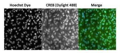
![Western Blot: CREB Antibody (LB9) [NB100-74393]](https://resources.rndsystems.com/images/products/CREB-Antibody-LB9-Western-Blot-NB100-74393-img0021.jpg "Western Blot: CREB Antibody (LB9) [NB100-74393]")
Western Blot: CREB Antibody (LB9) [NB100-74393]
Western Blot: CREB Antibody (LB9) [NB100-74393] - Proteins were transferred to a PVDF membrane and blocked with 5% Milk/TBST for at least 1 hour. Membranes were probed with a mouse monoclonal antibody recognizing CREB at a dilution of 1:500 overnight at 4C on a rocking platform. Membranes were washed in TBS-0.1%Tween 20 and probed with a goat anti-mouse-HRP secondary antibody at a dilution of 1:20,000 for at least one hour. Membranes were washed and chemiluminescent detection performed.![Western Blot: CREB Antibody (LB9) [NB100-74393]](https://resources.rndsystems.com/images/products/CREB-Antibody-LB9-Western-Blot-NB100-74393-img0022.jpg "Western Blot: CREB Antibody (LB9) [NB100-74393]")
Western Blot: CREB Antibody (LB9) [NB100-74393]
Western Blot: CREB Antibody (LB9) [NB100-74393] - Analysis of HepG2 cells![Immunohistochemistry-Paraffin: CREB Antibody (LB9) [NB100-74393]](https://resources.rndsystems.com/images/products/CREB-Antibody-LB9-Immunohistochemistry-Paraffin-NB100-74393-img0018.jpg "Immunohistochemistry-Paraffin: CREB Antibody (LB9) [NB100-74393]")
Immunohistochemistry-Paraffin: CREB Antibody (LB9) [NB100-74393]
Immunohistochemistry-Paraffin: CREB Antibody (LB9) [NB100-74393] - Cancer biopsies of deparaffinized human bladder and rectal tissue.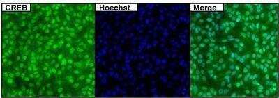
![Immunoprecipitation: CREB Antibody (LB9) [NB100-74393]](https://resources.rndsystems.com/images/products/CREB-Antibody-LB9-Immunoprecipitation-NB100-74393-img0023.jpg "Immunoprecipitation: CREB Antibody (LB9) [NB100-74393]")
Immunoprecipitation: CREB Antibody (LB9) [NB100-74393]
Immunoprecipitation: CREB Antibody (LB9) [NB100-74393] - Analysis of CREB was performed on untreated 293T cells. Antigen: antbody complex was formed by binding 500ug whole cell lysate with 2ug of mouse monoclonal antibody recognizing CREB overnight on a rocking platform at 4C. The immune-complex was then captured on 50ul Protein A/G Plus Agarose. Captured immune-complexes were then washed extensively and proteins eluted with 5X Reducing Sample Loading Dye. Samples were then resolved on a 4-20% Tris-HCl polyacrylamide gel. Proteins were transferred to PVDF membrane and blocked with 5% Milk/TBS-0.1%Tween for at least 1 hour. Membranes were then probed with a mouse monoclonal antibody recognizing CREB at a dilution of 1:1000 overnight rotating at 4C. Membranes were then washed in TBST and probed with a goat anti-mouse-HRP secondary antibody at a dilution of 1:20,000 for at least one hour. Membranes were washed and chemiluminescent detection was performed using Super Signal West Dura.Applications for CREB Antibody (LB9)
ELISA
Immunocytochemistry/ Immunofluorescence
Immunohistochemistry
Immunohistochemistry-Paraffin
Immunoprecipitation
Western Blot
Formulation, Preparation, and Storage
Purification
Formulation
Preservative
Concentration
Shipping
Stability & Storage
Background: CREB
Long Name
Alternate Names
Gene Symbol
Additional CREB Products
Product Documents for CREB Antibody (LB9)
Certificate of Analysis
To download a Certificate of Analysis, please enter a lot or batch number in the search box below.
Product Specific Notices for CREB Antibody (LB9)
This product is for research use only and is not approved for use in humans or in clinical diagnosis. Primary Antibodies are guaranteed for 1 year from date of receipt.
Citations for CREB Antibody (LB9)
 Powered by Bioz
Customer Reviews for CREB Antibody (LB9)
There are currently no reviews for this product. Be the first to review CREB Antibody (LB9) and earn rewards!
Have you used CREB Antibody (LB9)?
Submit a review and receive an Amazon gift card!
$25/€18/£15/$25CAN/¥2500 Yen for a review with an image
$10/€7/£6/$10CAN/¥1110 Yen for a review without an image
Submit a review
Protocols
Find general support by application which include: protocols, troubleshooting, illustrated assays, videos and webinars.
- Antigen Retrieval Protocol (PIER)
- Antigen Retrieval for Frozen Sections Protocol
- Appropriate Fixation of IHC/ICC Samples
- Cellular Response to Hypoxia Protocols
- ChIP Protocol Video
- Chromatin Immunoprecipitation (ChIP) Protocol
- Chromatin Immunoprecipitation Protocol
- Chromogenic IHC Staining of Formalin-Fixed Paraffin-Embedded (FFPE) Tissue Protocol
- Chromogenic Immunohistochemistry Staining of Frozen Tissue
- ClariTSA™ Fluorophore Kits
- Detection & Visualization of Antibody Binding
- ELISA Sample Preparation & Collection Guide
- ELISA Troubleshooting Guide
- Fluorescent IHC Staining of Frozen Tissue Protocol
- Graphic Protocol for Heat-induced Epitope Retrieval
- Graphic Protocol for the Preparation and Fluorescent IHC Staining of Frozen Tissue Sections
- Graphic Protocol for the Preparation and Fluorescent IHC Staining of Paraffin-embedded Tissue Sections
- Graphic Protocol for the Preparation of Gelatin-coated Slides for Histological Tissue Sections
- How to Run an R&D Systems DuoSet ELISA
- How to Run an R&D Systems Quantikine ELISA
- How to Run an R&D Systems Quantikine™ QuicKit™ ELISA
- ICC Cell Smear Protocol for Suspension Cells
- ICC Immunocytochemistry Protocol Videos
- ICC for Adherent Cells
- IHC Sample Preparation (Frozen sections vs Paraffin)
- Immunocytochemistry (ICC) Protocol
- Immunocytochemistry Troubleshooting
- Immunofluorescence of Organoids Embedded in Cultrex Basement Membrane Extract
- Immunofluorescent IHC Staining of Formalin-Fixed Paraffin-Embedded (FFPE) Tissue Protocol
- Immunohistochemistry (IHC) and Immunocytochemistry (ICC) Protocols
- Immunohistochemistry Frozen Troubleshooting
- Immunohistochemistry Paraffin Troubleshooting
- Immunoprecipitation Protocol
- Preparing Samples for IHC/ICC Experiments
- Preventing Non-Specific Staining (Non-Specific Binding)
- Primary Antibody Selection & Optimization
- Protocol for Heat-Induced Epitope Retrieval (HIER)
- Protocol for Making a 4% Formaldehyde Solution in PBS
- Protocol for VisUCyte™ HRP Polymer Detection Reagent
- Protocol for the Fluorescent ICC Staining of Cell Smears - Graphic
- Protocol for the Fluorescent ICC Staining of Cultured Cells on Coverslips - Graphic
- Protocol for the Preparation & Fixation of Cells on Coverslips
- Protocol for the Preparation and Chromogenic IHC Staining of Frozen Tissue Sections
- Protocol for the Preparation and Chromogenic IHC Staining of Frozen Tissue Sections - Graphic
- Protocol for the Preparation and Chromogenic IHC Staining of Paraffin-embedded Tissue Sections
- Protocol for the Preparation and Chromogenic IHC Staining of Paraffin-embedded Tissue Sections - Graphic
- Protocol for the Preparation and Fluorescent ICC Staining of Cells on Coverslips
- Protocol for the Preparation and Fluorescent ICC Staining of Non-adherent Cells
- Protocol for the Preparation and Fluorescent ICC Staining of Stem Cells on Coverslips
- Protocol for the Preparation and Fluorescent IHC Staining of Frozen Tissue Sections
- Protocol for the Preparation and Fluorescent IHC Staining of Paraffin-embedded Tissue Sections
- Protocol for the Preparation of Gelatin-coated Slides for Histological Tissue Sections
- Protocol for the Preparation of a Cell Smear for Non-adherent Cell ICC - Graphic
- Quantikine HS ELISA Kit Assay Principle, Alkaline Phosphatase
- Quantikine HS ELISA Kit Principle, Streptavidin-HRP Polymer
- R&D Systems Quality Control Western Blot Protocol
- Sandwich ELISA (Colorimetric) – Biotin/Streptavidin Detection Protocol
- Sandwich ELISA (Colorimetric) – Direct Detection Protocol
- TUNEL and Active Caspase-3 Detection by IHC/ICC Protocol
- The Importance of IHC/ICC Controls
- Troubleshooting Guide: ELISA
- Troubleshooting Guide: Immunohistochemistry
- Troubleshooting Guide: Western Blot Figures
- Western Blot Conditions
- Western Blot Protocol
- Western Blot Protocol for Cell Lysates
- Western Blot Troubleshooting
- Western Blot Troubleshooting Guide
- View all Protocols, Troubleshooting, Illustrated assays and Webinars
FAQs for CREB Antibody (LB9)
-
Q: May I know which creb1 antibody is good for ChIP? I found a paper using your rabbit polyclonal creb1 antibody for ChIP but they didn't indicate the catalog number.
A:
We are not aware of any of our Creb1 antibodies being used in ChIP at this time. If you can provide us with the PMID of the paper you are referring to I can contact the author. Otherwise, you can use our Innovators Reward Program to try an antibody in a novel species and application.
Associated Pathways


