![Immunohistochemistry-Paraffin: CXCR4 Antibody - BSA Free [NLS1380]](https://resources.rndsystems.com/images/products/CXCR4-Antibody-Immunohistochemistry-Paraffin-NLS1380-img0010.jpg "Immunohistochemistry-Paraffin: CXCR4 Antibody - BSA Free [NLS1380]")
Key Product Details
Validated by
Species Reactivity
Validated:
Predicted:
Applications
Label
Antibody Source
Format
Product Specifications
Immunogen
Epitope
Reactivity Notes
Localization
Specificity
Clonality
Host
Isotype
Description
Scientific Data Images for CXCR4 Antibody - BSA Free
Immunohistochemistry-Paraffin: CXCR4 Antibody - BSA Free [NLS1380]
Immunohistochemistry-Paraffin: CXCR4 Antibody [NLS1380] - Brain Cerebellum Purkinje Neuron![Immunohistochemistry-Paraffin: CXCR4 Antibody - BSA Free [NLS1380]](https://resources.rndsystems.com/images/products/CXCR4-Antibody-Immunohistochemistry-Paraffin-NLS1380-img0004.jpg "Immunohistochemistry-Paraffin: CXCR4 Antibody - BSA Free [NLS1380]")
Immunohistochemistry-Paraffin: CXCR4 Antibody - BSA Free [NLS1380]
Immunohistochemistry-Paraffin: CXCR4 Antibody [NLS1380] - Analysis of anti-CXCR4 antibody with human tonsil.![Immunohistochemistry-Paraffin: CXCR4 Antibody - BSA Free [NLS1380]](https://resources.rndsystems.com/images/products/CXCR4-Antibody-Immunohistochemistry-Paraffin-NLS1380-img0006.jpg "Immunohistochemistry-Paraffin: CXCR4 Antibody - BSA Free [NLS1380]")
Immunohistochemistry-Paraffin: CXCR4 Antibody - BSA Free [NLS1380]
Immunohistochemistry-Paraffin: CXCR4 Antibody [NLS1380] - Analysis of anti-CXCR4 antibody with human colon, carcinoma.![Immunohistochemistry-Paraffin: CXCR4 Antibody - BSA Free [NLS1380]](https://resources.rndsystems.com/images/products/CXCR4-Antibody-Immunohistochemistry-Paraffin-NLS1380-img0007.jpg "Immunohistochemistry-Paraffin: CXCR4 Antibody - BSA Free [NLS1380]")
Immunohistochemistry-Paraffin: CXCR4 Antibody - BSA Free [NLS1380]
Immunohistochemistry-Paraffin: CXCR4 Antibody [NLS1380] - Human Lymph Node, Non-Hodgkins Lymphoma formalin-fixed, paraffin-embedded tissue after heat-induced antigen retrieval.![Immunohistochemistry-Frozen: CXCR4 Antibody - BSA Free [NLS1380]](https://resources.rndsystems.com/images/products/CXCR4-Antibody-Immunohistochemistry-Frozen-NLS1380-img0008.jpg "Immunohistochemistry-Frozen: CXCR4 Antibody - BSA Free [NLS1380]")
Immunohistochemistry-Frozen: CXCR4 Antibody - BSA Free [NLS1380]
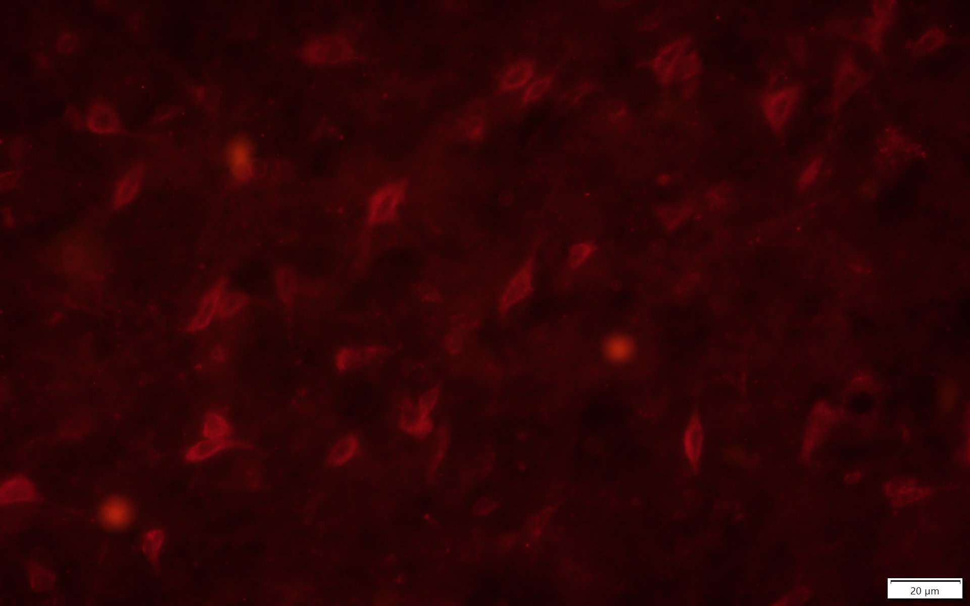
Immunohistochemistry-Frozen: CXCR4 Antibody [NLS1380] - Rat brain tissue sections. Rat Nucleus Accumbens treated with MDPV. Images taken with fluorescent microscope. Primary antibody was diluted to 1:200. Secondary antibody used was donkey anti-Rabbit Alexa Fluor 594 at 1:500. IHC-Fr image submitted by a verified customer review.![Immunohistochemistry-Paraffin: CXCR4 Antibody - BSA Free [NLS1380]](https://resources.rndsystems.com/images/products/CXCR4-Antibody-Immunohistochemistry-Paraffin-NLS1380-img0009.jpg "Immunohistochemistry-Paraffin: CXCR4 Antibody - BSA Free [NLS1380]")
Immunohistochemistry-Paraffin: CXCR4 Antibody - BSA Free [NLS1380]
Immunohistochemistry-Paraffin: CXCR4 Antibody [NLS1380] - Lung, small cell carcinomaApplications for CXCR4 Antibody - BSA Free
Immunohistochemistry-Paraffin
Reviewed Applications
Read 1 review rated 4 using NLS1380 in the following applications:
Formulation, Preparation, and Storage
Purification
Formulation
Format
Preservative
Concentration
Shipping
Stability & Storage
Background: CXCR4
Long Name
Alternate Names
Entrez Gene IDs
Gene Symbol
UniProt
Additional CXCR4 Products
Product Documents for CXCR4 Antibody - BSA Free
Certificate of Analysis
To download a Certificate of Analysis, please enter a lot or batch number in the search box below.
Product Specific Notices for CXCR4 Antibody - BSA Free
This product is for research use only and is not approved for use in humans or in clinical diagnosis. Primary Antibodies are guaranteed for 1 year from date of receipt.
Related Research Areas
Citations for CXCR4 Antibody - BSA Free
 Powered by Bioz
Customer Reviews for CXCR4 Antibody - BSA Free (1)
Have you used CXCR4 Antibody - BSA Free?
Submit a review and receive an Amazon gift card!
$25/€18/£15/$25CAN/¥2500 Yen for a review with an image
$10/€7/£6/$10CAN/¥1110 Yen for a review without an image
Submit a review
Customer Images
-
Application: Immunohistochemistry-FrozenSample Tested: Brain Tissue SectionsSpecies: RatVerified Customer | Posted 08/20/2019Rat Nucleus Accumbens treated with MDPV. Images taken with fluorescent microscope. Primary was diluted to 1:200. Secondary used was Donkey Anti-Rabbit 594 (Life Technologies) 1:500.

There are no reviews that match your criteria.
Protocols
View specific protocols for CXCR4 Antibody - BSA Free (NLS1380):
Immunohistochemistry
1. Prepare tissue with formalin fixation and by embedding it in paraffin wax.
2. Make 4 um sections and place on pre-cleaned and charged microscope slides.
3. Heat in a tissue-drying oven for 45 minutes @ 60 degrees Celcius.
4. Deparaffinize the tissues by wash drying the slides in 3 changes of xylene for 5 minutes each @ RT.
5. Rehydrate the tissues by washing the slides in 3 changes of 100% alcohol for 3 minutes each @ RT.
6. Wash the slides in 2 changes of 95% alcohol for 3 minutes each @ RT.
7. Wash the slides in 1 change of 80% alcohol for 3 minutes @ RT.
8. Rinse the slides in gentle running distilled water for 5 minutes @ RT.
9. Perform antigen retrieval by steaming the slides in 0.01M sodium citrate buffer (pH 6.0) @ 99-100 degrees Celcius
for 20 minutes.
10. Remove the slides from the heat and let stand in buffer @ RT for 20 minutes.
11. Rinse the slides in 1X TBS-T for 1 minute @ RT.
**Do not allow the tissues to dry at any time during the staining procedure**
12. Begin the immunostaining by applying a universal protein block for 20 minutes @ RT.
13. Drain protein block from the slides and apply the diluted primary antibody for 45 minutes @ RT.
14. Rinse the slide in 1X TBS-T for 1 minute @ RT.
15. Apply a biotinylated anti-rabbit IgG (H+L) secondary for 30 minutes @ RT.
16. Rinse the slide in 1X TBS-T for 1 minute @ RT.
17. Apply an alkaline phosphatase steptavidin for 30 minutes @ RT.
18. Rinse the slide in 1X TBS-T for 1 minute @ RT.
19. Apply an alkaline phosphatase chromagen substrate for 30 minutes @ RT.
20. Rinse the slide in distilled water for 1 minute @ RT.
**This method should only be used if the chromagen substrate is alcohol insoluble (ie: Vector Red, DAB)**
21. Dehydrate the tissue by washing the slides in 2 changes of 80% alcohol for 1 minute each @ RT.
22. Wash the slides in 2 changes of 95% alcohol for 1 minute each @ RT.
23. Wash the slides in 3 changes of 100% alcohol for 1 minute each @ RT.
24. Wash the slides in 3 changes of xylene for 1 minute each @ RT.
25. Apply cover slip.
Find general support by application which include: protocols, troubleshooting, illustrated assays, videos and webinars.
- Antigen Retrieval Protocol (PIER)
- Antigen Retrieval for Frozen Sections Protocol
- Appropriate Fixation of IHC/ICC Samples
- Cellular Response to Hypoxia Protocols
- Chromogenic IHC Staining of Formalin-Fixed Paraffin-Embedded (FFPE) Tissue Protocol
- Chromogenic Immunohistochemistry Staining of Frozen Tissue
- ClariTSA™ Fluorophore Kits
- Detection & Visualization of Antibody Binding
- Fluorescent IHC Staining of Frozen Tissue Protocol
- Graphic Protocol for Heat-induced Epitope Retrieval
- Graphic Protocol for the Preparation and Fluorescent IHC Staining of Frozen Tissue Sections
- Graphic Protocol for the Preparation and Fluorescent IHC Staining of Paraffin-embedded Tissue Sections
- Graphic Protocol for the Preparation of Gelatin-coated Slides for Histological Tissue Sections
- IHC Sample Preparation (Frozen sections vs Paraffin)
- Immunofluorescent IHC Staining of Formalin-Fixed Paraffin-Embedded (FFPE) Tissue Protocol
- Immunohistochemistry (IHC) and Immunocytochemistry (ICC) Protocols
- Immunohistochemistry Frozen Troubleshooting
- Immunohistochemistry Paraffin Troubleshooting
- Preparing Samples for IHC/ICC Experiments
- Preventing Non-Specific Staining (Non-Specific Binding)
- Primary Antibody Selection & Optimization
- Protocol for Heat-Induced Epitope Retrieval (HIER)
- Protocol for Making a 4% Formaldehyde Solution in PBS
- Protocol for VisUCyte™ HRP Polymer Detection Reagent
- Protocol for the Preparation & Fixation of Cells on Coverslips
- Protocol for the Preparation and Chromogenic IHC Staining of Frozen Tissue Sections
- Protocol for the Preparation and Chromogenic IHC Staining of Frozen Tissue Sections - Graphic
- Protocol for the Preparation and Chromogenic IHC Staining of Paraffin-embedded Tissue Sections
- Protocol for the Preparation and Chromogenic IHC Staining of Paraffin-embedded Tissue Sections - Graphic
- Protocol for the Preparation and Fluorescent IHC Staining of Frozen Tissue Sections
- Protocol for the Preparation and Fluorescent IHC Staining of Paraffin-embedded Tissue Sections
- Protocol for the Preparation of Gelatin-coated Slides for Histological Tissue Sections
- TUNEL and Active Caspase-3 Detection by IHC/ICC Protocol
- The Importance of IHC/ICC Controls
- Troubleshooting Guide: Immunohistochemistry
- View all Protocols, Troubleshooting, Illustrated assays and Webinars
FAQs for CXCR4 Antibody - BSA Free
-
Q: Which is your best CXCR4 for immunohistochemistry in paraffin tissues? I would like to detect it in paraffin embedded tissues from human breast cancer samples.
A:
CXCR4 antibody (NB100-74396) is our best selling product among all the CXCR4 antibodies and it has been validated for IHC-P in human cervical carcinoma tissue sections. NB100-74396 has been cited in at least 7 research publications. Additionally, here is a list of all of our CXCR4 antibodies that has been verified in IHC-P in human samples.


