Cyclin A2 Antibody - Azide and BSA Free
Novus Biologicals | Catalog # NBP1-31330
![Western Blot: Cyclin A2 Antibody [NBP1-31330]](https://resources.rndsystems.com/images/products/Cyclin-A2-Antibody-Western-Blot-NBP1-31330-img0021.jpg "Western Blot: Cyclin A2 Antibody [NBP1-31330]")
Loading...
Key Product Details
Validated by
Orthogonal Validation, Biological Validation
Species Reactivity
Validated:
Human, Mouse, Rat, Monkey
Cited:
Human, Mouse
Predicted:
Rhesus Macaque (98%). Backed by our 100% Guarantee.
Applications
Validated:
Immunohistochemistry, Immunohistochemistry-Paraffin, Western Blot, Immunocytochemistry/ Immunofluorescence, Immunoprecipitation
Cited:
Western Blot, Immunocytochemistry/ Immunofluorescence
Label
Unconjugated
Antibody Source
Polyclonal Rabbit IgG
Format
Azide and BSA Free
Loading...
Product Specifications
Immunogen
Recombinant protein encompassing a sequence within the center region of human Cyclin A2. The exact sequence is proprietary.
Reactivity Notes
Pig (89%), Rabbit (83%), Bovine (84%).
Localization
Nucleus, Cytoplasm
Clonality
Polyclonal
Host
Rabbit
Isotype
IgG
Theoretical MW
49 kDa.
Disclaimer note: The observed molecular weight of the protein may vary from the listed predicted molecular weight due to post translational modifications, post translation cleavages, relative charges, and other experimental factors.
Disclaimer note: The observed molecular weight of the protein may vary from the listed predicted molecular weight due to post translational modifications, post translation cleavages, relative charges, and other experimental factors.
Scientific Data Images for Cyclin A2 Antibody - Azide and BSA Free
![Immunohistochemistry-Paraffin: Cyclin A2 Antibody [NBP1-31330]](https://resources.rndsystems.com/images/products/Cyclin-A2-Antibody-Immunofluorescence-NBP1-31330-img0010.jpg "Immunohistochemistry-Paraffin: Cyclin A2 Antibody [NBP1-31330]")
Immunohistochemistry-Paraffin: Cyclin A2 Antibody [NBP1-31330]
Immunohistochemistry-Paraffin: Cyclin A2 Antibody [NBP1-31330] - 293T cells stained with Cyclin A2 antibody. Image from verified customer review.![Western Blot: Cyclin A2 Antibody [NBP1-31330]](https://resources.rndsystems.com/images/products/Cyclin-A2-Antibody-Western-Blot-NBP1-31330-img0006.jpg "Western Blot: Cyclin A2 Antibody [NBP1-31330]")
Western Blot: Cyclin A2 Antibody [NBP1-31330]
Western Blot: Cyclin A2 Antibody [NBP1-31330] - 30 ug Neuro2A whole cell lysate/extract 10% SDS-PAGE gel, antibody dilution 1:1000.![Western Blot: Cyclin A2 Antibody [NBP1-31330]](https://resources.rndsystems.com/images/products/Cyclin-A2-Antibody-Western-Blot-NBP1-31330-img0007.jpg "Western Blot: Cyclin A2 Antibody [NBP1-31330]")
Western Blot: Cyclin A2 Antibody [NBP1-31330]
Western Blot: Cyclin A2 Antibody [NBP1-31330] - 30 ug PC-12 whole cell lysate/extract 10% SDS-PAGE gel, antibody dilution 1:1000.![Western Blot: Cyclin A2 Antibody [NBP1-31330]](https://resources.rndsystems.com/images/products/Cyclin-A2-Antibody-Western-Blot-NBP1-31330-img0013.jpg "Western Blot: Cyclin A2 Antibody [NBP1-31330]")
Western Blot: Cyclin A2 Antibody [NBP1-31330]
Western Blot: Cyclin A2 Antibody [NBP1-31330] - Various whole cell extracts (30 ug) were separated by 10% SDS-PAGE, and the membrane was blotted with Cyclin A2 antibody diluted at a dilution of 1:1000.![Western Blot: Cyclin A2 Antibody [NBP1-31330]](https://resources.rndsystems.com/images/products/Cyclin-A2-Antibody-Western-Blot-NBP1-31330-img0014.jpg "Western Blot: Cyclin A2 Antibody [NBP1-31330]")
Western Blot: Cyclin A2 Antibody [NBP1-31330]
Western Blot: Cyclin A2 Antibody [NBP1-31330] - Untreated (-) and treated (+) HeLa whole cell extracts (30 ug) were separated by 10% SDS-PAGE, and the membrane was blotted with Cyclin A2 antibody. HRP-conjugated anti-rabbit IgG antibody was used to detect the primary antibody.![Western Blot: Cyclin A2 Antibody [NBP1-31330]](https://resources.rndsystems.com/images/products/Cyclin-A2-Antibody-Western-Blot-NBP1-31330-img0016.jpg "Western Blot: Cyclin A2 Antibody [NBP1-31330]")
Western Blot: Cyclin A2 Antibody [NBP1-31330]
Western Blot: Cyclin A2 Antibody [NBP1-31330] - Various whole cell extracts (30 ug) were separated by 10% SDS-PAGE, and the membrane was blotted with Cyclin A2 antibody diluted at 1:1000. The HRP-conjugated anti-rabbit IgG antibody (NBP2-19301) was used to detect the primary antibody.![Immunohistochemistry-Paraffin: Cyclin A2 Antibody [NBP1-31330]](https://resources.rndsystems.com/images/products/Cyclin-A2-Antibody-Immunohistochemistry-Paraffin-NBP1-31330-img0004.jpg "Immunohistochemistry-Paraffin: Cyclin A2 Antibody [NBP1-31330]")
Immunohistochemistry-Paraffin: Cyclin A2 Antibody [NBP1-31330]
Immunohistochemistry-Paraffin: Cyclin A2 Antibody [NBP1-31330] - NCIN87 Xenograft, using cyclin A antibody at 1:500 dilution. Antigen Retrieval: Trilogy™ (EDTA based, pH 8.0) buffer, 15min.![Immunoprecipitation: Cyclin A2 Antibody [NBP1-31330]](https://resources.rndsystems.com/images/products/Cyclin-A2-Antibody-Immunoprecipitation-NBP1-31330-img0009.jpg "Immunoprecipitation: Cyclin A2 Antibody [NBP1-31330]")
Immunoprecipitation: Cyclin A2 Antibody [NBP1-31330]
Immunoprecipitation: Cyclin A2 Antibody [NBP1-31330] - Sample: 1000 ug 293T whole cell lysate/extract A. 30 ug 293T whole cell lysate/extract, B. Control with 2. 5 ug of preimmune rabbit IgG, C. Immunoprecipitation of Cyclin A2 protein by 2. 5 ug of Cyclin A2 antibody 10% SDS-PAGE gel.
Western Blot: Cyclin A2 Antibody [NBP1-31330] -
Western Blot: Cyclin A2 Antibody [NBP1-31330] - Comparison of expression changes between young & old MRC-5 & HFF fibroblasts measured by RNA-seq, qRT-PCR, & Western Blots. (a) Four genes commonly downregulated & (b) 8 genes commonly upregulated in both cell lines. (a, b) The colors of the bars indicate the measurement technique (blue: RNA-seq; green: qRT-PCR; red: Western Blots/protein expression). Solid colored bars represent MRC-5 while shaded boxes represent HFF cells. The height of the bars corresponds to the logarithmic fold-change (FC) of expression between the first & the last PD investigated here (RNA-seq: log 2 RPKM FC; qRT-PCR: log 2− delta delta CT; protein: log 2 expression ratio). Error bars indicate standard deviation from the mean. Changes statistically different comparing young & old PD (RNA-seq: DESeq; rRT-PCR/Protein: Student's t-test; n = 3) are indicated with an asterisk: ∗p < 0.05, ∗∗p < 0.01, & ∗∗∗p < 0.001. (c) The blots show the protein expression levels in MRC-5 & HFF cells at young compared to old PDs. The up- or downregulation was signified by the presence or absence of bands in Western Blots. Image collected & cropped by CiteAb from the following publication (https://pubmed.ncbi.nlm.nih.gov/26339636), licensed under a CC-BY license. Not internally tested by Novus Biologicals.
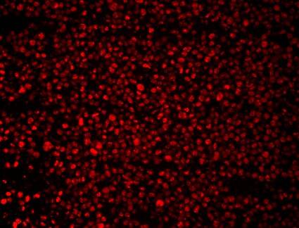
Immunocytochemistry/ Immunofluorescence: Cyclin A2 Antibody [NBP1-31330] -
Cyclin A2 antibody detects Cyclin A2 protein at nucleus by immunofluorescent analysis.Sample: HeLa cells were fixed in 4% paraformaldehyde at RT for 15 min.
Green: Cyclin A2 stained by Cyclin A2 antibody (NBP1-31330) diluted at 1:500.
Red: phalloidin, a cytoskeleton marker, diluted at 1:100.
Scale bar= 10 um.
Applications for Cyclin A2 Antibody - Azide and BSA Free
Application
Recommended Usage
Immunocytochemistry/ Immunofluorescence
1:100-1:1000
Immunohistochemistry
1:100-1:1000
Immunohistochemistry-Paraffin
1:100-1:1000
Immunoprecipitation
1:100-1:500
Western Blot
1:500-1:3000
Reviewed Applications
Read 3 reviews rated 4.7 using NBP1-31330 in the following applications:
Formulation, Preparation, and Storage
Purification
Antigen Affinity-purified
Formulation
PBS, 20% Glycerol
Format
Azide and BSA Free
Preservative
0.025% Proclin 300
Concentration
Concentrations vary lot to lot. See vial label for concentration. If unlisted please contact technical services.
Shipping
The product is shipped with polar packs. Upon receipt, store it immediately at the temperature recommended below.
Stability & Storage
Aliquot and store at -20C or -80C. Avoid freeze-thaw cycles.
Background: Cyclin A2
Alternate Names
CCNA, CCNA2
Gene Symbol
CCNA2
Additional Cyclin A2 Products
Product Documents for Cyclin A2 Antibody - Azide and BSA Free
Certificate of Analysis
To download a Certificate of Analysis, please enter a lot or batch number in the search box below.
Product Specific Notices for Cyclin A2 Antibody - Azide and BSA Free
This product is for research use only and is not approved for use in humans or in clinical diagnosis. Primary Antibodies are guaranteed for 1 year from date of receipt.
⚠ WARNING: This product can expose you to chemicals including mercury, which is known to the State of California to cause reproductive toxicity with developmental effects. For more information go to www.P65Warnings.ca.gov.Related Research Areas
Citations for Cyclin A2 Antibody - Azide and BSA Free
 Powered by Bioz
Customer Reviews for Cyclin A2 Antibody - Azide and BSA Free (3)
4.7 out of 5
3 Customer Ratings
Have you used Cyclin A2 Antibody - Azide and BSA Free?
Submit a review and receive an Amazon gift card!
$25/€18/£15/$25CAN/¥2500 Yen for a review with an image
$10/€7/£6/$10CAN/¥1110 Yen for a review without an image
Submit a review
Customer Images
-(01-ml)_NBP1-31330_10226.jpg)
Showing
1
-
3 of
3 reviews
Showing All
Filter By:
-
Application: Western BlotSample Tested: Human cancer cellSpecies: HumanVerified Customer | Posted 10/04/2015
-
Application: Immunohistochemistry-ParaffinSample Tested: 293T cell lineSpecies: HumanVerified Customer | Posted 10/02/2015Cyclin A2 on 293T

-
Application: Western BlotSample Tested: Whole cell lysate from HeLa cellsSpecies: HumanVerified Customer | Posted 09/19/2014Cyclin A2 WB in Hela cells
There are no reviews that match your criteria.
Protocols
Find general support by application which include: protocols, troubleshooting, illustrated assays, videos and webinars.
- Antigen Retrieval Protocol (PIER)
- Antigen Retrieval for Frozen Sections Protocol
- Appropriate Fixation of IHC/ICC Samples
- Cellular Response to Hypoxia Protocols
- Chromogenic IHC Staining of Formalin-Fixed Paraffin-Embedded (FFPE) Tissue Protocol
- Chromogenic Immunohistochemistry Staining of Frozen Tissue
- ClariTSA™ Fluorophore Kits
- Detection & Visualization of Antibody Binding
- Fluorescent IHC Staining of Frozen Tissue Protocol
- Graphic Protocol for Heat-induced Epitope Retrieval
- Graphic Protocol for the Preparation and Fluorescent IHC Staining of Frozen Tissue Sections
- Graphic Protocol for the Preparation and Fluorescent IHC Staining of Paraffin-embedded Tissue Sections
- Graphic Protocol for the Preparation of Gelatin-coated Slides for Histological Tissue Sections
- ICC Cell Smear Protocol for Suspension Cells
- ICC Immunocytochemistry Protocol Videos
- ICC for Adherent Cells
- IHC Sample Preparation (Frozen sections vs Paraffin)
- Immunocytochemistry (ICC) Protocol
- Immunocytochemistry Troubleshooting
- Immunofluorescence of Organoids Embedded in Cultrex Basement Membrane Extract
- Immunofluorescent IHC Staining of Formalin-Fixed Paraffin-Embedded (FFPE) Tissue Protocol
- Immunohistochemistry (IHC) and Immunocytochemistry (ICC) Protocols
- Immunohistochemistry Frozen Troubleshooting
- Immunohistochemistry Paraffin Troubleshooting
- Immunoprecipitation Protocol
- Preparing Samples for IHC/ICC Experiments
- Preventing Non-Specific Staining (Non-Specific Binding)
- Primary Antibody Selection & Optimization
- Protocol for Heat-Induced Epitope Retrieval (HIER)
- Protocol for Making a 4% Formaldehyde Solution in PBS
- Protocol for VisUCyte™ HRP Polymer Detection Reagent
- Protocol for the Fluorescent ICC Staining of Cell Smears - Graphic
- Protocol for the Fluorescent ICC Staining of Cultured Cells on Coverslips - Graphic
- Protocol for the Preparation & Fixation of Cells on Coverslips
- Protocol for the Preparation and Chromogenic IHC Staining of Frozen Tissue Sections
- Protocol for the Preparation and Chromogenic IHC Staining of Frozen Tissue Sections - Graphic
- Protocol for the Preparation and Chromogenic IHC Staining of Paraffin-embedded Tissue Sections
- Protocol for the Preparation and Chromogenic IHC Staining of Paraffin-embedded Tissue Sections - Graphic
- Protocol for the Preparation and Fluorescent ICC Staining of Cells on Coverslips
- Protocol for the Preparation and Fluorescent ICC Staining of Non-adherent Cells
- Protocol for the Preparation and Fluorescent ICC Staining of Stem Cells on Coverslips
- Protocol for the Preparation and Fluorescent IHC Staining of Frozen Tissue Sections
- Protocol for the Preparation and Fluorescent IHC Staining of Paraffin-embedded Tissue Sections
- Protocol for the Preparation of Gelatin-coated Slides for Histological Tissue Sections
- Protocol for the Preparation of a Cell Smear for Non-adherent Cell ICC - Graphic
- R&D Systems Quality Control Western Blot Protocol
- TUNEL and Active Caspase-3 Detection by IHC/ICC Protocol
- The Importance of IHC/ICC Controls
- Troubleshooting Guide: Immunohistochemistry
- Troubleshooting Guide: Western Blot Figures
- Western Blot Conditions
- Western Blot Protocol
- Western Blot Protocol for Cell Lysates
- Western Blot Troubleshooting
- Western Blot Troubleshooting Guide
- View all Protocols, Troubleshooting, Illustrated assays and Webinars
Loading...