Cytochrome c Antibody (7H8.2C12) - BSA Free
Novus Biologicals | Catalog # NB100-56503
Key Product Details
Validated by
Biological Validation
Species Reactivity
Validated:
Human, Mouse, Rat, Drosophila, Rabbit
Cited:
Human, Mouse, Rat, Insect - Drosophila, Rabbit
Applications
Validated:
Immunohistochemistry, Immunohistochemistry-Paraffin, Western Blot, Immunoblotting, Flow Cytometry, Flow (Cell Surface), Flow (Intracellular), Immunocytochemistry/ Immunofluorescence, Simple Western
Cited:
Western Blot, Flow Cytometry, Flow (Cell Surface), Immunocytochemistry/ Immunofluorescence, Flow Cytometry Control
Label
Unconjugated
Antibody Source
Monoclonal Mouse IgG2b Kappa Clone # 7H8.2C12
Format
BSA Free
Loading...
Product Specifications
Immunogen
Synthetic peptides corresponding to amino acids 1-80, 81-104 and 66-104 of pigeon CYT were used as the immunogen (Jemmerson et al. 1991). The antibody recognizes an epitope within amino acids 93-104 of pigeon cytochrome C based on competitive ELISA results (Jemmerson et al. 1991).
Marker
Mitochondria Marker
Specificity
This antibody recognizes total cytochrome C which includes both apocytochrome (i.e. cytochrome in the cytosol without heme attached) and holocytochrome (i.e cytochrome in the mitochondria with heme attached).
Clonality
Monoclonal
Host
Mouse
Isotype
IgG2b Kappa
Scientific Data Images for Cytochrome c Antibody (7H8.2C12) - BSA Free
![Western Blot: Cytochrome c Antibody (7H8.2C12)BSA Free [NB100-56503]](https://resources.rndsystems.com/images/products/Cytochrome-c-Antibody-7H8-2C12-Western-Blot-NB100-56503-img0015.jpg "Western Blot: Cytochrome c Antibody (7H8.2C12)BSA Free [NB100-56503]")
![Immunocytochemistry/ Immunofluorescence: Cytochrome c Antibody (7H8.2C12) - BSA Free [NB100-56503]](https://resources.rndsystems.com/images/products/Cytochrome-c-Antibody-7H8-2C12-Immunocytochemistry-Immunofluorescence-NB100-56503-img0010.jpg "Immunocytochemistry/ Immunofluorescence: Cytochrome c Antibody (7H8.2C12) - BSA Free [NB100-56503]")
Immunocytochemistry/ Immunofluorescence: Cytochrome c Antibody (7H8.2C12) - BSA Free [NB100-56503]
Immunocytochemistry/Immunofluorescence: Cytochrome c Antibody (7H8.2C12) [NB100-56503] - p73 was detected in immersion fixed Hela human cell line using NB100-56503 at 25 ug/ml for 3 hours at room temperature. Cells were stained using the NorthernLights(TM) 557-conjugated Anti-Mouse IgG Secondary Antibody (red; Catalog # NL007) and counterstained with DAPI (blue). Staining was observed in the cytoplasm and mitochondria.![Immunohistochemistry-Paraffin: Cytochrome c Antibody (7H8.2C12) - BSA Free [NB100-56503]](https://resources.rndsystems.com/images/products/Cytochrome-c-Antibody-7H8-2C12-Immunohistochemistry-Paraffin-NB100-56503-img0004.jpg "Immunohistochemistry-Paraffin: Cytochrome c Antibody (7H8.2C12) - BSA Free [NB100-56503]")
Immunohistochemistry-Paraffin: Cytochrome c Antibody (7H8.2C12) - BSA Free [NB100-56503]
Immunohistochemistry-Paraffin: Cytochrome c Antibody (7H8.2C12) [NB100-56503] - Cytochrome C was detected in immersion fixed paraffin-embedded sections of human heart using anti-human mouse monoclonal antibody (Catalog # NB100-56503) at 1:200 dilution overnight at 4C. Tissue was stained using the VisuCyte anti-mouse HRP polymer detection reagent (Catalog # VC001) with DAB chromogen (brown) and counterstained with hematoxylin (blue).Images may not be copied, printed or otherwise disseminated without express written permission of Novus Biologicals a bio-techne brand.
![Flow (Intracellular): Cytochrome c Antibody (7H8.2C12) - BSA Free [NB100-56503]](https://resources.rndsystems.com/images/products/Cytochrome-c-Antibody-7H8-2C12-Flow-Intracellular-NB100-56503-img0014.jpg "Flow (Intracellular): Cytochrome c Antibody (7H8.2C12) - BSA Free [NB100-56503]")
Flow (Intracellular): Cytochrome c Antibody (7H8.2C12) - BSA Free [NB100-56503]
Flow (Intracellular): Cytochrome c Antibody (7H8.2C12) [NB100-56503] - An intracellular stain was performed on HeLa cells with Cytochrome c (7H8.2C12) antibody NB100-56503PE (blue) and a matched isotype control (orange). Cells were fixed with 4% PFA and then permeablized with 0.1% saponin. Cells were incubated in an antibody dilution of 5 ug/mL for 30 minutes at room temperature. Both antibodies were conjugated to phycoerythrin.![Western Blot: Cytochrome c Antibody (7H8.2C12)BSA Free [NB100-56503]](https://resources.rndsystems.com/images/products/Cytochrome-c-Antibody-7H8-2C12-Western-Blot-NB100-56503-img0001.jpg "Western Blot: Cytochrome c Antibody (7H8.2C12)BSA Free [NB100-56503]")
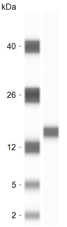
Western Blot: Cytochrome c Antibody (7H8.2C12)BSA Free [NB100-56503]
Western Blot: Cytochrome c Antibody (7H8.2C12) [NB100-56503] - Human HeLa lysate probed with Cytochrome C antibody at 0.1 ug/ml.![Western Blot: Cytochrome c Antibody (7H8.2C12)BSA Free [NB100-56503]](https://resources.rndsystems.com/images/products/Cytochrome-c-Antibody-7H8-2C12-Western-Blot-NB100-56503-img0003.jpg "Western Blot: Cytochrome c Antibody (7H8.2C12)BSA Free [NB100-56503]")
Western Blot: Cytochrome c Antibody (7H8.2C12)BSA Free [NB100-56503]
Western Blot: Cytochrome c Antibody (7H8.2C12) [NB100-56503] - Analysis using the biotin conjugate of NB100-56503. Detection of Cytochrome C in 15 ug of HeLa cell lysate using NB100-55775 at 1:1000. A 15 kDa band is detected.![Flow Cytometry: Cytochrome c Antibody (7H8.2C12) - BSA Free [NB100-56503]](https://resources.rndsystems.com/images/products/Cytochrome-c-Antibody-7H8-2C12-Flow-Cytometry-NB100-56503-img0005.jpg "Flow Cytometry: Cytochrome c Antibody (7H8.2C12) - BSA Free [NB100-56503]")
Flow Cytometry: Cytochrome c Antibody (7H8.2C12) - BSA Free [NB100-56503]
Flow Cytometry: Cytochrome c Antibody (7H8.2C12) [NB100-56503] - Detection of Cytochrome C in Human HeLa Cell Line by Flow Cytometry. Human HeLa cell line was stained with Mouse Anti- Cytochrome C Monoclonal Antibody (Catalog # NB100-56503, filled histogram), or Mouse IgG2B isotype control (Catalog # MAB0041, open histogram) followed by APC-conjugated Anti-Mouse IgG Secondary Antibody (Catalog # F0101B). To facilitate intracellular staining, cells were fixed with Flow Cytometry Fixation Buffer (Catalog # FC004) and permeabilized with Flow Cytometry Permeabilization/Wash Buffer I (Catalog # FC005).Images may not be copied, printed or otherwise disseminated without express written permission of Novus Biologicals a bio-techne brand.
![Flow Cytometry: Cytochrome c Antibody (7H8.2C12) - BSA Free [NB100-56503]](https://resources.rndsystems.com/images/products/Cytochrome-c-Antibody-7H8-2C12-Flow-Cytometry-NB100-56503-img0011.jpg "Flow Cytometry: Cytochrome c Antibody (7H8.2C12) - BSA Free [NB100-56503]")
Flow Cytometry: Cytochrome c Antibody (7H8.2C12) - BSA Free [NB100-56503]
Flow Cytometry: Cytochrome c Antibody (7H8.2C12) [NB100-56503] - Using the Alexa Fluor 488 direct conjugate, an intracellular stain was performed on HeLa cells with Cytochrome c (7H8.2C12) antibody NB100-56503AF488 (blue) and a matched isotype control NBP2-27231AF488 (orange). Cells were fixed with 4% PFA and then permeablized with 0.1% saponin. Cells were incubated in an antibody dilution of 10 ug/mL for 30 minutes at room temperature. Both antibodies were conjugated to Alexa Fluor 488.![Simple Western: Cytochrome c Antibody (7H8.2C12)BSA Free [NB100-56503]](https://resources.rndsystems.com/images/products/Cytochrome-c-Antibody-7H8-2C12-Simple-Western-NB100-56503-img0013.jpg "Simple Western: Cytochrome c Antibody (7H8.2C12)BSA Free [NB100-56503]")
Simple Western: Cytochrome c Antibody (7H8.2C12)BSA Free [NB100-56503]
Simple Western: Cytochrome c Antibody (7H8.2C12) [NB100-56503] - Lane view shows lysates of human heart tissue, loaded at 0.2 mg/mL. A specific band was detected for Cytochrome c at approximately 23 kDa (as indicated) using 2.5 ug/mL of Mouse Anti-Cytochrome c Monoclonal Antibody (Catalog # NB100-56503). This experiment was conducted under reducing conditions and using the 12-230 kDa separation system. [NB100-56503] -")
Simple Western: Cytochrome c Antibody (7H8.2C12) [NB100-56503] -
Simple Western: Cytochrome c Antibody (7H8.2C12) [NB100-56503] - Cytochrome C Antibodies (NB100-56503SS), ProteinSimple Western Blot on Jess Instrument; 1 microgram of human brain tissue lysate was tested with the antibodies diluted 20 times. Image from verified customer review. - BSA Free [NB100-56503] -")
Western Blot: Cytochrome c Antibody (7H8.2C12) - BSA Free [NB100-56503] -
Effects of resveratrol on the expression of SIRT1, PGC-1 alpha, HIF-1 alpha & apoptosis proteins in vivo. (A) Activation of the SIRT1–PGC-1 alpha –HIF-1 alpha signaling pathway in PC–AKI associated with DN in rabbits. Representative western blot images of proteins in the rabbits with DN treated with saline (Cont), resveratrol alone (Res), iohexol (PC–AKI), & co-treatment with resveratrol & iohexol (Res+PC–AKI). (B–H) Relative densitometry analysis of the ratios of SIRT1–PGC-1 alpha –HIF-1 alpha signaling proteins to beta -actin were expressed as mean ± standard error. *P < 0.05 vs. Cont; #P < 0.05 vs. PC–AKI. Cont, control; Res, resveratrol; PC–AKI, post-contrast acute kidney injury; SIRT1, silent information regulator l; HIF-1 alpha, hypoxia-inducible transcription factor-1 alpha ; DN, diabetic nephropathy; PGC-1 alpha, peroxisome proliferator-activated receptor gamma coactivator-1 alpha. Image collected & cropped by CiteAb from the following publication (https://pubmed.ncbi.nlm.nih.gov/31402864), licensed under a CC-BY license. Not internally tested by Novus Biologicals.Applications for Cytochrome c Antibody (7H8.2C12) - BSA Free
Application
Recommended Usage
Flow (Cell Surface)
reported in scientific literature (PMID 16467206)
Flow (Intracellular)
reported in scientific literature (Mohr et al (2004))
Flow Cytometry
1:20-1:2000
Immunoblotting
reported in scientific literature (PMID 16436379)
Immunocytochemistry/ Immunofluorescence
1:10-1:500. Use reported in scientific literature (Yamasaki et al (2006))
Immunohistochemistry
1:10-1:500
Immunohistochemistry-Paraffin
1:10-1:500. Use reported in scientific literature (Fujimara et al (1998))
Simple Western
2.5 ug/ml
Western Blot
0.05-0.5 ug/ml
Application Notes
An approx. 15 kDa band is observed. See Simple Western Antibody Database for Simple Western validation: tested in human heart tissue lysate; separated by Size-Jess/Wes, Sally Sue/Peggy Sue, antibody dilution of 2.5 ug/ml; matrix was 12-230 kDa.
Reviewed Applications
Read 1 review rated 5 using NB100-56503 in the following applications:
Flow Cytometry Panel Builder
Bio-Techne Knows Flow Cytometry
Save time and reduce costly mistakes by quickly finding compatible reagents using the Panel Builder Tool.
Advanced Features
- Spectra Viewer - Custom analysis of spectra from multiple fluorochromes
- Spillover Popups - Visualize the spectra of individual fluorochromes
- Antigen Density Selector - Match fluorochrome brightness with antigen density

Formulation, Preparation, and Storage
Purification
Protein G purified
Formulation
PBS
Format
BSA Free
Preservative
0.05% Sodium Azide
Concentration
1.0 mg/ml
Shipping
The product is shipped with polar packs. Upon receipt, store it immediately at the temperature recommended below.
Stability & Storage
Store at 4C short term. Aliquot and store at -20C long term. Avoid freeze-thaw cycles.
Background: Cytochrome c
Alternate Names
CYCS
Gene Symbol
CYCS
UniProt
Additional Cytochrome c Products
Product Documents for Cytochrome c Antibody (7H8.2C12) - BSA Free
Certificate of Analysis
To download a Certificate of Analysis, please enter a lot or batch number in the search box below.
Product Specific Notices for Cytochrome c Antibody (7H8.2C12) - BSA Free
This product is for research use only and is not approved for use in humans or in clinical diagnosis. Primary Antibodies are guaranteed for 1 year from date of receipt.
Related Research Areas
Citations for Cytochrome c Antibody (7H8.2C12) - BSA Free
 Powered by Bioz
Customer Reviews for Cytochrome c Antibody (7H8.2C12) - BSA Free (1)
5 out of 5
1 Customer Rating
Have you used Cytochrome c Antibody (7H8.2C12) - BSA Free?
Submit a review and receive an Amazon gift card!
$25/€18/£15/$25CAN/¥2500 Yen for a review with an image
$10/€7/£6/$10CAN/¥1110 Yen for a review without an image
Submit a review
Customer Images
Showing
1
-
1 of
1 review
Showing All
Filter By:
-
Application: Simple WesternSample Tested: human brain lysateSpecies: HumanVerified Customer | Posted 04/06/2023Cytochrome C Antibodies NB100-56503SS, ProteinSimple Western Blot on Jess Instrument; 1 microgram of human brain tissue lysate was tested with the antibodies diluted 20 times.

There are no reviews that match your criteria.
Protocols
Find general support by application which include: protocols, troubleshooting, illustrated assays, videos and webinars.
- 7-Amino Actinomycin D (7-AAD) Cell Viability Flow Cytometry Protocol
- Antigen Retrieval Protocol (PIER)
- Antigen Retrieval for Frozen Sections Protocol
- Appropriate Fixation of IHC/ICC Samples
- Cellular Response to Hypoxia Protocols
- Chromogenic IHC Staining of Formalin-Fixed Paraffin-Embedded (FFPE) Tissue Protocol
- Chromogenic Immunohistochemistry Staining of Frozen Tissue
- ClariTSA™ Fluorophore Kits
- Detection & Visualization of Antibody Binding
- Extracellular Membrane Flow Cytometry Protocol
- Flow Cytometry Protocol for Cell Surface Markers
- Flow Cytometry Protocol for Staining Membrane Associated Proteins
- Flow Cytometry Staining Protocols
- Flow Cytometry Troubleshooting Guide
- Fluorescent IHC Staining of Frozen Tissue Protocol
- Graphic Protocol for Heat-induced Epitope Retrieval
- Graphic Protocol for the Preparation and Fluorescent IHC Staining of Frozen Tissue Sections
- Graphic Protocol for the Preparation and Fluorescent IHC Staining of Paraffin-embedded Tissue Sections
- Graphic Protocol for the Preparation of Gelatin-coated Slides for Histological Tissue Sections
- ICC Cell Smear Protocol for Suspension Cells
- ICC Immunocytochemistry Protocol Videos
- ICC for Adherent Cells
- IHC Sample Preparation (Frozen sections vs Paraffin)
- Immunocytochemistry (ICC) Protocol
- Immunocytochemistry Troubleshooting
- Immunofluorescence of Organoids Embedded in Cultrex Basement Membrane Extract
- Immunofluorescent IHC Staining of Formalin-Fixed Paraffin-Embedded (FFPE) Tissue Protocol
- Immunohistochemistry (IHC) and Immunocytochemistry (ICC) Protocols
- Immunohistochemistry Frozen Troubleshooting
- Immunohistochemistry Paraffin Troubleshooting
- Intracellular Flow Cytometry Protocol Using Alcohol (Methanol)
- Intracellular Flow Cytometry Protocol Using Detergents
- Intracellular Nuclear Staining Flow Cytometry Protocol Using Detergents
- Intracellular Staining Flow Cytometry Protocol Using Alcohol Permeabilization
- Intracellular Staining Flow Cytometry Protocol Using Detergents to Permeabilize Cells
- Preparing Samples for IHC/ICC Experiments
- Preventing Non-Specific Staining (Non-Specific Binding)
- Primary Antibody Selection & Optimization
- Propidium Iodide Cell Viability Flow Cytometry Protocol
- Protocol for Heat-Induced Epitope Retrieval (HIER)
- Protocol for Liperfluo
- Protocol for Making a 4% Formaldehyde Solution in PBS
- Protocol for VisUCyte™ HRP Polymer Detection Reagent
- Protocol for the Characterization of Human Th22 Cells
- Protocol for the Characterization of Human Th9 Cells
- Protocol for the Fluorescent ICC Staining of Cell Smears - Graphic
- Protocol for the Fluorescent ICC Staining of Cultured Cells on Coverslips - Graphic
- Protocol for the Preparation & Fixation of Cells on Coverslips
- Protocol for the Preparation and Chromogenic IHC Staining of Frozen Tissue Sections
- Protocol for the Preparation and Chromogenic IHC Staining of Frozen Tissue Sections - Graphic
- Protocol for the Preparation and Chromogenic IHC Staining of Paraffin-embedded Tissue Sections
- Protocol for the Preparation and Chromogenic IHC Staining of Paraffin-embedded Tissue Sections - Graphic
- Protocol for the Preparation and Fluorescent ICC Staining of Cells on Coverslips
- Protocol for the Preparation and Fluorescent ICC Staining of Non-adherent Cells
- Protocol for the Preparation and Fluorescent ICC Staining of Stem Cells on Coverslips
- Protocol for the Preparation and Fluorescent IHC Staining of Frozen Tissue Sections
- Protocol for the Preparation and Fluorescent IHC Staining of Paraffin-embedded Tissue Sections
- Protocol for the Preparation of Gelatin-coated Slides for Histological Tissue Sections
- Protocol for the Preparation of a Cell Smear for Non-adherent Cell ICC - Graphic
- Protocol: Annexin V and PI Staining by Flow Cytometry
- Protocol: Annexin V and PI Staining for Apoptosis by Flow Cytometry
- R&D Systems Quality Control Western Blot Protocol
- TUNEL and Active Caspase-3 Detection by IHC/ICC Protocol
- The Importance of IHC/ICC Controls
- Troubleshooting Guide: Fluorokine Flow Cytometry Kits
- Troubleshooting Guide: Immunohistochemistry
- Troubleshooting Guide: Western Blot Figures
- Western Blot Conditions
- Western Blot Protocol
- Western Blot Protocol for Cell Lysates
- Western Blot Troubleshooting
- Western Blot Troubleshooting Guide
- View all Protocols, Troubleshooting, Illustrated assays and Webinars
Loading...
Associated Pathways

