DEC1 Antibody - BSA Free
Novus Biologicals | Catalog # NB100-1800
![Western Blot: DEC1 AntibodyBSA Free [NB100-1800]](https://resources.rndsystems.com/images/products/DEC1-Antibody---BSA-Free-Western-Blot-NB100-1800-img0011.jpg "Western Blot: DEC1 AntibodyBSA Free [NB100-1800]")
Key Product Details
Validated by
Knockout/Knockdown, Biological Validation
Species Reactivity
Validated:
Human, Mouse, Rat
Cited:
Human, Mouse
Applications
Validated:
Immunohistochemistry, Immunohistochemistry-Paraffin, Western Blot, Immunoblotting, ELISA, Flow Cytometry, Immunocytochemistry/ Immunofluorescence, Simple Western, Immunoprecipitation, Chromatin Immunoprecipitation, Chromatin Immunoprecipitation (ChIP), Chromatin Immunoprecipitation Sequencing, Knockdown Validated
Cited:
Immunohistochemistry, Immunohistochemistry-Paraffin, Western Blot, Flow Cytometry, Simple Western, Chromatin Immunoprecipitation (ChIP), Chemotaxis, Chip Cytometry, IF/IHC
Label
Unconjugated
Antibody Source
Polyclonal Rabbit IgG
Format
BSA Free
Loading...
Product Specifications
Immunogen
Synthetic peptide made to a C-terminal region of human DEC1 (between amino acids 350-412) [UniProt O14503]
Reactivity Notes
Mouse reactivity reported in scientific literature (PMID: 26834156).
Localization
Nucleus
Clonality
Polyclonal
Host
Rabbit
Isotype
IgG
Theoretical MW
46 kDa.
Disclaimer note: The observed molecular weight of the protein may vary from the listed predicted molecular weight due to post translational modifications, post translation cleavages, relative charges, and other experimental factors.
Disclaimer note: The observed molecular weight of the protein may vary from the listed predicted molecular weight due to post translational modifications, post translation cleavages, relative charges, and other experimental factors.
Scientific Data Images for DEC1 Antibody - BSA Free
Western Blot: DEC1 AntibodyBSA Free [NB100-1800]
Western Blot: DEC1 Antibody - BSA Free [NB100-1800] - Western Blot Image of anti-DEC1. Whole cell protein from A431 (lane 1) and PC3 (lane 2) was separated on a 12% gel by SDS-PAGE, transferred to PVDF membrane and blocked in 5% non-fat milk in TBST. The membrane was probed with 2 ug/ml anti-DEC1 in 1% milk, and detected with an anti-rabbit HRP secondary antibody using chemiluminescence.![Immunocytochemistry/ Immunofluorescence: DEC1 Antibody - BSA Free [NB100-1800]](https://resources.rndsystems.com/images/products/DEC1-Antibody---BSA-Free-Immunocytochemistry-Immunofluorescence-NB100-1800-img0009.jpg "Immunocytochemistry/ Immunofluorescence: DEC1 Antibody - BSA Free [NB100-1800]")
Immunocytochemistry/ Immunofluorescence: DEC1 Antibody - BSA Free [NB100-1800]
Immunocytochemistry/Immunofluorescence: DEC1 Antibody - BSA Free [NB100-1800] - HeLa cells were fixed for 10 minutes using 10% formalin and then permeabilized for 5 minutes using 1X TBS + 0.5% Triton-X100. The cells were incubated with anti-DEC1 (NB100-1800) at a 1:200 dilution overnight at 4C and detected with and anti-rabbit Dylight 488 (Green) at a 1:500 dilution. Alpha tubulin was used as a co-stain at a 1:1000 dilution and detected with and anti-mouse Dylight 550 (Red) at a 1:500 dilution. Nuclei were counterstained with DAPI (Blue). Cells were imaged using a 40X objective.![Immunohistochemistry: DEC1 Antibody - BSA Free [NB100-1800]](https://resources.rndsystems.com/images/products/DEC1-Antibody---BSA-Free-Immunohistochemistry-NB100-1800-img0017.jpg "Immunohistochemistry: DEC1 Antibody - BSA Free [NB100-1800]")
![Flow Cytometry: DEC1 Antibody - BSA Free [NB100-1800]](https://resources.rndsystems.com/images/products/DEC1-Antibody---BSA-Free-Flow-Cytometry-NB100-1800-img0016.jpg "Flow Cytometry: DEC1 Antibody - BSA Free [NB100-1800]")
Flow Cytometry: DEC1 Antibody - BSA Free [NB100-1800]
Flow Cytometry: DEC1 Antibody - BSA Free [NB100-1800] - An intracellular stain was performed on A431 cells with DEC1 Antibody NB100-1800AF647 (blue) and a matched isotype control (orange). Cells were fixed with 4% PFA and then permeabilized with 0.1% saponin. Cells were incubated in an antibody dilution of 2.5 ug/mL for 30 minutes at room temperature. Both antibodies were conjugated to Alexa Fluor 647.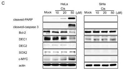
![Western Blot: DEC1 AntibodyBSA Free [NB100-1800]](https://resources.rndsystems.com/images/products/DEC1-Antibody---BSA-Free-Western-Blot-NB100-1800-img0001.jpg "Western Blot: DEC1 AntibodyBSA Free [NB100-1800]")
Western Blot: DEC1 AntibodyBSA Free [NB100-1800]
Western Blot: DEC1 Antibody - BSA Free [NB100-1800] - Western blot analysis of DEC1 in HeLa whole cell lysate.![Immunohistochemistry-Paraffin: DEC1 Antibody - BSA Free [NB100-1800]](https://resources.rndsystems.com/images/products/DEC1-Antibody---BSA-Free-Immunohistochemistry-Paraffin-NB100-1800-img0012.jpg "Immunohistochemistry-Paraffin: DEC1 Antibody - BSA Free [NB100-1800]")
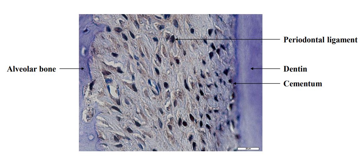
Immunohistochemistry-Paraffin: DEC1 Antibody - BSA Free [NB100-1800]
Immunohistochemistry-Paraffin: DEC1 Antibody - BSA Free [NB100-1800] - P. gingivalis-challenged rat maxilla using DEC1 antibody at 1:100. Heat mediated antigen retrieval (sodium citrate buffer pH 6.0) was performed. Counter-stained with hematoxylin. Image from verified customer review.![Immunoprecipitation: DEC1 Antibody - BSA Free [NB100-1800]](https://resources.rndsystems.com/images/products/DEC1-Antibody---BSA-Free-Immunoprecipitation-NB100-1800-img0007.jpg "Immunoprecipitation: DEC1 Antibody - BSA Free [NB100-1800]")
Immunoprecipitation: DEC1 Antibody - BSA Free [NB100-1800]
Immunoprecipitation: DEC1 Antibody - BSA Free [NB100-1800] - Samples: Whole cell lysate from HeLa (50 ug for WB) and 293T (1 mg/IP, 20% of IP loaded) cells. Antibodies: Affinity purified rabbit anti-DEC1 antibody BL2928 used for WB at 0.1 ug/ml (A) and 1 ug/ml (B) and for IP at 3 ug/mg lysate. DEC1 was also immunoprecipitated using rabbit anti-DEC1 antibodies BL2926 and BL2927, which recognize upstream epitopes. Detection: Chemiluminescence with exposure times of 3 minutes (A) and 30 seconds (B).![Simple Western: DEC1 AntibodyBSA Free [NB100-1800]](https://resources.rndsystems.com/images/products/DEC1-Antibody---BSA-Free-Simple-Western-NB100-1800-img0008.jpg "Simple Western: DEC1 AntibodyBSA Free [NB100-1800]")
Simple Western: DEC1 AntibodyBSA Free [NB100-1800]
Simple Western: DEC1 Antibody - BSA Free [NB100-1800] - Simple Western lane view shows a specific band for DEC1 in 1.0 mg/ml of HeLa lysate. This experiment was performed under reducing conditions using the 12-230 kDa separation system.![Immunoblotting: DEC1 Antibody - BSA Free [NB100-1800]](https://resources.rndsystems.com/images/products/DEC1-Antibody---BSA-Free-Immunoblotting-NB100-1800-img0013.jpg "Immunoblotting: DEC1 Antibody - BSA Free [NB100-1800]")
Immunoblotting: DEC1 Antibody - BSA Free [NB100-1800]
Immunoblotting: DEC1 Antibody - BSA Free [NB100-1800] - (B) HSC-2 cells were used to perform knockdown assays for three primary antibodies: HIF1A, DEC1, and DEC2 9. Real-time RT-PCR evaluated expression levels of MSH2, MBD4, MRE11A, BRCA1, and RAD51. Value displayed above the bar represents the ratio of expression under hypoxic conditions compared with that under normoxia. Citation: Nakamura H, Bono H, Hiyama K, Kawamoto T, Kato Y, Nakanishi T, et al. (2018) Differentiated embryo chondrocyte plays a crucial role in DNA damage response via transcriptional regulation under hypoxic conditions. PLoS ONE 13(2): e0192136. https://doi.org/10.1371/journal.pone.0192136![Immunoblotting: DEC1 Antibody - BSA Free [NB100-1800]](https://resources.rndsystems.com/images/products/DEC1-Antibody---BSA-Free-Immunoblotting-NB100-1800-img0014.jpg "Immunoblotting: DEC1 Antibody - BSA Free [NB100-1800]")
Immunoblotting: DEC1 Antibody - BSA Free [NB100-1800]
Immunoblotting: DEC1 Antibody - BSA Free [NB100-1800] - DNA-DRR genes promoter activities decreased with DEC. (B) The DEC1 or DEC2 expression plasmids (B), each reporter, and DEC1 or delta basic DEC1 expression plasmids (C) were co-transfected into HepG2. Under normoxic conditions they were then incubated for 36 hours. A dual-luciferase assay system determined luciferase activities. Citation: Nakamura H, Bono H, Hiyama K, Kawamoto T, Kato Y, Nakanishi T, et al. (2018) Differentiated embryo chondrocyte plays a crucial role in DNA damage response via transcriptional regulation under hypoxic conditions. PLoS ONE 13(2): e0192136. https://doi.org/10.1371/journal.pone.0192136![Immunoblotting: DEC1 Antibody - BSA Free [NB100-1800]](https://resources.rndsystems.com/images/products/DEC1-Antibody---BSA-Free-Immunoblotting-NB100-1800-img0015.jpg "Immunoblotting: DEC1 Antibody - BSA Free [NB100-1800]")
Immunoblotting: DEC1 Antibody - BSA Free [NB100-1800]
Immunoblotting: DEC1 Antibody - BSA Free [NB100-1800] - Knockdown assays in HSC-2 cells for HIF1A, DEC1, and DEC2. After transfecting and culturing non-specific siRNA or targeted siRNA into HSC-2, culturing them under normoxic and hypoxic conditions (24 hours), RT-PCR evaluated expression levels of the three primary antibodies. Citation: Nakamura H, Bono H, Hiyama K, Kawamoto T, Kato Y, Nakanishi T, et al. (2018) Differentiated embryo chondrocyte plays a crucial role in DNA damage response via transcriptional regulation under hypoxic conditions. PLoS ONE 13(2): e0192136. https://doi.org/10.1371/journal.pone.0192136
Detection of DEC1 in A431 Human Cell Line by Flow Cytometry.
A431 human skin carcinoma cell line was stained with Rabbit anti-DEC1 Affinity-purified Polyclonal Antibody conjugated to Phycoerythrin (Catalog # NB100-1800PE, blue histogram) or matched control antibody (Catalog # NBP2-24983, orange histogram).
Immunohistochemistry: DEC1 Antibody - BSA Free [NB100-1800] -
Immunohistochemistry: DEC1 Antibody - BSA Free [NB100-1800] - Dec1 expression is increased in the myocardial cells of cardiac hypertrophy & myocardial infarction. Immunohistochemical detection of Dec1 in human cardiac diseases. Representative image of Dec1 immunoreactivities from case 1 to case 8. Case 1 (control) had no significant findings (NS). Cases 2 to 6 are cardiac hypertrophy (CH). Case 7 is acute myocardial infarction (AMI), & case 8 is an old myocardial infarction (OMI). Magnification 400×. Image collected & cropped by CiteAb from the following publication (https://pubmed.ncbi.nlm.nih.gov/31597354), licensed under a CC-BY license. Not internally tested by Novus Biologicals.
Western Blot: DEC1 Antibody - BSA Free [NB100-1800] -
Western Blot: DEC1 Antibody - BSA Free [NB100-1800] - DEC1 has anti-apoptotic effects in HeLa cells under apoptosis. (A) Cell viability of HeLa, SiHa & Caski cells with (+) or without (−) cisplatin & empty vector, DEC1 or DEC2 plasmid treatment was determined. The absorbance (OD480/OD650) at 0 & 24 h is shown. The value of cell viability of control 0 h regarded as 10. Data are expressed as mean values ± SE (bars) of three independent samples. * p < 0.05, as determined using t-test. (B) Western blotting images of DEC1, DEC2, cleaved-PARP, cleaved-caspase 3, Bcl-2, beta -catenin, SOX2, c-MYC & actin treated with 50 μM cisplatin & empty vector, DEC1 or DEC2 plasmid in HeLa cells. Western blot analysis was repeated three times & similar results were obtained. (C) SOX2 & c-MYC mRNA expressions in HeLa & SiHa cells with cisplatin & empty vector, DEC1 or DEC2 plasmid treatment. Data are expressed as mean values ± SE (bars) of three independent samples. * p < 0.01, as determined using t-test. qPCR was repeated three times & similar results were obtained. Image collected & cropped by CiteAb from the following publication (https://pubmed.ncbi.nlm.nih.gov/33089188), licensed under a CC-BY license. Not internally tested by Novus Biologicals.
Immunohistochemistry: DEC1 Antibody - BSA Free [NB100-1800] -
Immunohistochemistry: DEC1 Antibody - BSA Free [NB100-1800] - Immunoreactivities of DEC1, DEC2, SOX2, c-MYC & vimentin in cervical cancer tissues. Representative images for the immunoreactivities of DEC1, DEC2, SOX2, c-MYC & vimentin in cervical cancer tissues. Each panel shows ×200 magnification. DEC1 immunoreactivities in (A) non-cancerous cells, (B) shallow cancer cells, & (C) deep cancer cells of case 14. DEC1 immunoreactivity in (D) SCC of case 1, & in (E) AC of case 5, respectively. DEC2 immunoreactivities in (F) non-cancerous cells, (G) & cancer cells of case 15. SOX2 immunoreactivities in (H) non-cancerous cells, (I) shallow cancer cells, & (J) deep cancer cells of case 14. c-MYC immunoreactivities in (K) non-cancerous cells, (L) & cancer cells of case 15. Vimentin immunoreactivities in (M) non-cancerous vascular cells, (N) & cancerous vascular cells of case 14. Black arrows in E-G show nuclear staining. The white arrow in E indicates non-cancerous cells. Image collected & cropped by CiteAb from the following publication (https://pubmed.ncbi.nlm.nih.gov/33089188), licensed under a CC-BY license. Not internally tested by Novus Biologicals.
Western Blot: DEC1 Antibody - BSA Free [NB100-1800] -
Western Blot: DEC1 Antibody - BSA Free [NB100-1800] - DEC1 expression decreases in HeLa cells under apoptosis. (A) Endogenous DEC1 protein expressions in HeLa & SiHa cells. Western blotting images of beta -catenin, DEC1, DEC2, SOX2, c-MYC & actin in HeLa & SiHa cells. The western blot analysis was repeated three times & similar results were obtained. (B) Cell viability of HeLa & SiHa cells with (+) or without (−) cisplatin treatment was determined. The absorbance (OD480/OD650) at 0 & 24 h is shown. The value of cell viability of control 0 h regarded as 10, which means each basal value without treatment. Data are expressed as mean values ± SE (bars) of three independent samples. * p <0.01, as determined using Dunnett’s test. Cis: Cisplatin treatment. (C) Western blotting images of cleaved-poly (ADP-ribose) polymerase (PARP), cleaved-caspase 3, Bcl-2, DEC1, DEC2, SOX2, c-MYC & actin treated with 10, 20, & 50 μM cisplatin or mock in HeLa & SiHa cells. The western blot analysis was repeated three times & similar results were obtained (D) DEC1, DEC2 & SOX2 mRNA expressions in HeLa & SiHa cells with (+) or without (−) cisplatin treatment. Data are expressed as mean values ± SE (bars) of three independent samples. * p < 0.01, as determined using t-test. qPCR was repeated three times & similar results were obtained. Image collected & cropped by CiteAb from the following publication (https://pubmed.ncbi.nlm.nih.gov/33089188), licensed under a CC-BY license. Not internally tested by Novus Biologicals.
Immunohistochemistry: DEC1 Antibody - BSA Free [NB100-1800] -
Immunohistochemistry: DEC1 Antibody - BSA Free [NB100-1800] - Immunoreactivities of DEC1, DEC2, SOX2, c-MYC & vimentin in cervical cancer tissues. Representative images for the immunoreactivities of DEC1, DEC2, SOX2, c-MYC & vimentin in cervical cancer tissues. Each panel shows ×200 magnification. DEC1 immunoreactivities in (A) non-cancerous cells, (B) shallow cancer cells, & (C) deep cancer cells of case 14. DEC1 immunoreactivity in (D) SCC of case 1, & in (E) AC of case 5, respectively. DEC2 immunoreactivities in (F) non-cancerous cells, (G) & cancer cells of case 15. SOX2 immunoreactivities in (H) non-cancerous cells, (I) shallow cancer cells, & (J) deep cancer cells of case 14. c-MYC immunoreactivities in (K) non-cancerous cells, (L) & cancer cells of case 15. Vimentin immunoreactivities in (M) non-cancerous vascular cells, (N) & cancerous vascular cells of case 14. Black arrows in E-G show nuclear staining. The white arrow in E indicates non-cancerous cells. Image collected & cropped by CiteAb from the following publication (https://pubmed.ncbi.nlm.nih.gov/33089188), licensed under a CC-BY license. Not internally tested by Novus Biologicals.
Detection of DEC1 in A431 Human Cell Line by Flow Cytometry.
An intracellular stain was performed on A431 human skin carcinoma cell line using Rabbit anti-DEC1 Affinity Purified Polyclonal Antibody conjugated to Alexa Fluor® 647 (Catalog # NB100-1800AF647, blue histogram) or matched control antibody (Catalog # NBP2-24981AF647, orange histogram) at 2.5 µg/mL for 30 minutes at RT.
Flow Cytometry: DEC1 Antibody - BSA Free [NB100-1800] -
TAC increases Dec1 expression. (A) The circadian expression of clock genes in WT mice treated with TAC (red dotted line) and sham treatment (black line). The mRNA levels of Dec1, Dec2, brain and muscle aryl hydrocarbon receptor nuclear translocator-like protein-1 (Bmal1), and period 2 (Per2) were analyzed by real-time PCR. Each right graph shows average of total mRNA expressions from zeitgeber time 2 (ZT2) to ZT22 in sham and TAC mice. The circadian expression of clock genes was assessed by analyzing one-way ANOVA. Multiple comparisons between sham and TAC groups were analyzed by two-way ANOVA with Tukey–Kramer post hoc test. Comparison of two groups was analyzed by a two-tailed Student’s t-test. The number of mice was four or five mice per group per time point. Data are the means +/- SEM. ** p < 0.01; NS: not significant; ZT: zeitgeber time with light on at 8:00 a.m. (ZT0) and light off at 8:00 p.m. (ZT12). (B) Immunohistochemical detection of Dec1 in myocardial and stromal cells. Representative images of one WT heart treated with TAC and sham at four weeks. The black square shows representative large images, magnification 400×. Image collected and cropped by CiteAb from the following open publication (https://pubmed.ncbi.nlm.nih.gov/31597354), licensed under a CC-BY license. Not internally tested by Novus Biologicals.Applications for DEC1 Antibody - BSA Free
Application
Recommended Usage
Chromatin Immunoprecipitation
reported in scientific literature (PMID 29715265)
Chromatin Immunoprecipitation (ChIP)
reported in scientific literature (PMID 31061528)
Chromatin Immunoprecipitation Sequencing
reported in scientific literature (PMID 31061528)
ELISA
reported in scientific literature (PMID 31061528)
Flow Cytometry
2-5 ug/ml
Immunoblotting
1:500-1:1500
Immunocytochemistry/ Immunofluorescence
1:500 - 1:1000
Immunohistochemistry
1:200 - 1:500
Immunohistochemistry-Paraffin
1:200 - 1:500
Simple Western
1:50
Western Blot
1:500-1:1500
Application Notes
In Western Blot, a band is seen approx. 49 kDa. In ICC/IF punctate nuclear staining is observed.
In Simple Western only 10 - 15 uL of the recommended dilution is used per data point.
See Simple Western Antibody Database for Simple Western validation: separated by Size, antibody dilution of 1:50. Separated by Size-Wes, Sally Sue/Peggy Sue.
In Simple Western only 10 - 15 uL of the recommended dilution is used per data point.
See Simple Western Antibody Database for Simple Western validation: separated by Size, antibody dilution of 1:50. Separated by Size-Wes, Sally Sue/Peggy Sue.
Reviewed Applications
Read 2 reviews rated 5 using NB100-1800 in the following applications:
Flow Cytometry Panel Builder
Bio-Techne Knows Flow Cytometry
Save time and reduce costly mistakes by quickly finding compatible reagents using the Panel Builder Tool.
Advanced Features
- Spectra Viewer - Custom analysis of spectra from multiple fluorochromes
- Spillover Popups - Visualize the spectra of individual fluorochromes
- Antigen Density Selector - Match fluorochrome brightness with antigen density

Formulation, Preparation, and Storage
Purification
Immunogen affinity purified
Formulation
PBS
Format
BSA Free
Preservative
0.02% Sodium Azide
Concentration
1 mg/ml
Shipping
The product is shipped with polar packs. Upon receipt, store it immediately at the temperature recommended below.
Stability & Storage
Store at 4C short term. Aliquot and store at -20C long term. Avoid freeze-thaw cycles.
Background: DEC1
Alternate Names
basic helix-loop-helix domain containing, class B, 2, basic helix-loop-helix family, member e40, bHLHb2, bHLHe40, Class B basic helix-loop-helix protein 2, class E basic helix-loop-helix protein 40, DEC1HLHB2, differentially expressed in chondrocytes 1, Differentially expressed in chondrocytes protein 1, differentiated embryo chondrocyte expressed gene 1, Enhancer-of-split and hairy-related protein 2, SHARP2, SHARP-2, Stimulated by retinoic acid gene 13 protein, STRA13FLJ99214, Stra14
Entrez Gene IDs
8553 (Human)
Gene Symbol
BHLHE40
UniProt
Additional DEC1 Products
Product Documents for DEC1 Antibody - BSA Free
Certificate of Analysis
To download a Certificate of Analysis, please enter a lot or batch number in the search box below.
Product Specific Notices for DEC1 Antibody - BSA Free
This product is for research use only and is not approved for use in humans or in clinical diagnosis. Primary Antibodies are guaranteed for 1 year from date of receipt.
Citations for DEC1 Antibody - BSA Free
 Powered by Bioz
Customer Reviews for DEC1 Antibody - BSA Free (2)
5 out of 5
2 Customer Ratings
Have you used DEC1 Antibody - BSA Free?
Submit a review and receive an Amazon gift card!
$25/€18/£15/$25CAN/¥2500 Yen for a review with an image
$10/€7/£6/$10CAN/¥1110 Yen for a review without an image
Submit a review
Customer Images
Showing
1
-
2 of
2 reviews
Showing All
Filter By:
-
Application: Immunohistochemistry-ParaffinSample Tested: Porphyromonas gingivalis-challenged rat maxillaSpecies: RatVerified Customer | Posted 06/28/2018Immunohistochemistry analysis of P. gingivalis-challenged rat maxilla using DEC1 antibody at 1/100. Heat mediated antigen retrieval (sodium citrate buffer pH 6.0) was performed. Counter-stained with hematoxylin.

-
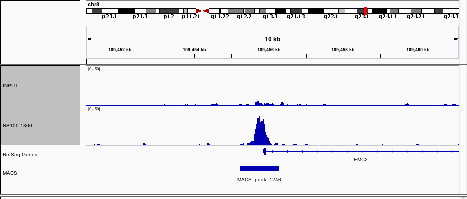
Application: Chromatin ImmunoprecipitationSample Tested: Renal cell lineSpecies: HumanVerified Customer | Posted 07/07/2016ChIP-seq tracks comparing Input and NB-100-1800
There are no reviews that match your criteria.
Protocols
View specific protocols for DEC1 Antibody - BSA Free (NB100-1800):
Culture cells to appropriate density in 35 mm culture dishes or 6-well plates.
1. Remove culture medium and add 10% formalin to the dish. Fix at room temperature for 30 minutes.
2. Remove the formalin and add ice cold methanol. Incubate for 5-10 minutes.
3. Remove methanol and add washing solution (i.e. PBS). Be sure to not let the specimen dry out. Wash three times for 10 minutes.
4. To block nonspecific antibody binding incubate in 10% normal goat serum from 1 hour to overnight at room temperature.
5. Add primary antibody at appropriate dilution and incubate at room temperature from 2 hours to overnight at room temperature.
6. Remove primary antibody and replace with washing solution. Wash three times for 10 minutes.
7. Add secondary antibody at appropriate dilution. Incubate for 1 hour at room temperature.
8. Remove antibody and replace with wash solution, then wash for 10 minutes. Add Hoechst 33258 to wash solution at 1:25,0000 and incubate for 10 minutes. Wash a third time for 10 minutes.
9. Cells can be viewed directly after washing. The plates can also be stored in PBS containing Azide covered in Parafilm (TM). Cells can also be cover-slipped using Fluoromount, with appropriate sealing.
*The above information is only intended as a guide. The researcher should determine what protocol best meets their needs. Please follow safe laboratory procedures.
Western Blot Protocol
1. Perform SDS-PAGE on samples to be analyzed, loading 25 ug of total protein per lane.
2. Transfer proteins to membrane according to the instructions provided by the manufacturer of the membrane and transfer apparatus.
3. Stain according to standard Ponceau S procedure (or similar product) to assess transfer success, and mark molecular weight standards where appropriate.
4. Rinse the blot.
5. Block the membrane using standard blocking buffer for at least 1 hour.
6. Wash the membrane in wash buffer three times for 10 minutes each.
7. Dilute anti-DEC1 primary antibody in blocking buffer and incubate 1 hour at room temperature.
8. Wash the membrane in wash buffer three times for 10 minutes each.
9. Apply the diluted HRP conjugated secondary antibody in blocking buffer (as per manufacturers instructions) and incubate 1 hour at room temperature.
10. Wash the blot in wash buffer three times for 10 minutes each (this step can be repeated as required to reduce background).
11. Apply the detection reagent of choice in accordance with the manufacturers instructions.
Note: Tween-20 can be added to the blocking or antibody dilution buffer at a final concentration of 0.05-0.2%.
Find general support by application which include: protocols, troubleshooting, illustrated assays, videos and webinars.
- 7-Amino Actinomycin D (7-AAD) Cell Viability Flow Cytometry Protocol
- Antigen Retrieval Protocol (PIER)
- Antigen Retrieval for Frozen Sections Protocol
- Appropriate Fixation of IHC/ICC Samples
- Cellular Response to Hypoxia Protocols
- ChIP Protocol Video
- Chromatin Immunoprecipitation (ChIP) Protocol
- Chromatin Immunoprecipitation Protocol
- Chromogenic IHC Staining of Formalin-Fixed Paraffin-Embedded (FFPE) Tissue Protocol
- Chromogenic Immunohistochemistry Staining of Frozen Tissue
- ClariTSA™ Fluorophore Kits
- Detection & Visualization of Antibody Binding
- ELISA Sample Preparation & Collection Guide
- ELISA Troubleshooting Guide
- Extracellular Membrane Flow Cytometry Protocol
- Flow Cytometry Protocol for Cell Surface Markers
- Flow Cytometry Protocol for Staining Membrane Associated Proteins
- Flow Cytometry Staining Protocols
- Flow Cytometry Troubleshooting Guide
- Fluorescent IHC Staining of Frozen Tissue Protocol
- Graphic Protocol for Heat-induced Epitope Retrieval
- Graphic Protocol for the Preparation and Fluorescent IHC Staining of Frozen Tissue Sections
- Graphic Protocol for the Preparation and Fluorescent IHC Staining of Paraffin-embedded Tissue Sections
- Graphic Protocol for the Preparation of Gelatin-coated Slides for Histological Tissue Sections
- How to Run an R&D Systems DuoSet ELISA
- How to Run an R&D Systems Quantikine ELISA
- How to Run an R&D Systems Quantikine™ QuicKit™ ELISA
- ICC Cell Smear Protocol for Suspension Cells
- ICC Immunocytochemistry Protocol Videos
- ICC for Adherent Cells
- IHC Sample Preparation (Frozen sections vs Paraffin)
- Immunocytochemistry (ICC) Protocol
- Immunocytochemistry Troubleshooting
- Immunofluorescence of Organoids Embedded in Cultrex Basement Membrane Extract
- Immunofluorescent IHC Staining of Formalin-Fixed Paraffin-Embedded (FFPE) Tissue Protocol
- Immunohistochemistry (IHC) and Immunocytochemistry (ICC) Protocols
- Immunohistochemistry Frozen Troubleshooting
- Immunohistochemistry Paraffin Troubleshooting
- Immunoprecipitation Protocol
- Intracellular Flow Cytometry Protocol Using Alcohol (Methanol)
- Intracellular Flow Cytometry Protocol Using Detergents
- Intracellular Nuclear Staining Flow Cytometry Protocol Using Detergents
- Intracellular Staining Flow Cytometry Protocol Using Alcohol Permeabilization
- Intracellular Staining Flow Cytometry Protocol Using Detergents to Permeabilize Cells
- Preparing Samples for IHC/ICC Experiments
- Preventing Non-Specific Staining (Non-Specific Binding)
- Primary Antibody Selection & Optimization
- Propidium Iodide Cell Viability Flow Cytometry Protocol
- Protocol for Heat-Induced Epitope Retrieval (HIER)
- Protocol for Liperfluo
- Protocol for Making a 4% Formaldehyde Solution in PBS
- Protocol for VisUCyte™ HRP Polymer Detection Reagent
- Protocol for the Characterization of Human Th22 Cells
- Protocol for the Characterization of Human Th9 Cells
- Protocol for the Fluorescent ICC Staining of Cell Smears - Graphic
- Protocol for the Fluorescent ICC Staining of Cultured Cells on Coverslips - Graphic
- Protocol for the Preparation & Fixation of Cells on Coverslips
- Protocol for the Preparation and Chromogenic IHC Staining of Frozen Tissue Sections
- Protocol for the Preparation and Chromogenic IHC Staining of Frozen Tissue Sections - Graphic
- Protocol for the Preparation and Chromogenic IHC Staining of Paraffin-embedded Tissue Sections
- Protocol for the Preparation and Chromogenic IHC Staining of Paraffin-embedded Tissue Sections - Graphic
- Protocol for the Preparation and Fluorescent ICC Staining of Cells on Coverslips
- Protocol for the Preparation and Fluorescent ICC Staining of Non-adherent Cells
- Protocol for the Preparation and Fluorescent ICC Staining of Stem Cells on Coverslips
- Protocol for the Preparation and Fluorescent IHC Staining of Frozen Tissue Sections
- Protocol for the Preparation and Fluorescent IHC Staining of Paraffin-embedded Tissue Sections
- Protocol for the Preparation of Gelatin-coated Slides for Histological Tissue Sections
- Protocol for the Preparation of a Cell Smear for Non-adherent Cell ICC - Graphic
- Protocol: Annexin V and PI Staining by Flow Cytometry
- Protocol: Annexin V and PI Staining for Apoptosis by Flow Cytometry
- Quantikine HS ELISA Kit Assay Principle, Alkaline Phosphatase
- Quantikine HS ELISA Kit Principle, Streptavidin-HRP Polymer
- R&D Systems Quality Control Western Blot Protocol
- Sandwich ELISA (Colorimetric) – Biotin/Streptavidin Detection Protocol
- Sandwich ELISA (Colorimetric) – Direct Detection Protocol
- TUNEL and Active Caspase-3 Detection by IHC/ICC Protocol
- The Importance of IHC/ICC Controls
- Troubleshooting Guide: ELISA
- Troubleshooting Guide: Fluorokine Flow Cytometry Kits
- Troubleshooting Guide: Immunohistochemistry
- Troubleshooting Guide: Western Blot Figures
- Western Blot Conditions
- Western Blot Protocol
- Western Blot Protocol for Cell Lysates
- Western Blot Troubleshooting
- Western Blot Troubleshooting Guide
- View all Protocols, Troubleshooting, Illustrated assays and Webinars
Loading...