Desmin Antibody - C-terminus
Novus Biologicals | Catalog # NBP1-45143
![Western Blot: Desmin Antibody [NBP1-45143]](https://resources.rndsystems.com/images/products/Desmin-Antibody-NBP1-45143-img0002.jpg "Western Blot: Desmin Antibody [NBP1-45143]")
Loading...
Key Product Details
Species Reactivity
Validated:
Human
Cited:
Human
Applications
Validated:
Immunohistochemistry, Immunohistochemistry-Paraffin, Western Blot, ELISA
Cited:
Western Blot
Label
Unconjugated
Antibody Source
Polyclonal Goat IgG
Loading...
Product Specifications
Immunogen
Synthetic peptide sequence C-RDGEVVSEATQQQHE from the C-Terminal region of Desmin (NP_001918.3)
Reactivity Notes
Predicted cross-reactivities: Mouse, Porcine, Rat, Dog, Bovine
Specificity
Goat anti Human Desmin recognizes an epitope within the C-terminal (CT) region of human.
Clonality
Polyclonal
Host
Goat
Isotype
IgG
Theoretical MW
55 kDa.
Disclaimer note: The observed molecular weight of the protein may vary from the listed predicted molecular weight due to post translational modifications, post translation cleavages, relative charges, and other experimental factors.
Disclaimer note: The observed molecular weight of the protein may vary from the listed predicted molecular weight due to post translational modifications, post translation cleavages, relative charges, and other experimental factors.
Scientific Data Images for Desmin Antibody - C-terminus
Western Blot: Desmin Antibody [NBP1-45143]
Western Blot: Desmin Antibody [NBP1-45143] - Analysis of human (A), mouse (B), rat (C) and pig (D) skeletal muscle lysate (35ug protein in RIPA buffer) probed with Goat anti Human desmin antibody (NBP1-45143) at 1.0ugml-1. Primary incubation was 1 hour. Signal was detected by chemiluminescence![Immunohistochemistry-Paraffin: Desmin Antibody [NBP1-45143]](https://resources.rndsystems.com/images/products/Desmin-Antibody-Immunohistochemistry-Paraffin-NBP1-45143-img0003.jpg "Immunohistochemistry-Paraffin: Desmin Antibody [NBP1-45143]")
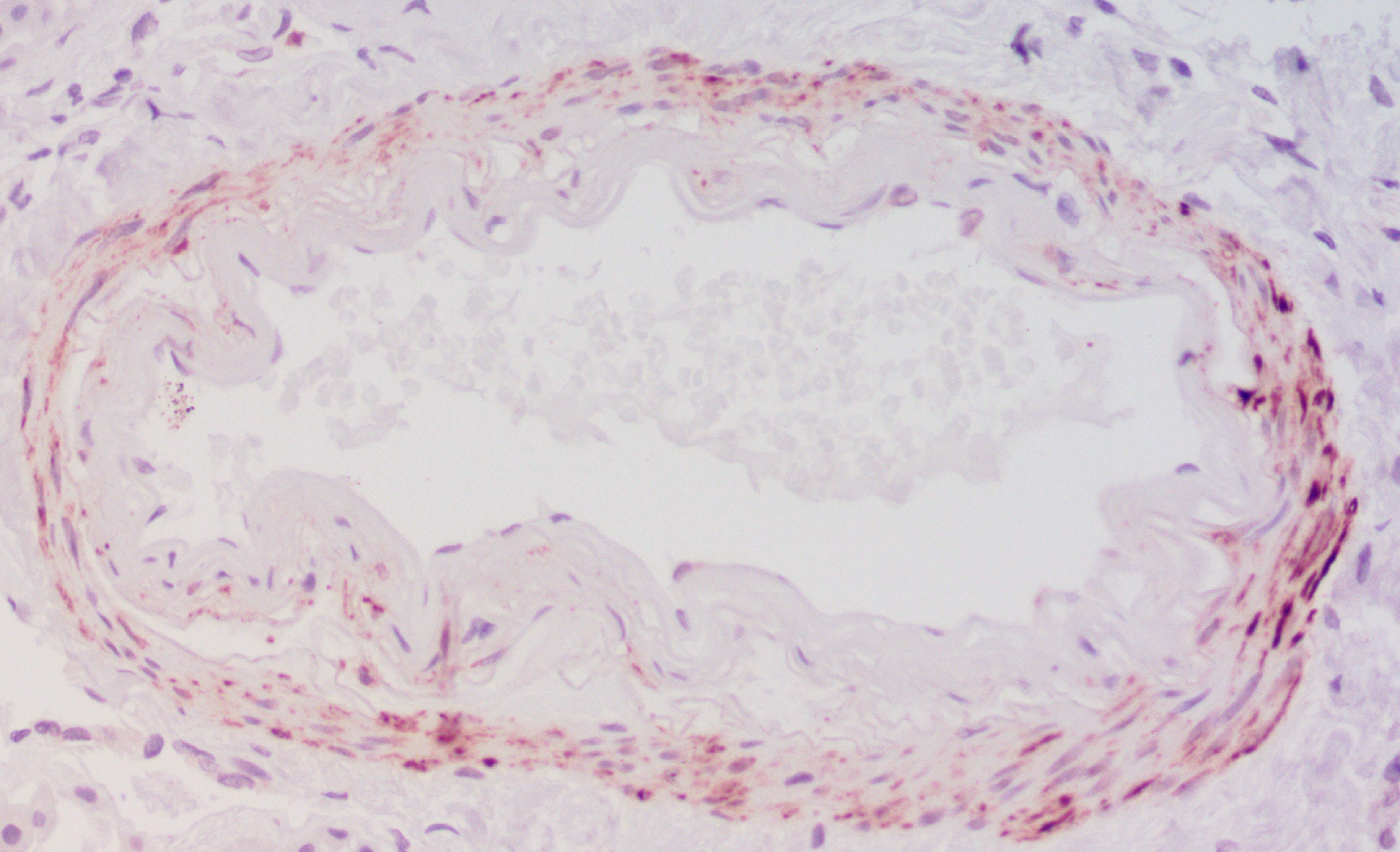
Immunohistochemistry-Paraffin: Desmin Antibody [NBP1-45143]
Immunohistochemistry-Paraffin: Desmin Antibody [NBP1-45143] - Staining showing strong specific labeling in pericytes around blood vessels in the brain. Antigen retrieval at pH6 (citra) was performed and Desmin was used at 1:500 dilution. Hematoxyllin was used as a counterstain to mark nuclei. Image from verified customer review.![Western Blot: Desmin Antibody [NBP1-45143]](https://resources.rndsystems.com/images/products/Desmin-Antibody-NBP1-45143-img0001.jpg "Western Blot: Desmin Antibody [NBP1-45143]")
Western Blot: Desmin Antibody [NBP1-45143]
Western Blot: Desmin Antibody [NBP1-45143] - Human skeletal muscle cell lysate probed with Goat anti Human Desmin C-Terminal (NBP1-45143)Applications for Desmin Antibody - C-terminus
Application
Recommended Usage
ELISA
1:2000
Immunohistochemistry
3-6 ug/ml
Immunohistochemistry-Paraffin
3-6 ug/ml
Western Blot
0.1-0.3 ug/ml
Application Notes
This product requires heat-mediated antigen retrieval prior to staining of paraffin sections. Sodium citrate buffer pH6.0 is recommended for this purpose. Goat anti Human Desmin detects a band of approximately 55kDa in human skeletal muscle cell lysates.
Reviewed Applications
Read 1 review rated 5 using NBP1-45143 in the following applications:
Formulation, Preparation, and Storage
Purification
Immunogen affinity purified
Formulation
TRIS buffered saline, 0.5% Bovine Serum Albumin
Preservative
0.02% Sodium Azide
Concentration
0.5 mg/ml
Shipping
The product is shipped with polar packs. Upon receipt, store it immediately at the temperature recommended below.
Stability & Storage
Store at 4C short term. Aliquot and store at -20C long term. Avoid freeze-thaw cycles.
Background: Desmin
Additional Desmin Products
Product Documents for Desmin Antibody - C-terminus
Certificate of Analysis
To download a Certificate of Analysis, please enter a lot or batch number in the search box below.
Product Specific Notices for Desmin Antibody - C-terminus
This product is for research use only and is not approved for use in humans or in clinical diagnosis. Primary Antibodies are guaranteed for 1 year from date of receipt.
Related Research Areas
Citations for Desmin Antibody - C-terminus
 Powered by Bioz
Customer Reviews for Desmin Antibody - C-terminus (1)
5 out of 5
1 Customer Rating
Have you used Desmin Antibody - C-terminus?
Submit a review and receive an Amazon gift card!
$25/€18/£15/$25CAN/¥2500 Yen for a review with an image
$10/€7/£6/$10CAN/¥1110 Yen for a review without an image
Submit a review
Customer Images
Showing
1
-
1 of
1 review
Showing All
Filter By:
-
Application: Immunohistochemistry-ParaffinSample Tested: brain and spinal cordSpecies: HumanVerified Customer | Posted 03/01/2019Desmin staining showing strong specific labeling in pericytes around blood vessels in the brain. Antigen retrieval at pH6 (citra) was performed and Desmin was used at 1:500 dilution. Hematoxyllin was used as a counterstain to mark nucleiAntigen retrieval at pH6 fro 20 minutes in a steamer; Used at 1:500 concentration. Novared (Vector labs) was used as a chromogen to develop the signal.

There are no reviews that match your criteria.
Protocols
Find general support by application which include: protocols, troubleshooting, illustrated assays, videos and webinars.
- Antigen Retrieval Protocol (PIER)
- Antigen Retrieval for Frozen Sections Protocol
- Appropriate Fixation of IHC/ICC Samples
- Cellular Response to Hypoxia Protocols
- Chromogenic IHC Staining of Formalin-Fixed Paraffin-Embedded (FFPE) Tissue Protocol
- Chromogenic Immunohistochemistry Staining of Frozen Tissue
- ClariTSA™ Fluorophore Kits
- Detection & Visualization of Antibody Binding
- ELISA Sample Preparation & Collection Guide
- ELISA Troubleshooting Guide
- Fluorescent IHC Staining of Frozen Tissue Protocol
- Graphic Protocol for Heat-induced Epitope Retrieval
- Graphic Protocol for the Preparation and Fluorescent IHC Staining of Frozen Tissue Sections
- Graphic Protocol for the Preparation and Fluorescent IHC Staining of Paraffin-embedded Tissue Sections
- Graphic Protocol for the Preparation of Gelatin-coated Slides for Histological Tissue Sections
- How to Run an R&D Systems DuoSet ELISA
- How to Run an R&D Systems Quantikine ELISA
- How to Run an R&D Systems Quantikine™ QuicKit™ ELISA
- IHC Sample Preparation (Frozen sections vs Paraffin)
- Immunofluorescent IHC Staining of Formalin-Fixed Paraffin-Embedded (FFPE) Tissue Protocol
- Immunohistochemistry (IHC) and Immunocytochemistry (ICC) Protocols
- Immunohistochemistry Frozen Troubleshooting
- Immunohistochemistry Paraffin Troubleshooting
- Preparing Samples for IHC/ICC Experiments
- Preventing Non-Specific Staining (Non-Specific Binding)
- Primary Antibody Selection & Optimization
- Protocol for Heat-Induced Epitope Retrieval (HIER)
- Protocol for Making a 4% Formaldehyde Solution in PBS
- Protocol for VisUCyte™ HRP Polymer Detection Reagent
- Protocol for the Preparation & Fixation of Cells on Coverslips
- Protocol for the Preparation and Chromogenic IHC Staining of Frozen Tissue Sections
- Protocol for the Preparation and Chromogenic IHC Staining of Frozen Tissue Sections - Graphic
- Protocol for the Preparation and Chromogenic IHC Staining of Paraffin-embedded Tissue Sections
- Protocol for the Preparation and Chromogenic IHC Staining of Paraffin-embedded Tissue Sections - Graphic
- Protocol for the Preparation and Fluorescent IHC Staining of Frozen Tissue Sections
- Protocol for the Preparation and Fluorescent IHC Staining of Paraffin-embedded Tissue Sections
- Protocol for the Preparation of Gelatin-coated Slides for Histological Tissue Sections
- Quantikine HS ELISA Kit Assay Principle, Alkaline Phosphatase
- Quantikine HS ELISA Kit Principle, Streptavidin-HRP Polymer
- R&D Systems Quality Control Western Blot Protocol
- Sandwich ELISA (Colorimetric) – Biotin/Streptavidin Detection Protocol
- Sandwich ELISA (Colorimetric) – Direct Detection Protocol
- TUNEL and Active Caspase-3 Detection by IHC/ICC Protocol
- The Importance of IHC/ICC Controls
- Troubleshooting Guide: ELISA
- Troubleshooting Guide: Immunohistochemistry
- Troubleshooting Guide: Western Blot Figures
- Western Blot Conditions
- Western Blot Protocol
- Western Blot Protocol for Cell Lysates
- Western Blot Troubleshooting
- Western Blot Troubleshooting Guide
- View all Protocols, Troubleshooting, Illustrated assays and Webinars
Loading...
Associated Pathways
