DGAT1 Antibody - BSA Free
Novus Biologicals | Catalog # NB110-41487
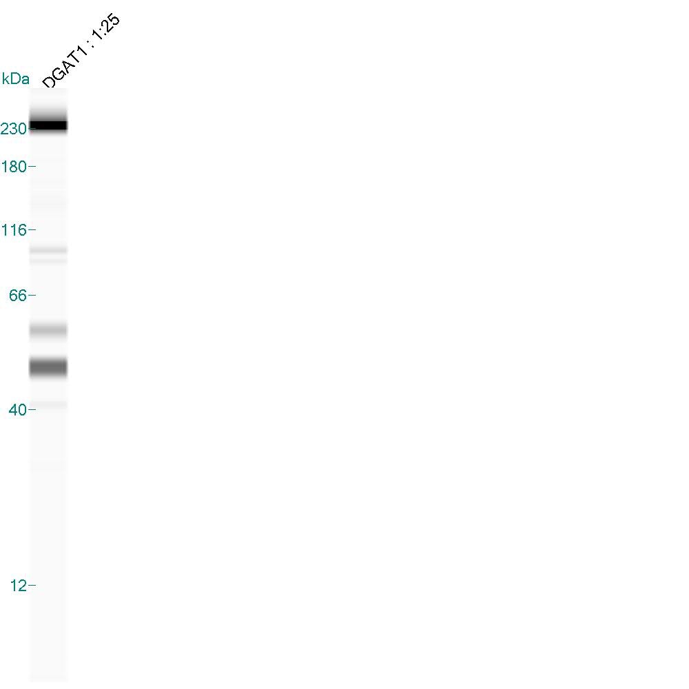
![Western Blot: DGAT1 AntibodyBSA Free [NB110-41487]](https://resources.rndsystems.com/images/products/DGAT1-Antibody-Western-Blot-NB110-41487-img0002.jpg "Western Blot: DGAT1 AntibodyBSA Free [NB110-41487]")
Key Product Details
Species Reactivity
Validated:
Cited:
Predicted:
Applications
Validated:
Cited:
Label
Antibody Source
Format
Product Specifications
Immunogen
Reactivity Notes
Localization
Clonality
Host
Isotype
Theoretical MW
Disclaimer note: The observed molecular weight of the protein may vary from the listed predicted molecular weight due to post translational modifications, post translation cleavages, relative charges, and other experimental factors.
Scientific Data Images for DGAT1 Antibody - BSA Free
Western Blot: DGAT1 AntibodyBSA Free [NB110-41487]
Western Blot: DGAT1 Antibody [NB110-41487] - Detection of DGAT1 in HepG2 lysate.![Immunocytochemistry/ Immunofluorescence: DGAT1 Antibody - BSA Free [NB110-41487]](https://resources.rndsystems.com/images/products/DGAT1-Antibody-Immunocytochemistry-Immunofluorescence-NB110-41487-img0003.jpg "Immunocytochemistry/ Immunofluorescence: DGAT1 Antibody - BSA Free [NB110-41487]")
Immunocytochemistry/ Immunofluorescence: DGAT1 Antibody - BSA Free [NB110-41487]
Immunocytochemistry/Immunofluorescence: DGAT1 Antibody [NB110-41487] - HepG2 cells were fixed for 10 minutes using 10% formalin and then permeabilized for 5 minutes using 1X PBS + 0.05% Triton X-100. The cells were incubated with anti-DGAT1 at 5 ug/ml overnight at 4C and detected with an anti-rabbit DyLight 488 (Green) at a 1:500 dilution. Nuclei were counterstained with DAPI (Blue). Cells were imaged using a 40X objective.![Western Blot: DGAT1 AntibodyBSA Free [NB110-41487]](https://resources.rndsystems.com/images/products/DGAT1-Antibody-Western-Blot-NB110-41487-img0004.jpg "Western Blot: DGAT1 AntibodyBSA Free [NB110-41487]")
Western Blot: DGAT1 AntibodyBSA Free [NB110-41487]
DGAT1-Antibody-Western-Blot-NB110-41487-img0004.jpgApplications for DGAT1 Antibody - BSA Free
Immunoblotting
Immunocytochemistry/ Immunofluorescence
Simple Western
Western Blot
Reviewed Applications
Read 1 review rated 3 using NB110-41487 in the following applications:
Formulation, Preparation, and Storage
Purification
Formulation
Format
Preservative
Concentration
Shipping
Stability & Storage
Background: DGAT1
Long Name
Alternate Names
Gene Symbol
Additional DGAT1 Products
Product Documents for DGAT1 Antibody - BSA Free
Certificate of Analysis
To download a Certificate of Analysis, please enter a lot or batch number in the search box below.
Product Specific Notices for DGAT1 Antibody - BSA Free
This product is for research use only and is not approved for use in humans or in clinical diagnosis. Primary Antibodies are guaranteed for 1 year from date of receipt.
Related Research Areas
Citations for DGAT1 Antibody - BSA Free
 Powered by Bioz
Customer Reviews for DGAT1 Antibody - BSA Free (1)
Have you used DGAT1 Antibody - BSA Free?
Submit a review and receive an Amazon gift card!
$25/€18/£15/$25CAN/¥2500 Yen for a review with an image
$10/€7/£6/$10CAN/¥1110 Yen for a review without an image
Submit a review
Customer Images
-
Application: Simple WesternSample Tested: Mouse skeletal muscle homogenateSpecies: MouseVerified Customer | Posted 07/22/20150.2 ug/uL mouse skeletal muscle tissue homogenate. Specific band ~50 kDa.

There are no reviews that match your criteria.
Protocols
View specific protocols for DGAT1 Antibody - BSA Free (NB110-41487):
Immunocytochemistry Protocol
Culture cells to appropriate density in 35 mm culture dishes or 6-well plates.
1. Remove culture medium and add 10% formalin to the dish. Fix at room temperature for 30 minutes.
2. Remove the formalin and add ice cold methanol. Incubate for 5-10 minutes.
3. Remove methanol and add washing solution (i.e. PBS). Be sure to not let the specimen dry out. Wash three times for 10 minutes.
4. To block nonspecific antibody binding incubate in 10% normal goat serum from 1 hour to overnight at room temperature.
5. Add primary antibody at appropriate dilution and incubate at room temperature from 2 hours to overnight at room temperature.
6. Remove primary antibody and replace with washing solution. Wash three times for 10 minutes.
7. Add secondary antibody at appropriate dilution. Incubate for 1 hour at room temperature.
8. Remove antibody and replace with wash solution, then wash for 10 minutes. Add Hoechst 33258 to wash solution at 1:25,0000 and incubate for 10 minutes. Wash a third time for 10 minutes.
9. Cells can be viewed directly after washing. The plates can also be stored in PBS containing Azide covered in Parafilm (TM). Cells can also be cover-slipped using Fluoromount, with appropriate sealing.
*The above information is only intended as a guide. The researcher should determine what protocol best meets their needs. Please follow safe laboratory procedures.
Western Blot Protocol
1. Perform SDS-PAGE on samples to be analyzed, loading 40 ug of total protein per lane.
2. Transfer proteins to membrane according to the instructions provided by the manufacturer of the membrane and transfer apparatus.
3. Stain according to standard Ponceau S procedure (or similar product) to assess transfer success, and mark molecular weight standards where appropriate.
4. Rinse the blot.
5. Block the membrane using standard blocking buffer for at least 1 hour.
6. Wash the membrane in wash buffer three times for 10 minutes each.
7. Dilute primary antibody in blocking buffer and incubate 1 hour at room temperature.
8. Wash the membrane in wash buffer three times for 10 minutes each.
9. Apply the diluted HRP conjugated secondary antibody in blocking buffer (as per manufacturers instructions) and incubate 1 hour at room temperature.
10. Wash the blot in wash buffer three times for 10 minutes each (this step can be repeated as required to reduce background).
11. Apply the detection reagent of choice in accordance with the manufacturers instructions.
Note: Tween-20 can be added to the blocking or antibody dilution buffer at a final concentration of 0.05-0.2%.
*The above information is only intended as a guide. The researcher should determine what protocol best meets their needs. Please follow safe laboratory procedures.
Find general support by application which include: protocols, troubleshooting, illustrated assays, videos and webinars.
- Appropriate Fixation of IHC/ICC Samples
- Cellular Response to Hypoxia Protocols
- ClariTSA™ Fluorophore Kits
- Detection & Visualization of Antibody Binding
- ICC Cell Smear Protocol for Suspension Cells
- ICC Immunocytochemistry Protocol Videos
- ICC for Adherent Cells
- Immunocytochemistry (ICC) Protocol
- Immunocytochemistry Troubleshooting
- Immunofluorescence of Organoids Embedded in Cultrex Basement Membrane Extract
- Immunohistochemistry (IHC) and Immunocytochemistry (ICC) Protocols
- Preparing Samples for IHC/ICC Experiments
- Preventing Non-Specific Staining (Non-Specific Binding)
- Primary Antibody Selection & Optimization
- Protocol for VisUCyte™ HRP Polymer Detection Reagent
- Protocol for the Fluorescent ICC Staining of Cell Smears - Graphic
- Protocol for the Fluorescent ICC Staining of Cultured Cells on Coverslips - Graphic
- Protocol for the Preparation and Fluorescent ICC Staining of Cells on Coverslips
- Protocol for the Preparation and Fluorescent ICC Staining of Non-adherent Cells
- Protocol for the Preparation and Fluorescent ICC Staining of Stem Cells on Coverslips
- Protocol for the Preparation of a Cell Smear for Non-adherent Cell ICC - Graphic
- R&D Systems Quality Control Western Blot Protocol
- TUNEL and Active Caspase-3 Detection by IHC/ICC Protocol
- The Importance of IHC/ICC Controls
- Troubleshooting Guide: Western Blot Figures
- Western Blot Conditions
- Western Blot Protocol
- Western Blot Protocol for Cell Lysates
- Western Blot Troubleshooting
- Western Blot Troubleshooting Guide
- View all Protocols, Troubleshooting, Illustrated assays and Webinars
FAQs for DGAT1 Antibody - BSA Free
-
Q: Do you offer the blocking peptide for NB110-41487?
A: Unfortunately, we do not offer the blocking peptide for NB110-41487. I apologize for any inconvenience.
-
Q: Have you ever tested if DGAT1 NB110-41487 work for rat liver tissues?
A: We have not specifically tested rat liver tissue, but rat is a species covered by our guarantee for this product.
-
Q: I would like to order your anti-DGAT1 antibody NB110-41487. The immunogen for this is a synthetic peptide designed within residues 200-300 of human DGAT1. I would like to use it to detect the homolog of DGAT1 in Drosophila. I know that the sequence is proprietary but could you tell me if the sequence is conserved in Drosophila (that means that the peptide is designed within residues 243-288 of human DGAT1) so that it could work in flies.
A: Unfortunately, the Drosophila and human DGAT1 sequences are too divergent to recommend that NB110-41487 may cross-react with fly.
-
Q: Do you offer the blocking peptide for NB110-41487?
A: Unfortunately, we do not offer the blocking peptide for NB110-41487. I apologize for any inconvenience.
-
Q: Have you ever tested if DGAT1 NB110-41487 work for rat liver tissues?
A: We have not specifically tested rat liver tissue, but rat is a species covered by our guarantee for this product.
-
Q: I would like to order your anti-DGAT1 antibody NB110-41487. The immunogen for this is a synthetic peptide designed within residues 200-300 of human DGAT1. I would like to use it to detect the homolog of DGAT1 in Drosophila. I know that the sequence is proprietary but could you tell me if the sequence is conserved in Drosophila (that means that the peptide is designed within residues 243-288 of human DGAT1) so that it could work in flies.
A: Unfortunately, the Drosophila and human DGAT1 sequences are too divergent to recommend that NB110-41487 may cross-react with fly.