EGLN3/PHD3 Antibody (EG188e/d5) - BSA Free
Novus Biologicals | Catalog # NBP1-30440
Key Product Details
Validated by
Species Reactivity
Validated:
Cited:
Predicted:
Applications
Validated:
Cited:
Label
Antibody Source
Format
Product Specifications
Immunogen
Localization
Clonality
Host
Isotype
Theoretical MW
Disclaimer note: The observed molecular weight of the protein may vary from the listed predicted molecular weight due to post translational modifications, post translation cleavages, relative charges, and other experimental factors.
Scientific Data Images for EGLN3/PHD3 Antibody (EG188e/d5) - BSA Free
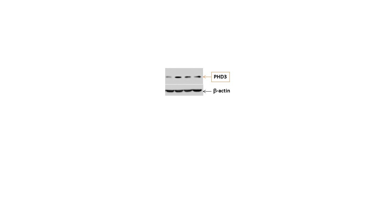
![Western Blot: EGLN3/PHD3 Antibody (EG188e/d5)BSA Free [NBP1-30440]](https://resources.rndsystems.com/images/products/EGLN3-PHD3-Antibody-EG188e-d5-Western-Blot-NBP1-30440-img0007.jpg "Western Blot: EGLN3/PHD3 Antibody (EG188e/d5)BSA Free [NBP1-30440]")
![Immunohistochemistry: EGLN3/PHD3 Antibody (EG188e/d5) - BSA Free [NBP1-30440]](https://resources.rndsystems.com/images/products/EGLN3-PHD3-Antibody-EG188e-d5-Immunohistochemistry-NBP1-30440-img0005.jpg "Immunohistochemistry: EGLN3/PHD3 Antibody (EG188e/d5) - BSA Free [NBP1-30440]")
Immunohistochemistry: EGLN3/PHD3 Antibody (EG188e/d5) - BSA Free [NBP1-30440]
Immunohistochemistry: EGLN3/PHD3 Antibody (EG188e/d5) [NBP1-30440] - Analysis of HIF Prolyl Hydroxylase 3 in human renal cancer using DAB with hematoxylin counterstain.![Western Blot: EGLN3/PHD3 Antibody (EG188e/d5)BSA Free [NBP1-30440]](https://resources.rndsystems.com/images/products/EGLN3-PHD3-Antibody-EG188e-d5-Western-Blot-NBP1-30440-img0004.jpg "Western Blot: EGLN3/PHD3 Antibody (EG188e/d5)BSA Free [NBP1-30440]")
![Simple Western: EGLN3/PHD3 Antibody (EG188e/d5)BSA Free [NBP1-30440]](https://resources.rndsystems.com/images/products/EGLN3-PHD3-Antibody-EG188e-d5-Simple-Western-NBP1-30440-img0008.jpg "Simple Western: EGLN3/PHD3 Antibody (EG188e/d5)BSA Free [NBP1-30440]")
Simple Western: EGLN3/PHD3 Antibody (EG188e/d5)BSA Free [NBP1-30440]
Simple Western: EGLN3/PHD3 Antibody (EG188e/d5) [NBP1-30440] - Lane view shows a specific band for PHD3/HIF Prolyl Hydroxylase 3 in 0.5 mg/ml of Hypoxic HeLa lysate. This experiment was performed under reducing conditions using the 12-230 kDa separation system.Applications for EGLN3/PHD3 Antibody (EG188e/d5) - BSA Free
Immunohistochemistry
Immunohistochemistry-Paraffin
Simple Western
Western Blot
In Simple Western only 10 - 15 uL of the recommended dilution is used per data point.
See Simple Western Antibody Database for Simple Western validation: Tested in Hypoxic HeLa lysate 0.5 mg/mL, separated by Size, antibody dilution of 1:1000, apparent MW was 32 kDa. Separated by Size-Wes, Sally Sue/Peggy Sue.
Reviewed Applications
Read 4 reviews rated 4.8 using NBP1-30440 in the following applications:
Formulation, Preparation, and Storage
Purification
Formulation
Format
Preservative
Concentration
Shipping
Stability & Storage
Background: EGLN3/PHD3
Long Name
Alternate Names
Entrez Gene IDs
Gene Symbol
UniProt
Additional EGLN3/PHD3 Products
Product Documents for EGLN3/PHD3 Antibody (EG188e/d5) - BSA Free
Certificate of Analysis
To download a Certificate of Analysis, please enter a lot or batch number in the search box below.
Product Specific Notices for EGLN3/PHD3 Antibody (EG188e/d5) - BSA Free
This product is for research use only and is not approved for use in humans or in clinical diagnosis. Primary Antibodies are guaranteed for 1 year from date of receipt.
Related Research Areas
Citations for EGLN3/PHD3 Antibody (EG188e/d5) - BSA Free
 Powered by Bioz
Customer Reviews for EGLN3/PHD3 Antibody (EG188e/d5) - BSA Free (4)
Have you used EGLN3/PHD3 Antibody (EG188e/d5) - BSA Free?
Submit a review and receive an Amazon gift card!
$25/€18/£15/$25CAN/¥2500 Yen for a review with an image
$10/€7/£6/$10CAN/¥1110 Yen for a review without an image
Submit a review
Customer Images
-
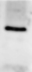
Application: Western BlotSample Tested: HBE-16Species: HumanVerified Customer | Posted 04/16/2019PHD3

-
Application: Western BlotSample Tested: MCF-7 whole cell lysateSpecies: HumanVerified Customer | Posted 11/20/2015EGLN3 in MCF7 cells
-
Application: Western BlotSample Tested: SK-N-BE(2) whole cell lysateSpecies: OtherVerified Customer | Posted 10/30/2014
-
Application: Western BlotSample Tested: PC12 whole cell lysateSpecies: OtherVerified Customer | Posted 08/27/2014
There are no reviews that match your criteria.
Protocols
View specific protocols for EGLN3/PHD3 Antibody (EG188e/d5) - BSA Free (NBP1-30440):
Immunohistochemistry-Paraffin Embedded Sections
Antigen Unmasking:
Bring slides to a boil in 10 mM sodium citrate buffer (pH 6.0) then maintain at a sub-boiling temperature for 10 minutes. Cool slides on bench-top for 30 minutes.
Staining:
1. Wash sections in deionized water three times for 5 minutes each.
2. Wash sections in wash buffer for 5 minutes.
3. Block each section with 100-400 ul blocking solution for 1 hour at room temperature.
4. Remove blocking solution and add 100-400 ul diluted primary antibody. Incubate overnight at 4C.
5. Remove antibody solution and wash sections in wash buffer three times for 5 minutes each.
6. Add 100-400 ul biotinylated diluted secondary antibody. Incubate 30 minutes at room temperature.
7. Remove secondary antibody solution and wash sections three times with wash buffer for 5 minutes each.
8. Add 100-400 ul Streptavidin-HRP reagent to each section and incubate for 30 minutes at room temperature.
9. Wash sections three times in wash buffer for 5 minutes each.
10. Add 100-400 ul DAB substrate to each section and monitor staining closely.
11. As soon as the sections develop, immerse slides in deionized water.
12. Counterstain sections in hematoxylin.
13. Wash sections in deionized water two times for 5 minutes each.
14. Dehydrate sections.
15. Mount coverslips.
Western Blot Protocol
1. Perform SDS-PAGE (4-12% MOPS) on samples to be analyzed, loading 40 ug of total protein per lane.
2. Transfer proteins to Nitrocellulose according to the instructions provided by the manufacturer of the transfer apparatus.
3. Rinse membrane with dH2O and then stain the blot using Ponceau S for 1-2 minutes to access the transfer of proteins onto the nitrocellulose membrane. Rinse the blot in water to remove excess stain and mark the lane locations and locations of molecular weight markers using a pencil.
4. Rinse the blot in TBS for approximately 5 minutes.
5. Block the membrane using 5% NFDM + 1% BSA in TBS + Tween, 1 hour at RT.
6. Rinse the membrane in dH2O and then wash the membrane in wash buffer [TBS + 0.1% Tween] 3 times for 10 minutes each.
7. Dilute the mouse anti-PHD3 primary antibody (NBP1-30328) in blocking buffer and incubate 1 hour at room temperature.
8. Rinse the membrane in dH2O and then wash the membrane in wash buffer [TBS + 0.1% Tween] 3 times for 10 minutes each.
9. Apply the diluted mouse-IgG HRP-conjugated secondary antibody in blocking buffer (as per manufacturers instructions) and incubate 1 hour at room temperature.
10. Wash the blot in wash buffer [TBS + 0.1% Tween] 3 times for 10 minutes each (this step can be repeated as required to reduce background).
11. Apply the detection reagent of choice in accordance with the manufacturers instructions (Pierce ECL).
**Note: Tween-20 can be added to the blocking or antibody dilution buffer at a final concentration of 0.05-0.2%, provided it does not interfere with antibody-antigen binding.
Find general support by application which include: protocols, troubleshooting, illustrated assays, videos and webinars.
- Antigen Retrieval Protocol (PIER)
- Antigen Retrieval for Frozen Sections Protocol
- Appropriate Fixation of IHC/ICC Samples
- Cellular Response to Hypoxia Protocols
- Chromogenic IHC Staining of Formalin-Fixed Paraffin-Embedded (FFPE) Tissue Protocol
- Chromogenic Immunohistochemistry Staining of Frozen Tissue
- ClariTSA™ Fluorophore Kits
- Detection & Visualization of Antibody Binding
- Fluorescent IHC Staining of Frozen Tissue Protocol
- Graphic Protocol for Heat-induced Epitope Retrieval
- Graphic Protocol for the Preparation and Fluorescent IHC Staining of Frozen Tissue Sections
- Graphic Protocol for the Preparation and Fluorescent IHC Staining of Paraffin-embedded Tissue Sections
- Graphic Protocol for the Preparation of Gelatin-coated Slides for Histological Tissue Sections
- IHC Sample Preparation (Frozen sections vs Paraffin)
- Immunofluorescent IHC Staining of Formalin-Fixed Paraffin-Embedded (FFPE) Tissue Protocol
- Immunohistochemistry (IHC) and Immunocytochemistry (ICC) Protocols
- Immunohistochemistry Frozen Troubleshooting
- Immunohistochemistry Paraffin Troubleshooting
- Preparing Samples for IHC/ICC Experiments
- Preventing Non-Specific Staining (Non-Specific Binding)
- Primary Antibody Selection & Optimization
- Protocol for Heat-Induced Epitope Retrieval (HIER)
- Protocol for Making a 4% Formaldehyde Solution in PBS
- Protocol for VisUCyte™ HRP Polymer Detection Reagent
- Protocol for the Preparation & Fixation of Cells on Coverslips
- Protocol for the Preparation and Chromogenic IHC Staining of Frozen Tissue Sections
- Protocol for the Preparation and Chromogenic IHC Staining of Frozen Tissue Sections - Graphic
- Protocol for the Preparation and Chromogenic IHC Staining of Paraffin-embedded Tissue Sections
- Protocol for the Preparation and Chromogenic IHC Staining of Paraffin-embedded Tissue Sections - Graphic
- Protocol for the Preparation and Fluorescent IHC Staining of Frozen Tissue Sections
- Protocol for the Preparation and Fluorescent IHC Staining of Paraffin-embedded Tissue Sections
- Protocol for the Preparation of Gelatin-coated Slides for Histological Tissue Sections
- R&D Systems Quality Control Western Blot Protocol
- TUNEL and Active Caspase-3 Detection by IHC/ICC Protocol
- The Importance of IHC/ICC Controls
- Troubleshooting Guide: Immunohistochemistry
- Troubleshooting Guide: Western Blot Figures
- Western Blot Conditions
- Western Blot Protocol
- Western Blot Protocol for Cell Lysates
- Western Blot Troubleshooting
- Western Blot Troubleshooting Guide
- View all Protocols, Troubleshooting, Illustrated assays and Webinars
FAQs for EGLN3/PHD3 Antibody (EG188e/d5) - BSA Free
-
Q: I received an inquiry about Anti-PHD3/HIF Prolyl Hydroxylase 3 Antibody (EG188e/d5), #NBP1-30440. Could you let me know if this antibody cross-reacts with PHD1 or PHD2?
A: We have not tested our PHD3 antibody for cross reactivity with PHD1 or PHD2, therefore we are unaware of any possible cross reactivity.
Associated Pathways
