FIH-1/HIF-1AN Antibody (162c) - BSA Free
Novus Biologicals | Catalog # NBP1-30333
Key Product Details
Species Reactivity
Validated:
Human
Cited:
Human
Applications
Validated:
Immunohistochemistry, Immunohistochemistry-Paraffin, Western Blot, Immunocytochemistry/ Immunofluorescence
Cited:
Immunohistochemistry-Paraffin, Western Blot, Immunocytochemistry/ Immunofluorescence, IF/IHC
Label
Unconjugated
Antibody Source
Monoclonal Mouse IgG1 Clone # 162c
Format
BSA Free
Loading...
Product Specifications
Immunogen
Full length human Factor Inhibiting HIF-1 protein [Swiss-Prot# Q9NWT6]
Localization
Nuclear
Clonality
Monoclonal
Host
Mouse
Isotype
IgG1
Theoretical MW
40 kDa.
Disclaimer note: The observed molecular weight of the protein may vary from the listed predicted molecular weight due to post translational modifications, post translation cleavages, relative charges, and other experimental factors.
Disclaimer note: The observed molecular weight of the protein may vary from the listed predicted molecular weight due to post translational modifications, post translation cleavages, relative charges, and other experimental factors.
Scientific Data Images for FIH-1/HIF-1AN Antibody (162c) - BSA Free
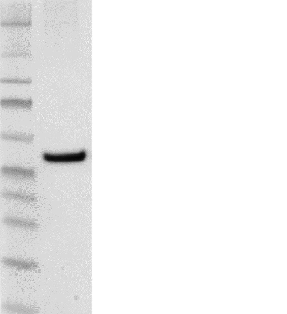
![Western Blot: FIH-1/HIF-1AN Antibody (162c) [NBP1-30333]](https://resources.rndsystems.com/images/products/Factor-Inhibiting-HIF-1-Antibody-162c-Western-Blot-NBP1-30333-img0006.jpg "Western Blot: FIH-1/HIF-1AN Antibody (162c) [NBP1-30333]")
Western Blot: FIH-1/HIF-1AN Antibody (162c) [NBP1-30333]
Western Blot: Factor Inhibiting HIF-1 Antibody (162c) [NBP1-30333] - Analysis in A431 cell lysates using NBP1-30333.![Immunocytochemistry/ Immunofluorescence: FIH-1/HIF-1AN Antibody (162c) [NBP1-30333]](https://resources.rndsystems.com/images/products/Factor-Inhibiting-HIF-1-Antibody-162c-Immunocytochemistry-Immunofluorescence-NBP1-30333-img0005.jpg "Immunocytochemistry/ Immunofluorescence: FIH-1/HIF-1AN Antibody (162c) [NBP1-30333]")
Immunocytochemistry/ Immunofluorescence: FIH-1/HIF-1AN Antibody (162c) [NBP1-30333]
Immunocytochemistry/Immunofluorescence: Factor Inhibiting HIF-1 Antibody (162c) [NBP1-30333] - FIH (162C) antibody was tested in A431 cells with FITC (green). Nuclei and alpha-tubulin were counterstained with Dapi (blue) and Dylight 550 (red).![Immunohistochemistry: FIH-1/HIF-1AN Antibody (162c) [NBP1-30333]](https://resources.rndsystems.com/images/products/Factor-Inhibiting-HIF-1-Antibody-162c-Immunohistochemistry-NBP1-30333-img0007.jpg "Immunohistochemistry: FIH-1/HIF-1AN Antibody (162c) [NBP1-30333]")
Immunohistochemistry: FIH-1/HIF-1AN Antibody (162c) [NBP1-30333]
Immunohistochemistry: Factor Inhibiting HIF-1 Antibody (162c) [NBP1-30333] - Analysis of FIH in human renal cancer using DAB with hematoxylin counterstain. - BSA Free [NBP1-30333] -")
Western Blot: FIH-1/HIF-1AN Antibody (162c) - BSA Free [NBP1-30333] -
Hypoxia sensitivity of the FIH-dependent FIH-OTUB1 heterodimer formation. (A) Heterodimer (HD) formation was sensitive to hypoxia (Hx, 0.2% O2), DMOG and DFX, but not to the PHD-specific inhibitor FG-4592. (B) Immunoblotting of cell lysates with the indicated ectopic expressions. The H199A FIH mutant is catalytically inactive and was incapable of forming the HD. (C) Hypoxia sensitivity of HD formation in comparison with HIF-1 alpha and HIF-2 alpha stabilization following 24 h of incubation at the indicated O2 levels. (D) Quantification of the experiment described in (C) and calculation of the oxygen sensitivity of HD formation and HIF-1 alpha and HIF-2 alpha stabilization based on this quantification. Nx, normoxia; M, monomer. Data are shown as mean +/- SEM from four independent experiments or are representative for (A, B) three or (C) four independent experiments. Image collected and cropped by CiteAb from the following open publication (https://pubmed.ncbi.nlm.nih.gov/31299612), licensed under a CC-BY license. Not internally tested by Novus Biologicals. - BSA Free [NBP1-30333] -")
Western Blot: FIH-1/HIF-1AN Antibody (162c) - BSA Free [NBP1-30333] -
Characterization of the FIH-OTUB1 conjugation. (A) Immunoblot analysis of endogenous FIH IP detected the unexpected protein signal (“X"), which was insensitive to 858 mM beta -mercaptoethanol ( beta ME) and 10 mM DTT. The same antibody was used for the FIH IP and subsequent FIH immunoblotting and the anti-beta -actin antibody was derived from the same species as the anti-FIH antibody, leading to the detection of fragments of the IP antibodies (ab; highlighted by open arrows). Black arrows highlight specific signals of the indicated proteins. (B) The intensity of X detected by immunoblotting of cell lysates was proportional to FIH and wildtype OTUB1 (WT) levels, as seen with ectopic FLAG-OTUB1 expression, knockdown and ectopic mutant OTUB1 (N22A) expression. Protein signal X was insensitive to 100 mM iodoacetamide, the only agent present that disrupts disulfide bonds. (C) Investigation of the resistance of the FIH-OTUB1 HD interaction to a chaotropic agent by urea-PAGE followed by immunoblotting. (D) Investigation of the effect of treatments disrupting thioesters and oxyesters using purified HD. The samples were analysed by Coomassie-stained SDS-PAGE, which included boiling in the presence of 858 mM beta ME during sample preparation. (E) Residues of the FIH interaction site of OTUB1 were mutated and their relevance for HD formation investigated by ectopic expression of the mutated proteins and anti-FLAG-IP. All samples were boiled in the presence of 858 mM beta ME or 10 mM DTT as indicated followed by immunoblotting exp, exposure; HD, heterodimer; M, monomer; ect, ectopic (plasmid-based) expression; end, endogenous protein. The data represent (A) two, (C, D, E (IPs)) three, (B) four or (E (lysates)) six independent experiments. Image collected and cropped by CiteAb from the following open publication (https://pubmed.ncbi.nlm.nih.gov/31299612), licensed under a CC-BY license. Not internally tested by Novus Biologicals. - BSA Free [NBP1-30333] -")
Western Blot: FIH-1/HIF-1AN Antibody (162c) - BSA Free [NBP1-30333] -
Co-translational formation of the extraordinarily stable FIH-OTUB1 HD. (A) Following ectopic expression of the indicated proteins, the FIH-OTUB1 heterodimer (HD) was allowed to form for 24 h prior to the analysis of the HD stability in hypoxia when FIH is inhibited and no additional HD can be formed. (B) HD formation kinetics and (C) HD formation during translation inhibition by cycloheximide (CHX) according to the experimental setups described in Fig. S3B. Re-Ox CHX, addition of CHX at the same time as re-oxygenation was started; Re-Ox CHX pre, pre-incubation of cells with CHX for 1 h prior to the start of re-oxygenation. (D) Bicistronic expression of the indicated His-OTUB1-MBP-FIH in E.coli followed by immunoblot analysis. Time points indicate the time after induction of protein production by addition of isopropyl-beta -D-thiogalactoside (IPTG). Nx, normoxia; M, monomer. Data are representative for three independent experiments throughout. Image collected and cropped by CiteAb from the following open publication (https://pubmed.ncbi.nlm.nih.gov/31299612), licensed under a CC-BY license. Not internally tested by Novus Biologicals.Applications for FIH-1/HIF-1AN Antibody (162c) - BSA Free
Application
Recommended Usage
Immunocytochemistry/ Immunofluorescence
1:25
Immunohistochemistry
1:400
Immunohistochemistry-Paraffin
1:400
Western Blot
5 ug/ml
Application Notes
This Factor Inhibiting HIF-1 antibody is useful for Immunocytochemistry/Immunofluorescence, Immunohistochemistry paraffin embedded sections and Western blot analysis where a band can be seen at 40 kDa. Prior to immunostaining paraffin tissues, antigen retrieval with sodium citrate buffer (pH 6.0) is recommended. The observed molecular weight of the protein may vary from the listed predicted molecular weight due to post translational modifications, post translation cleavages, relative charges, and other experimental factors.
Reviewed Applications
Read 1 review rated 5 using NBP1-30333 in the following applications:
Formulation, Preparation, and Storage
Purification
Protein G purified
Formulation
Tris-Glycine and 0.15M NaCl
Format
BSA Free
Preservative
0.05% Sodium Azide
Concentration
1 mg/ml
Shipping
The product is shipped with polar packs. Upon receipt, store it immediately at the temperature recommended below.
Stability & Storage
Store at 4C short term. Aliquot and store at -20C long term. Avoid freeze-thaw cycles.
Background: FIH-1/HIF-1AN
Long Name
Factor Inhibiting HIF-1
Alternate Names
FIH1, HIF-1AN, HIF1AN, Peptide-aspartate beta-dioxygenase
Entrez Gene IDs
55662 (Human)
Gene Symbol
HIF1AN
UniProt
Additional FIH-1/HIF-1AN Products
Product Documents for FIH-1/HIF-1AN Antibody (162c) - BSA Free
Certificate of Analysis
To download a Certificate of Analysis, please enter a lot or batch number in the search box below.
Product Specific Notices for FIH-1/HIF-1AN Antibody (162c) - BSA Free
This product is for research use only and is not approved for use in humans or in clinical diagnosis. Primary Antibodies are guaranteed for 1 year from date of receipt.
Related Research Areas
Citations for FIH-1/HIF-1AN Antibody (162c) - BSA Free
 Powered by Bioz
Customer Reviews for FIH-1/HIF-1AN Antibody (162c) - BSA Free (1)
5 out of 5
1 Customer Rating
Have you used FIH-1/HIF-1AN Antibody (162c) - BSA Free?
Submit a review and receive an Amazon gift card!
$25/€18/£15/$25CAN/¥2500 Yen for a review with an image
$10/€7/£6/$10CAN/¥1110 Yen for a review without an image
Submit a review
Customer Images
Showing
1
-
1 of
1 review
Showing All
Filter By:
-
Application: Western BlotSample Tested: RPMI 8226 Cells, whole cell lysateSpecies: HumanVerified Customer | Posted 10/23/2015FIH expression in RPMI8226 cells

There are no reviews that match your criteria.
Protocols
View specific protocols for FIH-1/HIF-1AN Antibody (162c) - BSA Free (NBP1-30333):
FIH-1/HIF-1AN Antibody (162c):
Immunocytochemistry Protocol
Culture cells to appropriate density in 35 mm culture dishes or 6-well plates.
1. Remove culture medium and add 10% formalin to the dish. Fix at room temperature for 30 minutes.
2. Remove the formalin and add ice cold methanol. Incubate for 5-10 minutes.
3. Remove methanol and add washing solution (i.e. PBS). Be sure to not let the specimen dry out. Wash three times for 10 minutes.
4. To block nonspecific antibody binding incubate in 10% normal goat serum from 1 hour to overnight at room temperature.
5. Add primary antibody at appropriate dilution and incubate at room temperature from 2 hours to overnight at room temperature.
6. Remove primary antibody and replace with washing solution. Wash three times for 10 minutes.
7. Add secondary antibody at appropriate dilution. Incubate for 1 hour at room temperature.
8. Remove antibody and replace with wash solution, then wash for 10 minutes. Add Hoechst 33258 to wash solution at 1:25,0000 and incubate for 10 minutes. Wash a third time for 10 minutes.
9. Cells can be viewed directly after washing. The plates can also be stored in PBS containing Azide covered in Parafilm (TM). Cells can also be cover-slipped using Fluoromount, with appropriate sealing.
*The above information is only intended as a guide. The researcher should determine what protocol best meets their needs. Please follow safe laboratory procedures.
FIH-1/HIF-1AN Antibody (162c):
Immunohistochemistry-Paraffin Embedded Sections
Antigen Unmasking:
Bring slides to a boil in 10 mM sodium citrate buffer (pH 6.0) then maintain at a sub-boiling temperature for 10 minutes. Cool slides on bench-top for 30 minutes.
Staining:
1. Wash sections in deionized water three times for 5 minutes each.
2. Wash sections in wash buffer for 5 minutes.
3. Block each section with 100-400 ul blocking solution for 1 hour at room temperature.
4. Remove blocking solution and add 100-400 ul diluted primary antibody. Incubate overnight at 4C.
5. Remove antibody solution and wash sections in wash buffer three times for 5 minutes each.
6. Add 100-400 ul biotinylated diluted secondary antibody. Incubate 30 minutes at room temperature.
7. Remove secondary antibody solution and wash sections three times with wash buffer for 5 minutes each.
8. Add 100-400 ul Streptavidin-HRP reagent to each section and incubate for 30 minutes at room temperature.
9. Wash sections three times in wash buffer for 5 minutes each.
10. Add 100-400 ul DAB substrate to each section and monitor staining closely.
11. As soon as the sections develop, immerse slides in deionized water.
12. Counterstain sections in hematoxylin.
13. Wash sections in deionized water two times for 5 minutes each.
14. Dehydrate sections.
15. Mount coverslips.
FIH-1/HIF-1AN Antibody (162c):
Western Blot Protocol
1. Perform SDS-PAGE (4-12% MOPS) on samples to be analyzed, loading 25 ug of total protein per lane.
2. Transfer proteins to Nitrocellulose according to the instructions provided by the manufacturer of the transfer apparatus.
3. Rinse membrane with dH2O and then stain the blot using Ponceau S for 1-2 minutes to access the transfer of proteins onto the nitrocellulose membrane. Rinse the blot in water to remove excess stain and mark the lane locations and locations of molecular weight markers using a pencil.
4. Rinse the blot in TBS for approximately 5 minutes.
5. Block the membrane using 5% NFDM + 1% BSA in TBS + Tween, 1 hour at RT.
6. Rinse the membrane in dH2O and then wash the membrane in wash buffer [TBS + 0.1% Tween] 3 times for 10 minutes each.
7. Dilute the mouse anti-FIH primary antibody (NBP1-30333) in blocking buffer and incubate 1 hour at room temperature.
8. Rinse the membrane in dH2O and then wash the membrane in wash buffer [TBS + 0.1% Tween] 3 times for 10 minutes each.
9. Apply the diluted mouset-IgG HRP-conjugated secondary antibody in blocking buffer (as per manufacturers instructions) and incubate 1 hour at room temperature.
10. Wash the blot in wash buffer [TBS + 0.1% Tween] 3 times for 10 minutes each (this step can be repeated as required to reduce background).
11. Apply the detection reagent of choice in accordance with the manufacturers instructions (Pierce ECL).
**Note: Tween-20 can be added to the blocking or antibody dilution buffer at a final concentration of 0.05-0.2%, provided it does not interfere with antibody-antigen binding.
Find general support by application which include: protocols, troubleshooting, illustrated assays, videos and webinars.
- Antigen Retrieval Protocol (PIER)
- Antigen Retrieval for Frozen Sections Protocol
- Appropriate Fixation of IHC/ICC Samples
- Cellular Response to Hypoxia Protocols
- Chromogenic IHC Staining of Formalin-Fixed Paraffin-Embedded (FFPE) Tissue Protocol
- Chromogenic Immunohistochemistry Staining of Frozen Tissue
- ClariTSA™ Fluorophore Kits
- Detection & Visualization of Antibody Binding
- Fluorescent IHC Staining of Frozen Tissue Protocol
- Graphic Protocol for Heat-induced Epitope Retrieval
- Graphic Protocol for the Preparation and Fluorescent IHC Staining of Frozen Tissue Sections
- Graphic Protocol for the Preparation and Fluorescent IHC Staining of Paraffin-embedded Tissue Sections
- Graphic Protocol for the Preparation of Gelatin-coated Slides for Histological Tissue Sections
- ICC Cell Smear Protocol for Suspension Cells
- ICC Immunocytochemistry Protocol Videos
- ICC for Adherent Cells
- IHC Sample Preparation (Frozen sections vs Paraffin)
- Immunocytochemistry (ICC) Protocol
- Immunocytochemistry Troubleshooting
- Immunofluorescence of Organoids Embedded in Cultrex Basement Membrane Extract
- Immunofluorescent IHC Staining of Formalin-Fixed Paraffin-Embedded (FFPE) Tissue Protocol
- Immunohistochemistry (IHC) and Immunocytochemistry (ICC) Protocols
- Immunohistochemistry Frozen Troubleshooting
- Immunohistochemistry Paraffin Troubleshooting
- Preparing Samples for IHC/ICC Experiments
- Preventing Non-Specific Staining (Non-Specific Binding)
- Primary Antibody Selection & Optimization
- Protocol for Heat-Induced Epitope Retrieval (HIER)
- Protocol for Making a 4% Formaldehyde Solution in PBS
- Protocol for VisUCyte™ HRP Polymer Detection Reagent
- Protocol for the Fluorescent ICC Staining of Cell Smears - Graphic
- Protocol for the Fluorescent ICC Staining of Cultured Cells on Coverslips - Graphic
- Protocol for the Preparation & Fixation of Cells on Coverslips
- Protocol for the Preparation and Chromogenic IHC Staining of Frozen Tissue Sections
- Protocol for the Preparation and Chromogenic IHC Staining of Frozen Tissue Sections - Graphic
- Protocol for the Preparation and Chromogenic IHC Staining of Paraffin-embedded Tissue Sections
- Protocol for the Preparation and Chromogenic IHC Staining of Paraffin-embedded Tissue Sections - Graphic
- Protocol for the Preparation and Fluorescent ICC Staining of Cells on Coverslips
- Protocol for the Preparation and Fluorescent ICC Staining of Non-adherent Cells
- Protocol for the Preparation and Fluorescent ICC Staining of Stem Cells on Coverslips
- Protocol for the Preparation and Fluorescent IHC Staining of Frozen Tissue Sections
- Protocol for the Preparation and Fluorescent IHC Staining of Paraffin-embedded Tissue Sections
- Protocol for the Preparation of Gelatin-coated Slides for Histological Tissue Sections
- Protocol for the Preparation of a Cell Smear for Non-adherent Cell ICC - Graphic
- R&D Systems Quality Control Western Blot Protocol
- TUNEL and Active Caspase-3 Detection by IHC/ICC Protocol
- The Importance of IHC/ICC Controls
- Troubleshooting Guide: Immunohistochemistry
- Troubleshooting Guide: Western Blot Figures
- Western Blot Conditions
- Western Blot Protocol
- Western Blot Protocol for Cell Lysates
- Western Blot Troubleshooting
- Western Blot Troubleshooting Guide
- View all Protocols, Troubleshooting, Illustrated assays and Webinars
Loading...
Associated Pathways
