FIZZ1/RELM alpha Antibody - BSA Free
Novus Biologicals | Catalog # NBP2-29355
![Immunocytochemistry/ Immunofluorescence: FIZZ1/RELM alpha Antibody - BSA Free [NBP2-29355]](https://resources.rndsystems.com/images/products/FIZZ1-RELM-alpha-Antibody-Immunocytochemistry-Immunofluorescence-NBP2-29355-img0003.jpg "Immunocytochemistry/ Immunofluorescence: FIZZ1/RELM alpha Antibody - BSA Free [NBP2-29355]")
Key Product Details
Validated by
Biological Validation
Species Reactivity
Validated:
Mouse
Cited:
Mouse
Applications
Validated:
Immunohistochemistry, Immunohistochemistry-Paraffin, Immunohistochemistry-Frozen, Western Blot, Flow Cytometry, Immunocytochemistry/ Immunofluorescence
Cited:
Western Blot
Label
Unconjugated
Antibody Source
Polyclonal Rabbit IgG
Format
BSA Free
Loading...
Product Specifications
Immunogen
A synthetic peptide made to amino acids sequence within the residues 10-50 of the mouse RELM alpha protein.
Reactivity Notes
Mouse
Localization
Secreted
Clonality
Polyclonal
Host
Rabbit
Isotype
IgG
Theoretical MW
11.9 kDa.
Disclaimer note: The observed molecular weight of the protein may vary from the listed predicted molecular weight due to post translational modifications, post translation cleavages, relative charges, and other experimental factors.
Disclaimer note: The observed molecular weight of the protein may vary from the listed predicted molecular weight due to post translational modifications, post translation cleavages, relative charges, and other experimental factors.
Scientific Data Images for FIZZ1/RELM alpha Antibody - BSA Free
Immunocytochemistry/ Immunofluorescence: FIZZ1/RELM alpha Antibody - BSA Free [NBP2-29355]
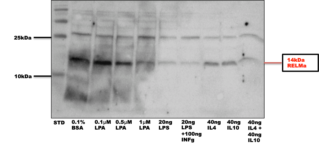
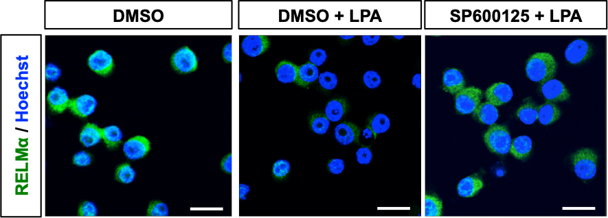
Immunocytochemistry/Immunofluorescence: FIZZ1/RELM alpha Antibody [NBP2-29355] - Staining in Mouse BV2 microglia cells, FIZZ1/RELM staining in green and nuclear staining in blue. Image from a verified customer review. Cell treatment with DMSO, DMSO+lipopolysaccharide, SMSO+SP600125 (a JNK inhibitor).![Western Blot: FIZZ1/RELM alpha AntibodyBSA Free [NBP2-29355]](https://resources.rndsystems.com/images/products/FIZZ1-RELM-alpha-Antibody-Western-Blot-NBP2-29355-img0004.jpg "Western Blot: FIZZ1/RELM alpha AntibodyBSA Free [NBP2-29355]")
![Western Blot: FIZZ1/RELM alpha AntibodyBSA Free [NBP2-29355]](https://resources.rndsystems.com/images/products/RELM-alpha-Antibody-Western-Blot-NBP2-29355-img0001.jpg "Western Blot: FIZZ1/RELM alpha AntibodyBSA Free [NBP2-29355]")
Western Blot: FIZZ1/RELM alpha AntibodyBSA Free [NBP2-29355]
Western Blot: RELM alpha Antibody [NBP2-29355] - WB detection of secreted Mouse Resistin-Like Molecule alpha (RELM alpha) from media of transfected 293T cells with #NBP2-29355 antibody used at a dilution of 1:500![Western Blot: FIZZ1/RELM alpha AntibodyBSA Free [NBP2-29355]](https://resources.rndsystems.com/images/products/FIZZ1-RELM-alpha-Antibody-Western-Blot-NBP2-29355-img0002.jpg "Western Blot: FIZZ1/RELM alpha AntibodyBSA Free [NBP2-29355]")
Western Blot: FIZZ1/RELM alpha AntibodyBSA Free [NBP2-29355]
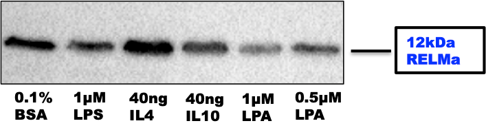
Western Blot: FIZZ1/RELM alpha Antibody [NBP2-29355] - Functional analysis mouse BV2 cell lysate. Image from a verified customer review.Applications for FIZZ1/RELM alpha Antibody - BSA Free
Application
Recommended Usage
Flow Cytometry
1:10-1:1000
Immunocytochemistry/ Immunofluorescence
1:10-1:500
Immunohistochemistry
1:10-1:500
Immunohistochemistry-Frozen
1:10-1:500
Immunohistochemistry-Paraffin
1:10-1:500
Western Blot
1:500 -1:1000
Application Notes
In Western blot this RELM antibody detects a specific target band between 10-15 kDa position (monomer form) under complete reducing conditions. Larger bands representing dimers or multimers may also be seen if WB run under non-reducing conditions. The observed molecular weight of the protein may vary from the listed predicted molecular weight due to post translational modifications, post translation cleavages, relative charges, and other experimental factors. ICC/IF and WB reported in a customer review.
Reviewed Applications
Read 3 reviews rated 4.3 using NBP2-29355 in the following applications:
Flow Cytometry Panel Builder
Bio-Techne Knows Flow Cytometry
Save time and reduce costly mistakes by quickly finding compatible reagents using the Panel Builder Tool.
Advanced Features
- Spectra Viewer - Custom analysis of spectra from multiple fluorochromes
- Spillover Popups - Visualize the spectra of individual fluorochromes
- Antigen Density Selector - Match fluorochrome brightness with antigen density

Formulation, Preparation, and Storage
Purification
Immunogen affinity purified
Formulation
PBS
Format
BSA Free
Preservative
0.02% Sodium Azide
Concentration
1 mg/ml
Shipping
The product is shipped with polar packs. Upon receipt, store it immediately at the temperature recommended below.
Stability & Storage
Store at 4C short term. Aliquot and store at -20C long term. Avoid freeze-thaw cycles.
Background: FIZZ1/RELM alpha
Long Name
Found in Inflammatory Zone 1/Resistin-like Molecule alpha
Alternate Names
RELM alpha, RETNLA, RETNLB
Entrez Gene IDs
57262 (Mouse)
Gene Symbol
RETNLA
UniProt
Additional FIZZ1/RELM alpha Products
Product Documents for FIZZ1/RELM alpha Antibody - BSA Free
Certificate of Analysis
To download a Certificate of Analysis, please enter a lot or batch number in the search box below.
Product Specific Notices for FIZZ1/RELM alpha Antibody - BSA Free
This product is for research use only and is not approved for use in humans or in clinical diagnosis. Primary Antibodies are guaranteed for 1 year from date of receipt.
Related Research Areas
Citations for FIZZ1/RELM alpha Antibody - BSA Free
 Powered by Bioz
Customer Reviews for FIZZ1/RELM alpha Antibody - BSA Free (3)
4.3 out of 5
3 Customer Ratings
Have you used FIZZ1/RELM alpha Antibody - BSA Free?
Submit a review and receive an Amazon gift card!
$25/€18/£15/$25CAN/¥2500 Yen for a review with an image
$10/€7/£6/$10CAN/¥1110 Yen for a review without an image
Submit a review
Customer Images
Showing
1
-
3 of
3 reviews
Showing All
Filter By:
-
Application: Western BlotSample Tested: BV2 microglia cellsSpecies: MouseVerified Customer | Posted 06/10/2019The antibody was used in order to study the effects of lysophosphatidic acid (LPA) on microglia polarization state. It was used in a dilution of 1:500. Using a dilution 1:1000 didn't work so well.

-
Application: ImmunocytochemistrySample Tested: BV2 microglia cellsSpecies: MouseVerified Customer | Posted 06/07/2019The antibody was used at a concentration 1:100. Having used the antibody for both ICC and WB, I would say that for ICC it worked perfectly. In WB there were some problems with the detection but still a very good antibody to use for both methods.
-
Application: Western BlotSample Tested: Murine microglia cell lysate and BV2 microglia cell lysateSpecies: MouseVerified Customer | Posted 06/07/2019The antibody was used in a concentration 1:500 in order to detect changes in expression levels in BV2 microglia cells.
There are no reviews that match your criteria.
Protocols
Find general support by application which include: protocols, troubleshooting, illustrated assays, videos and webinars.
- 7-Amino Actinomycin D (7-AAD) Cell Viability Flow Cytometry Protocol
- Antigen Retrieval Protocol (PIER)
- Antigen Retrieval for Frozen Sections Protocol
- Appropriate Fixation of IHC/ICC Samples
- Cellular Response to Hypoxia Protocols
- Chromogenic IHC Staining of Formalin-Fixed Paraffin-Embedded (FFPE) Tissue Protocol
- Chromogenic Immunohistochemistry Staining of Frozen Tissue
- ClariTSA™ Fluorophore Kits
- Detection & Visualization of Antibody Binding
- Extracellular Membrane Flow Cytometry Protocol
- Flow Cytometry Protocol for Cell Surface Markers
- Flow Cytometry Protocol for Staining Membrane Associated Proteins
- Flow Cytometry Staining Protocols
- Flow Cytometry Troubleshooting Guide
- Fluorescent IHC Staining of Frozen Tissue Protocol
- Graphic Protocol for Heat-induced Epitope Retrieval
- Graphic Protocol for the Preparation and Fluorescent IHC Staining of Frozen Tissue Sections
- Graphic Protocol for the Preparation and Fluorescent IHC Staining of Paraffin-embedded Tissue Sections
- Graphic Protocol for the Preparation of Gelatin-coated Slides for Histological Tissue Sections
- ICC Cell Smear Protocol for Suspension Cells
- ICC Immunocytochemistry Protocol Videos
- ICC for Adherent Cells
- IHC Sample Preparation (Frozen sections vs Paraffin)
- Immunocytochemistry (ICC) Protocol
- Immunocytochemistry Troubleshooting
- Immunofluorescence of Organoids Embedded in Cultrex Basement Membrane Extract
- Immunofluorescent IHC Staining of Formalin-Fixed Paraffin-Embedded (FFPE) Tissue Protocol
- Immunohistochemistry (IHC) and Immunocytochemistry (ICC) Protocols
- Immunohistochemistry Frozen Troubleshooting
- Immunohistochemistry Paraffin Troubleshooting
- Intracellular Flow Cytometry Protocol Using Alcohol (Methanol)
- Intracellular Flow Cytometry Protocol Using Detergents
- Intracellular Nuclear Staining Flow Cytometry Protocol Using Detergents
- Intracellular Staining Flow Cytometry Protocol Using Alcohol Permeabilization
- Intracellular Staining Flow Cytometry Protocol Using Detergents to Permeabilize Cells
- Preparing Samples for IHC/ICC Experiments
- Preventing Non-Specific Staining (Non-Specific Binding)
- Primary Antibody Selection & Optimization
- Propidium Iodide Cell Viability Flow Cytometry Protocol
- Protocol for Heat-Induced Epitope Retrieval (HIER)
- Protocol for Liperfluo
- Protocol for Making a 4% Formaldehyde Solution in PBS
- Protocol for VisUCyte™ HRP Polymer Detection Reagent
- Protocol for the Characterization of Human Th22 Cells
- Protocol for the Characterization of Human Th9 Cells
- Protocol for the Fluorescent ICC Staining of Cell Smears - Graphic
- Protocol for the Fluorescent ICC Staining of Cultured Cells on Coverslips - Graphic
- Protocol for the Preparation & Fixation of Cells on Coverslips
- Protocol for the Preparation and Chromogenic IHC Staining of Frozen Tissue Sections
- Protocol for the Preparation and Chromogenic IHC Staining of Frozen Tissue Sections - Graphic
- Protocol for the Preparation and Chromogenic IHC Staining of Paraffin-embedded Tissue Sections
- Protocol for the Preparation and Chromogenic IHC Staining of Paraffin-embedded Tissue Sections - Graphic
- Protocol for the Preparation and Fluorescent ICC Staining of Cells on Coverslips
- Protocol for the Preparation and Fluorescent ICC Staining of Non-adherent Cells
- Protocol for the Preparation and Fluorescent ICC Staining of Stem Cells on Coverslips
- Protocol for the Preparation and Fluorescent IHC Staining of Frozen Tissue Sections
- Protocol for the Preparation and Fluorescent IHC Staining of Paraffin-embedded Tissue Sections
- Protocol for the Preparation of Gelatin-coated Slides for Histological Tissue Sections
- Protocol for the Preparation of a Cell Smear for Non-adherent Cell ICC - Graphic
- Protocol: Annexin V and PI Staining by Flow Cytometry
- Protocol: Annexin V and PI Staining for Apoptosis by Flow Cytometry
- R&D Systems Quality Control Western Blot Protocol
- TUNEL and Active Caspase-3 Detection by IHC/ICC Protocol
- The Importance of IHC/ICC Controls
- Troubleshooting Guide: Fluorokine Flow Cytometry Kits
- Troubleshooting Guide: Immunohistochemistry
- Troubleshooting Guide: Western Blot Figures
- Western Blot Conditions
- Western Blot Protocol
- Western Blot Protocol for Cell Lysates
- Western Blot Troubleshooting
- Western Blot Troubleshooting Guide
- View all Protocols, Troubleshooting, Illustrated assays and Webinars
Loading...