![Western Blot: FoxP3 Antibody [NB600-242]](https://resources.rndsystems.com/images/products/FoxP3-Antibody-Western-Blot-NB600-242-img0010.jpg "Western Blot: FoxP3 Antibody [NB600-242]")
Key Product Details
Species Reactivity
Validated:
Cited:
Applications
Validated:
Cited:
Label
Antibody Source
Product Specifications
Immunogen
Reactivity Notes
Localization
Specificity
Clonality
Host
Isotype
Scientific Data Images for FoxP3 Antibody
Western Blot: FoxP3 Antibody [NB600-242]
Western Blot: FoxP3 Antibody [NB600-242] - (1 ug/mL) staining of Human Muscle (A), (2 ug/mL) MOLT4 (B) and (1 ug/mL) negative control Pancreas (C) lysate (35 ug protein in RIPA buffer). Detected by chemiluminescence.![Immunocytochemistry: FoxP3 Antibody [NB600-242]](https://resources.rndsystems.com/images/products/FoxP3-Antibody-Immunocytochemistry-Immunofluorescence-NB600-242-img0008.jpg "Immunocytochemistry: FoxP3 Antibody [NB600-242]")
Immunocytochemistry: FoxP3 Antibody [NB600-242]
Immunocytochemistry: FoxP3 Antibody [NB600-242] - (Treg) human blood cells gathered by cytospin and detected by FITC (A) and in phase contrast (B).![Immunohistochemistry: FoxP3 Antibody [NB600-242]](https://resources.rndsystems.com/images/products/FoxP3-Antibody-Immunohistochemistry-NB600-242-img0012.jpg "Immunohistochemistry: FoxP3 Antibody [NB600-242]")
Immunohistochemistry: FoxP3 Antibody [NB600-242]
Immunohistochemistry: FoxP3 Antibody [NB600-242] - (8ug/ml) staining of paraffin embedded Human Spleen. Heat induced antigen retrieval with citrate buffer Ph 6, HRP-staining.![Flow Cytometry: FoxP3 Antibody [NB600-242]](https://resources.rndsystems.com/images/products/FoxP3-Antibody-Flow-Cytometry-NB600-242-img0009.jpg "Flow Cytometry: FoxP3 Antibody [NB600-242]")
Flow Cytometry: FoxP3 Antibody [NB600-242]
Flow Cytometry: FoxP3 Antibody [NB600-242] - Analysis of paraformaldehyde fixed Jurkat cells (blue line), permeabilized with 0.5% Triton. Primary incubation 1hr (10 ug/mL) followed by Alexa Fluor 488 secondary antibody (4 ug/mL). IgG control: Unimmunized goat IgG (black line) followed by Alexa Fluor 488 secondary antibody.![Immunofluorescence: FoxP3 Antibody [NB600-242]](https://resources.rndsystems.com/images/products/FoxP3-Antibody-Immunocytochemistry-Immunofluorescence-NB600-242-img0011.jpg "Immunofluorescence: FoxP3 Antibody [NB600-242]")
Immunofluorescence: FoxP3 Antibody [NB600-242]
Immunofluorescence: FoxP3 Antibody [NB600-242] - Paraformaldehyde fixed U2OS cells, permeabilized with 0.15% Triton. Primary incubation 1hr (10 ug/mL) followed by Alexa Fluor 488 secondary antibody (4 ug/mL), showing strong localization to nucleoplasm. The nuclear stain is DAPI (blue). Negative control: Unimmunized goat IgG (10 ug/mL) followed by Alexa Fluor 488 secondary antibody (4 ug/mL).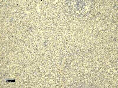
Applications for FoxP3 Antibody
Flow Cytometry
Immunohistochemistry-Paraffin
Peptide ELISA
Western Blot
Reviewed Applications
Read 1 review rated 5 using NB600-242 in the following applications:
Flow Cytometry Panel Builder
Bio-Techne Knows Flow Cytometry
Save time and reduce costly mistakes by quickly finding compatible reagents using the Panel Builder Tool.
Advanced Features
- Spectra Viewer - Custom analysis of spectra from multiple fluorochromes
- Spillover Popups - Visualize the spectra of individual fluorochromes
- Antigen Density Selector - Match fluorochrome brightness with antigen density

Formulation, Preparation, and Storage
Purification
Formulation
Preservative
Concentration
Shipping
Stability & Storage
Background: FoxP3
Long Name
Alternate Names
Entrez Gene IDs
Gene Symbol
UniProt
Additional FoxP3 Products
Product Documents for FoxP3 Antibody
Certificate of Analysis
To download a Certificate of Analysis, please enter a lot or batch number in the search box below.
Product Specific Notices for FoxP3 Antibody
This product is for research use only and is not approved for use in humans or in clinical diagnosis. Primary Antibodies are guaranteed for 1 year from date of receipt.
Related Research Areas
Citations for FoxP3 Antibody
 Powered by Bioz
Customer Reviews for FoxP3 Antibody (1)
Have you used FoxP3 Antibody?
Submit a review and receive an Amazon gift card!
$25/€18/£15/$25CAN/¥2500 Yen for a review with an image
$10/€7/£6/$10CAN/¥1110 Yen for a review without an image
Submit a review
Customer Images
-
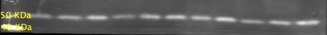
Application: Western BlotSample Tested: PC12 cell whole lysateSpecies: RatVerified Customer | Posted 10/26/2018Whole cell lysate of PC12 cells transfected with the FOXP3 plasmid. Band size is around 50 KDa.

There are no reviews that match your criteria.
Protocols
Find general support by application which include: protocols, troubleshooting, illustrated assays, videos and webinars.
- 7-Amino Actinomycin D (7-AAD) Cell Viability Flow Cytometry Protocol
- Antigen Retrieval Protocol (PIER)
- Antigen Retrieval for Frozen Sections Protocol
- Appropriate Fixation of IHC/ICC Samples
- Cellular Response to Hypoxia Protocols
- Chromogenic IHC Staining of Formalin-Fixed Paraffin-Embedded (FFPE) Tissue Protocol
- Chromogenic Immunohistochemistry Staining of Frozen Tissue
- ClariTSA™ Fluorophore Kits
- Detection & Visualization of Antibody Binding
- ELISA Sample Preparation & Collection Guide
- ELISA Troubleshooting Guide
- Extracellular Membrane Flow Cytometry Protocol
- Flow Cytometry Protocol for Cell Surface Markers
- Flow Cytometry Protocol for Staining Membrane Associated Proteins
- Flow Cytometry Staining Protocols
- Flow Cytometry Troubleshooting Guide
- Fluorescent IHC Staining of Frozen Tissue Protocol
- Graphic Protocol for Heat-induced Epitope Retrieval
- Graphic Protocol for the Preparation and Fluorescent IHC Staining of Frozen Tissue Sections
- Graphic Protocol for the Preparation and Fluorescent IHC Staining of Paraffin-embedded Tissue Sections
- Graphic Protocol for the Preparation of Gelatin-coated Slides for Histological Tissue Sections
- How to Run an R&D Systems DuoSet ELISA
- How to Run an R&D Systems Quantikine ELISA
- How to Run an R&D Systems Quantikine™ QuicKit™ ELISA
- ICC Cell Smear Protocol for Suspension Cells
- ICC Immunocytochemistry Protocol Videos
- ICC for Adherent Cells
- IHC Sample Preparation (Frozen sections vs Paraffin)
- Immunocytochemistry (ICC) Protocol
- Immunocytochemistry Troubleshooting
- Immunofluorescence of Organoids Embedded in Cultrex Basement Membrane Extract
- Immunofluorescent IHC Staining of Formalin-Fixed Paraffin-Embedded (FFPE) Tissue Protocol
- Immunohistochemistry (IHC) and Immunocytochemistry (ICC) Protocols
- Immunohistochemistry Frozen Troubleshooting
- Immunohistochemistry Paraffin Troubleshooting
- Intracellular Flow Cytometry Protocol Using Alcohol (Methanol)
- Intracellular Flow Cytometry Protocol Using Detergents
- Intracellular Nuclear Staining Flow Cytometry Protocol Using Detergents
- Intracellular Staining Flow Cytometry Protocol Using Alcohol Permeabilization
- Intracellular Staining Flow Cytometry Protocol Using Detergents to Permeabilize Cells
- Preparing Samples for IHC/ICC Experiments
- Preventing Non-Specific Staining (Non-Specific Binding)
- Primary Antibody Selection & Optimization
- Propidium Iodide Cell Viability Flow Cytometry Protocol
- Protocol for Heat-Induced Epitope Retrieval (HIER)
- Protocol for Liperfluo
- Protocol for Making a 4% Formaldehyde Solution in PBS
- Protocol for VisUCyte™ HRP Polymer Detection Reagent
- Protocol for the Characterization of Human Th22 Cells
- Protocol for the Characterization of Human Th9 Cells
- Protocol for the Fluorescent ICC Staining of Cell Smears - Graphic
- Protocol for the Fluorescent ICC Staining of Cultured Cells on Coverslips - Graphic
- Protocol for the Preparation & Fixation of Cells on Coverslips
- Protocol for the Preparation and Chromogenic IHC Staining of Frozen Tissue Sections
- Protocol for the Preparation and Chromogenic IHC Staining of Frozen Tissue Sections - Graphic
- Protocol for the Preparation and Chromogenic IHC Staining of Paraffin-embedded Tissue Sections
- Protocol for the Preparation and Chromogenic IHC Staining of Paraffin-embedded Tissue Sections - Graphic
- Protocol for the Preparation and Fluorescent ICC Staining of Cells on Coverslips
- Protocol for the Preparation and Fluorescent ICC Staining of Non-adherent Cells
- Protocol for the Preparation and Fluorescent ICC Staining of Stem Cells on Coverslips
- Protocol for the Preparation and Fluorescent IHC Staining of Frozen Tissue Sections
- Protocol for the Preparation and Fluorescent IHC Staining of Paraffin-embedded Tissue Sections
- Protocol for the Preparation of Gelatin-coated Slides for Histological Tissue Sections
- Protocol for the Preparation of a Cell Smear for Non-adherent Cell ICC - Graphic
- Protocol: Annexin V and PI Staining by Flow Cytometry
- Protocol: Annexin V and PI Staining for Apoptosis by Flow Cytometry
- Quantikine HS ELISA Kit Assay Principle, Alkaline Phosphatase
- Quantikine HS ELISA Kit Principle, Streptavidin-HRP Polymer
- R&D Systems Quality Control Western Blot Protocol
- Sandwich ELISA (Colorimetric) – Biotin/Streptavidin Detection Protocol
- Sandwich ELISA (Colorimetric) – Direct Detection Protocol
- TUNEL and Active Caspase-3 Detection by IHC/ICC Protocol
- The Importance of IHC/ICC Controls
- Troubleshooting Guide: ELISA
- Troubleshooting Guide: Fluorokine Flow Cytometry Kits
- Troubleshooting Guide: Immunohistochemistry
- Troubleshooting Guide: Western Blot Figures
- Western Blot Conditions
- Western Blot Protocol
- Western Blot Protocol for Cell Lysates
- Western Blot Troubleshooting
- Western Blot Troubleshooting Guide
- View all Protocols, Troubleshooting, Illustrated assays and Webinars
FAQs for FoxP3 Antibody
-
Q: Does FoxP3 antibodies comes in lyophilized form?
A: FoxP3 antibodies, aka JM2 IPEX, and Scurfin antibodies, have the following products in lyophilized form: NBP1-18319, AF3240.
-
Q: What is the immunogen sequence of this FoxP3 antibody?
A: A sequence within the C-terminal region (SQRPSRCSNPTPGP) of FoxP3.
-
Q: What the theoretical molecular weight for FoxP3 antibodies?
A: The TMW of FoxP3 antibodies is approximately 47 - 52 kDa.
-
Q: Does FoxP3 antibodies comes in lyophilized form?
A: FoxP3 antibodies, aka JM2 IPEX, and Scurfin antibodies, have the following products in lyophilized form: NBP1-18319, AF3240.
-
Q: What is the immunogen sequence of this FoxP3 antibody?
A: A sequence within the C-terminal region (SQRPSRCSNPTPGP) of FoxP3.
-
Q: What the theoretical molecular weight for FoxP3 antibodies?
A: The TMW of FoxP3 antibodies is approximately 47 - 52 kDa.
Associated Pathways

