GAPDH Antibody (6C5cc) - BSA Free
Novus Biologicals | Catalog # NB600-502
![Western Blot: GAPDH Antibody (6C5cc) [NB600-502]](https://resources.rndsystems.com/images/products/GAPDH-Antibody-6C5-Western-Blot-NB600-502-img0007.jpg "Western Blot: GAPDH Antibody (6C5cc) [NB600-502]")
Key Product Details
Species Reactivity
Validated:
Human, Mouse, Rat, Porcine, Amphibian, Bovine (Negative), Canine, Chinese Hamster, Feline, Fish, Goat (Negative), Rabbit
Cited:
Human, Mouse, Rat, Amphibian, Hamster - Cricetulus (Chinese Hamster), Rabbit
Applications
Validated:
Immunohistochemistry, Immunohistochemistry-Paraffin, Western Blot, ELISA, Immunoassay, Immunocytochemistry/ Immunofluorescence, Simple Western, Immunoprecipitation
Cited:
Immunohistochemistry-Paraffin, Western Blot, Block/Neutralize, Immunocytochemistry/ Immunofluorescence, Simple Western, Immunoprecipitation
Label
Unconjugated
Antibody Source
Monoclonal Mouse IgG1 Clone # 6C5cc
Format
BSA Free
Loading...
Product Specifications
Immunogen
Hybridoma clone has been derived from hybridization of Sp2/0 myeloma cells with spleen cells of Balb/c mice immunized with Rabbit GAPDH.
Reactivity Notes
Please note that this antibody is reactive to Mouse and derived from the same host, Mouse. Additional Mouse on Mouse blocking steps may be required for IHC and ICC experiments. Please contact Technical Support for more information. Chinese Hamster reactivity reported in scientific literature (PMID: 26115091).
Localization
Cytoplasmic, Nucleus, Membrane.
Clonality
Monoclonal
Host
Mouse
Isotype
IgG1
Theoretical MW
36 kDa.
Disclaimer note: The observed molecular weight of the protein may vary from the listed predicted molecular weight due to post translational modifications, post translation cleavages, relative charges, and other experimental factors.
Disclaimer note: The observed molecular weight of the protein may vary from the listed predicted molecular weight due to post translational modifications, post translation cleavages, relative charges, and other experimental factors.
Scientific Data Images for GAPDH Antibody (6C5cc) - BSA Free
Western Blot: GAPDH Antibody (6C5cc) [NB600-502]
GAPDH-Antibody-6C5-Western-Blot-NB600-502-img0007.jpg![Western Blot: GAPDH Antibody (6C5cc) [NB600-502]](https://resources.rndsystems.com/images/products/GAPDH-Antibody-6C5-Western-Blot-NB600-502-img0001.jpg "Western Blot: GAPDH Antibody (6C5cc) [NB600-502]")
Western Blot: GAPDH Antibody (6C5cc) [NB600-502]
Western Blot: GAPDH Antibody (6C5) [NB600-502] - Western blot analysis of GAPDH expression in 1) HeLa, 2) NTERA-2, 3) A-431, 4) HepG2, 5) MCF-7, 6) NIH 3T3, 7) PC-12 and 8) COS-7 whole cell lysates. Theoretical molecular weight: 36 kDa.![Western Blot: GAPDH Antibody (6C5cc) [NB600-502]](https://resources.rndsystems.com/images/products/GAPDH-Antibody-6C5-Western-Blot-NB600-502-img0006.jpg "Western Blot: GAPDH Antibody (6C5cc) [NB600-502]")
Western Blot: GAPDH Antibody (6C5cc) [NB600-502]
Western Blot: GAPDH Antibody (6C5) [NB600-502] - Western blot of FGF23 in the kidney. GAPDH antibody (6C5) served as an internal control. Each value represents the mean +/- SEM. Citation: Sugiura H, Matsushita A, Futaya M, Teraoka A, Akiyama K-i, Usui N, et al. (2018) Fibroblast growth factor 23 is upregulated in the kidney in a chronic kidney disease rat model. PLoS ONE 13(3): e0191706. https://doi.org/10.1371/journal.pone.0191706![Simple Western: GAPDH Antibody (6C5cc) [NB600-502]](https://resources.rndsystems.com/images/products/GAPDH-Antibody-6C5-Simple-Western-NB600-502-img0003.jpg "Simple Western: GAPDH Antibody (6C5cc) [NB600-502]")
Simple Western: GAPDH Antibody (6C5cc) [NB600-502]
Simple Western: GAPDH Antibody (6C5) [NB600-502] - Simple Western lane view shows a specific band for GAPDH in 0.5 mg/ml of HeLa lysate. This experiment was performed under reducing conditions using the 12-230 kDa separation system. Note: band observed higher than predicted 36 kDa molecular weight.![Simple Western: GAPDH Antibody (6C5cc) [NB600-502]](https://resources.rndsystems.com/images/products/GAPDH-Antibody-6C5-Simple-Western-NB600-502-img0005.jpg "Simple Western: GAPDH Antibody (6C5cc) [NB600-502]")
Simple Western: GAPDH Antibody (6C5cc) [NB600-502]
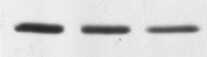
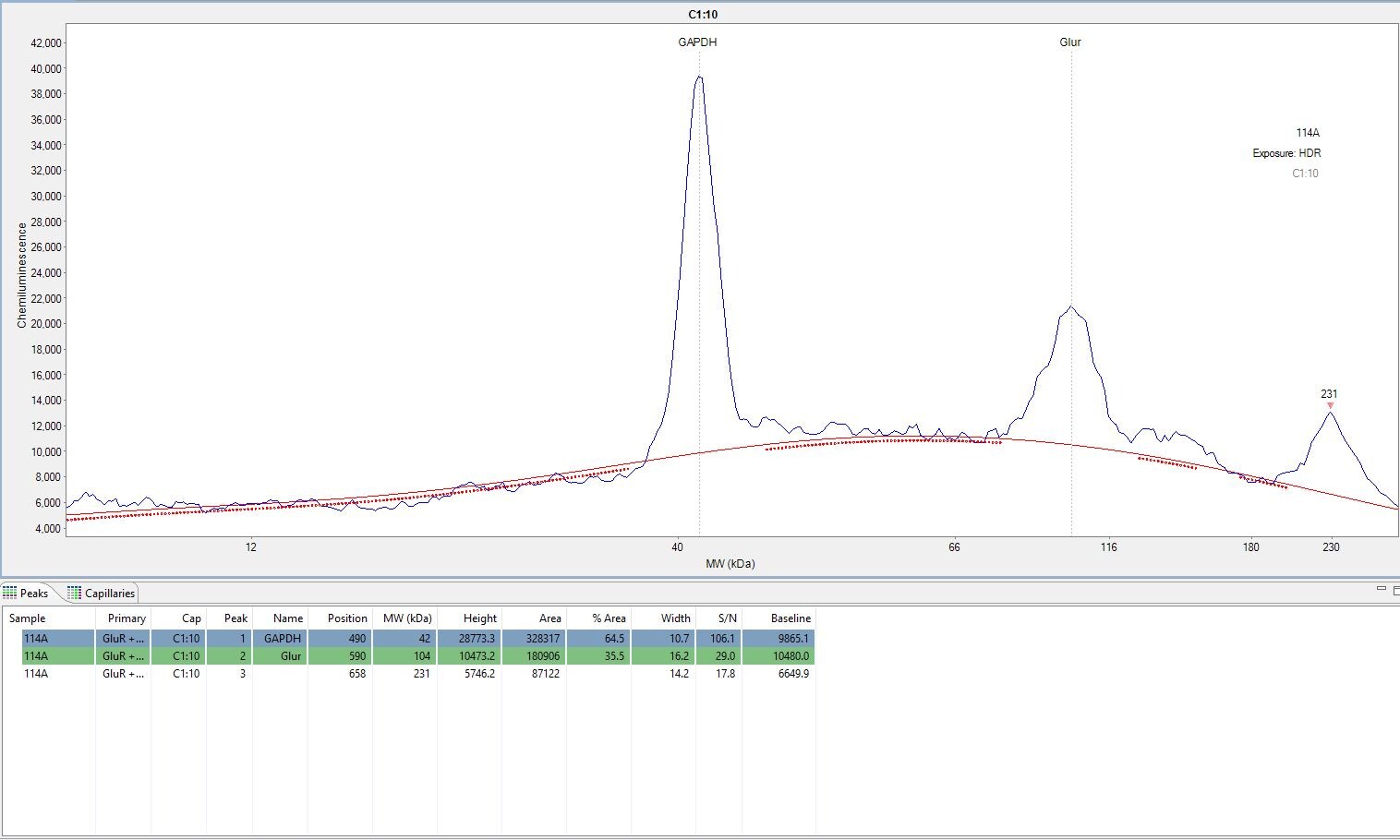
Simple Western: GAPDH Antibody (6C5) [NB600-502] - Anaylsis in rat brain and spinal cord. This image includes the GAPDH at the predicted kDa as well as a glucocorticoid receptor antibody (peak 102). Verified customer review. [NB600-502] -")
Western Blot: GAPDH Antibody (6C5cc) [NB600-502] -
Western Blot: GAPDH Antibody (6C5cc) [NB600-502] - Western blot analysis for whole cell & nuclear signal transduction proteins. (A) Phosphorylated FAK (Y397) was significantly decreased at 24 h & significantly increased at 48 h as compared to sham. (B) Nuclear localization of corresponding signal transduction molecules, p38, & p65, were decreased at 24 h & increased at 48 h as compared to sham. *p < 0.05, #p = 0.056, Data are represented as mean ± SEM, whole cell: n = 9–10/group; nuclear: n = 7–9/group. Image collected & cropped by CiteAb from the following publication (https://pubmed.ncbi.nlm.nih.gov/30853931), licensed under a CC-BY license. Not internally tested by Novus Biologicals. [NB600-502] -")
Western Blot: GAPDH Antibody (6C5cc) [NB600-502] -
Western Blot: GAPDH Antibody (6C5cc) [NB600-502] - Upregulation of innate immune response genes in BRCA2-deficient human cells. a Venn diagram of common genes upregulated in H1299 & MDA-MB-231 human cells, upon chronic (28 days) DOX-induced BRCA2 depletion. b Gene set enrichment analysis (Gene Ontology—Biological Process database) of the 28 genes upregulated upon chronic BRCA2 inactivation in both H1299 & MDA-MB-231 cells. c, d Whole-cell extracts prepared after 4 & 28 days of DOX treatment were immunoblotted as indicated. GAPDH was used as a loading control. Phosphorylation site are indicated in red Image collected & cropped by CiteAb from the following publication (https://pubmed.ncbi.nlm.nih.gov/31316060), licensed under a CC-BY license. Not internally tested by Novus Biologicals. [NB600-502] -")
Western Blot: GAPDH Antibody (6C5cc) [NB600-502] -
Western Blot: GAPDH Antibody (6C5cc) [NB600-502] - FGF23 expression in the partial nephrectomy rat model.(a) Serum FGF23 concentration. (b) FGF23 mRNA expression in the kidney. Sham group was used as a normalization control. (c) Western blot of FGF23 in the kidney. (d) Histology in the kidney. HE: hematoxylin-eosin staining. MT: Masson’s trichrome staining to evaluate fibrosis. VK: Von Kossa staining to evaluate calcification. *P<0.05, **P<0.01, ***P<0.001; sham group (n = 6), partial nephrectomy mild group (PN mild) (n = 6), partial nephrectomy severe group (PN severe) (n = 6). Image collected & cropped by CiteAb from the following publication (https://pubmed.ncbi.nlm.nih.gov/29518087), licensed under a CC-BY license. Not internally tested by Novus Biologicals. [NB600-502] -")
Western Blot: GAPDH Antibody (6C5cc) [NB600-502] -
Western Blot: GAPDH Antibody (6C5cc) [NB600-502] - FGF23 expression in hemi-nephrectomized rats fed a high-P diet.(a) Serum FGF23 concentration. (b) FGF23 mRNA expression in the bone. NP sham group was used as a normalization control. (c) FGF23 mRNA expression in the kidney. NP sham group was used as a normalization control. (d) Western blot of FGF23 in the kidney. GAPDH was used as an internal control. Each value shown represents the mean ± SEM; *P<0.05, **P<0.01; NP sham group (n = 8), NP Nx group (n = 7), HP sham group (n = 7) & HP Nx group (n = 9). Image collected & cropped by CiteAb from the following publication (https://pubmed.ncbi.nlm.nih.gov/29518087), licensed under a CC-BY license. Not internally tested by Novus Biologicals. [NB600-502] -")
Western Blot: GAPDH Antibody (6C5cc) [NB600-502] -
Western Blot: GAPDH Antibody (6C5cc) [NB600-502] - Time course of innate immune response activation in BRCA2-deficient cells. a H1299 cells expressing a DOX-inducible BRCA2 shRNA were grown in the presence or absence of DOX for 28 days. Cells collected every 2 days were subjected to quantitative RT-PCR analyses using primers specific for indicated genes. mRNA levels were expressed relative to the gene encoding beta -actin & to day 2 (2− delta delta CT). n = 3 independent experiments, each performed in triplicates. Each dot represents a single replicate. b Whole-cell extracts prepared at indicated times after DOX addition were immunoblotted as shown. GAPDH was used as a loading control. Phosphorylation sites are indicated in red Image collected & cropped by CiteAb from the following publication (https://pubmed.ncbi.nlm.nih.gov/31316060), licensed under a CC-BY license. Not internally tested by Novus Biologicals. [NB600-502] -")
Western Blot: GAPDH Antibody (6C5cc) [NB600-502] -
Western Blot: GAPDH Antibody (6C5cc) [NB600-502] - Decreased proliferation rates in ALDH2‐deficient human fibroblasts upon HR abrogationA–CHuman fibroblasts carrying either WT ALDH2 or the E487K homozygous ALDH2 mutation were transfected with control (SCR) or siRNAs against BRCA1 (A), BRCA2 (B), or RAD51 (C). Cells were processed for proliferation assays 48 h after transfection. siRNAs were retransfected at an interval of 4 days. Graphs are representative of three independent experiments, each performed with three technical replicas. Error bars represent SD of three technical replica values obtained from a single experiment. Cell extracts were prepared 48 h after transfection & immunoblotted as shown. SMC1 or GAPDH were used as loading controls. Image collected & cropped by CiteAb from the following publication (https://pubmed.ncbi.nlm.nih.gov/28729482), licensed under a CC-BY license. Not internally tested by Novus Biologicals. [NB600-502] -")
Western Blot: GAPDH Antibody (6C5cc) [NB600-502] -
Western Blot: GAPDH Antibody (6C5cc) [NB600-502] - Upregulation of innate immune response genes in BRCA2-deficient human cells. a Venn diagram of common genes upregulated in H1299 & MDA-MB-231 human cells, upon chronic (28 days) DOX-induced BRCA2 depletion. b Gene set enrichment analysis (Gene Ontology—Biological Process database) of the 28 genes upregulated upon chronic BRCA2 inactivation in both H1299 & MDA-MB-231 cells. c, d Whole-cell extracts prepared after 4 & 28 days of DOX treatment were immunoblotted as indicated. GAPDH was used as a loading control. Phosphorylation site are indicated in red Image collected & cropped by CiteAb from the following publication (https://pubmed.ncbi.nlm.nih.gov/31316060), licensed under a CC-BY license. Not internally tested by Novus Biologicals. [NB600-502] -")
Western Blot: GAPDH Antibody (6C5cc) [NB600-502] -
Western Blot: GAPDH Antibody (6C5cc) [NB600-502] - CtIP is not required for degradation of the single-stranded telomeric overhang generated by TRF2 inactivationImmortalized MEFs were infected with retroviruses expressing the indicated shRNAs, followed by selection with puromycin for 72 h. Cell extracts were prepared 48 h later & analysed by Western blotting as indicated. SMC1 & GAPDH were used as loading controls. *non-specific band.MboI- & AluI-digested DNA from cells treated as in (A) was resolved by pulsed-field gel electrophoresis & probed with end-labelled (AACCCT)4 probe. Representative pulsed-field gel samples run under native & denatured conditions are shown.Quantification of the 3′ overhang in cells treated as in (B). For each sample, the ss/total DNA ratios were expressed relative to the GFP shRNA-treated control. Error bars represent SD of two independent experiments. Image collected & cropped by CiteAb from the following publication (https://pubmed.ncbi.nlm.nih.gov/25582120), licensed under a CC-BY license. Not internally tested by Novus Biologicals. [NB600-502] -")
Western Blot: GAPDH Antibody (6C5cc) [NB600-502] -
Western Blot: GAPDH Antibody (6C5cc) [NB600-502] - ISG induction in HR-deficient human cells & tumors & olaparib impact on this process. a Upregulation of innate immune response genes in BRCA2-deleted ovarian tumors (n = 4) versus tumors with median BRCA2 mRNA expression (n = 145). b Upregulation of innate immune response genes in RAD51-deleted ovarian tumors (n = 11) versus tumors with median RAD51 mRNA expression (n = 145). Dots in graphs represent individual tumors. Middle line (white), median; box limits 25 & 75 percentiles; whiskers, minimum & maximum values. c H1299 cells expressing a DOX-inducible BRCA2 shRNA were grown in the presence or absence of DOX for 4 days. Then, olaparib (1 or 10 µM) was added for 72 h, followed by quantitative RT-PCR analyses. Primers specific for the indicated genes were used. mRNA levels were expressed relative to the gene encoding beta -actin & to untreated (−DOX) control cells (2− delta delta CT). Error bars represent SD of n = 3 independent experiments. *, p < 0.05 (unpaired two-tailed t test). d Whole-cell extracts from cells treated as in (c) were immunoblotted as shown. GAPDH & SMC1 were used as loading controls. Phosphorylation sites are indicated in red. e Cells treated as in (c) were pulse-labeled with EdU for 30 min. Frequency of cells in G1, S, & G2/M stages of the cell cycle were determined using FACS analyses of EdU incorporation & propidium iodide staining. Error bars represent SD of n = 3 independent experiments Image collected & cropped by CiteAb from the following publication (https://pubmed.ncbi.nlm.nih.gov/31316060), licensed under a CC-BY license. Not internally tested by Novus Biologicals. [NB600-502] -")
Western Blot: GAPDH Antibody (6C5cc) [NB600-502] -
Western Blot: GAPDH Antibody (6C5cc) [NB600-502] - BRCA1 & CtIP mediate Ku- & LIG4-independent telomere fusionsImmortalized Brca1F/− MEFs were infected with retroviruses expressing the indicated shRNAs and/or Cre recombinase, followed by selection with puromycin for 72 h. Mitotic chromosomes isolated 48 h later were fixed & stained with a Cy3-conjugated (CCCTAA)3-PNA probe. The frequency of end-to-end chromosome-type fusions is represented as a percentage of fusions observed after TRF2 depletion. A minimum of 2,000 chromosomes were scored for each sample. Error bars represent SD of three independent experiments. P-values were calculated using an unpaired two-tailed t-test. NS, P > 0.05.Immortalized Brca1F/C61G MEFs were infected with retroviruses expressing the indicated shRNAs and/or Cre recombinase, followed by selection with puromycin for 72 h. The frequency of end-to-end chromosome-type fusions was analysed as in (A).MEFs of the indicated genotypes were infected with retroviruses expressing TRF2 and/or CtIP shRNAs, followed by selection with puromycin for 72 h. Cell extracts were prepared 48 h later & analysed by Western blotting as indicated. GAPDH was used as a loading control. *non-specific band. Cells treated as in (C) & (E) were arrested in mitosis with colcemid, & mitotic chromosomes isolated 48 h later were fixed & stained with a Cy3-conjugated (CCCTAA)3-PNA probe (D, F). The frequency of end-to-end chromosome-type fusions is represented as a percentage of fusions observed after TRF2 depletion. Error bars represent SD of two independent experiments. P-values were calculated using an unpaired two-tailed t-test. *P ≤ 0.05. Image collected & cropped by CiteAb from the following publication (https://pubmed.ncbi.nlm.nih.gov/25582120), licensed under a CC-BY license. Not internally tested by Novus Biologicals.Applications for GAPDH Antibody (6C5cc) - BSA Free
Application
Recommended Usage
Immunocytochemistry/ Immunofluorescence
1:10-1:2000
Simple Western
1:100
Western Blot
0.5-1 ug/ml
Application Notes
Use in IHC-P reported in scientific literature (PMID:34496231). Since GAPDH is expressed in all cells it is for example becoming the marker of choice for a loading control in Western Blotting.
See Simple Western Antibody Database for Simple Western validation: tested in HeLa lysate (0.5 mg/ml); antibody dilution of 1:100; separated by size; detects a band at 42 kDa
See Simple Western Antibody Database for Simple Western validation: tested in HeLa lysate (0.5 mg/ml); antibody dilution of 1:100; separated by size; detects a band at 42 kDa
Reviewed Applications
Read 4 reviews rated 4.5 using NB600-502 in the following applications:
Formulation, Preparation, and Storage
Purification
Protein A purified
Formulation
PBS (pH 7.4)
Format
BSA Free
Preservative
0.09% Sodium Azide
Concentration
Concentrations vary lot to lot. See vial label for concentration. If unlisted please contact technical services.
Shipping
The product is shipped with polar packs. Upon receipt, store it immediately at the temperature recommended below.
Stability & Storage
Store at 4C. Do not freeze.
Background: GAPDH
References
1) Barber RD, Harmer DW, Coleman RA, Clark BJ. (2005) GAPDH as a housekeeping gene: analysis of GAPDH mRNA expression in a panel of 72 human tissues. Physiol Genomics. 21(3):389-95. PMID: 15769908
2) Jia Y, Takimoto K. (2006) Mitogen-activated protein kinases control cardiac KChIP2 gene expression. Circ Res. 98(3):386-93. PMID: 16385079
3) Godsel LM, Hsieh SN, Amargo EV, Bass AE, Pascoe-McGillicuddy LT, Huen AC, Thorne ME, Gaudry CA, Park JK, Myung K, Goldman RD, Chew TL, Green KJ. (2005) Desmoplakin assembly dynamics in four dimensions: multiple phases differentially regulated by intermediate filaments and actin. J Cell Biol. 171(6):1045-59. PMID: 16365169
4) Sirover MA1. (1999) New insights into an old protein: the functional diversity of mammalian glyceraldehyde-3-phosphate dehydrogenase. Biochim Biophys Acta. 1432(2): 159-84. PMID: 10407139
5) Tristan C, Shahani N, Sedlak TW, Sawa A. (2011) The diverse functions of GAPDH: views from different subcellular compartments. Cell Signal. 23(2):317-23. PMID: 20727968
Long Name
Glyceraldehyde-3-phosphate Dehydrogenase
Alternate Names
G3PDH, anti-GAPDH 6c5, gapdh 6c5
Gene Symbol
GAPDH
UniProt
Additional GAPDH Products
Product Documents for GAPDH Antibody (6C5cc) - BSA Free
Certificate of Analysis
To download a Certificate of Analysis, please enter a lot or batch number in the search box below.
Product Specific Notices for GAPDH Antibody (6C5cc) - BSA Free
This product is for research use only and is not approved for use in humans or in clinical diagnosis. Primary Antibodies are guaranteed for 1 year from date of receipt.
Related Research Areas
Citations for GAPDH Antibody (6C5cc) - BSA Free
 Powered by Bioz
Customer Reviews for GAPDH Antibody (6C5cc) - BSA Free (4)
4.5 out of 5
4 Customer Ratings
Have you used GAPDH Antibody (6C5cc) - BSA Free?
Submit a review and receive an Amazon gift card!
$25/€18/£15/$25CAN/¥2500 Yen for a review with an image
$10/€7/£6/$10CAN/¥1110 Yen for a review without an image
Submit a review
Customer Images

-(01-mg)_NB600-502_8346.jpg)
Showing
1
-
4 of
4 reviews
Showing All
Filter By:
-
Application: Western BlotSample Tested: Epidermoid carcinoma cell line A-431Species: HumanVerified Customer | Posted 02/05/2022Human Epidermoid carcinoma cell line A-431

-
Application: Simple WesternSample Tested: brain and spinal cordSpecies: RatVerified Customer | Posted 06/04/2018This image includes the GAPDH at the predicted kDa as well as a glucocorticoid receptor antibody (peak 102)We really like how reliable this antibody is however we have noticed that the baseline on our Wes plates has been slightly elevated.
-
Verified Customer | Posted 05/30/2018Very consistent results however the baseline for the Wes was elevated (see attached photo)
-
Application: Western BlotSample Tested: 293T and HepG2 lysatesSpecies: HumanVerified Customer | Posted 06/17/2014Western blot with total lysate from 293T and HepG2 cell lines
There are no reviews that match your criteria.
Protocols
Find general support by application which include: protocols, troubleshooting, illustrated assays, videos and webinars.
- Antigen Retrieval Protocol (PIER)
- Antigen Retrieval for Frozen Sections Protocol
- Appropriate Fixation of IHC/ICC Samples
- Cellular Response to Hypoxia Protocols
- Chromogenic IHC Staining of Formalin-Fixed Paraffin-Embedded (FFPE) Tissue Protocol
- Chromogenic Immunohistochemistry Staining of Frozen Tissue
- ClariTSA™ Fluorophore Kits
- Detection & Visualization of Antibody Binding
- ELISA Sample Preparation & Collection Guide
- ELISA Troubleshooting Guide
- Fluorescent IHC Staining of Frozen Tissue Protocol
- Graphic Protocol for Heat-induced Epitope Retrieval
- Graphic Protocol for the Preparation and Fluorescent IHC Staining of Frozen Tissue Sections
- Graphic Protocol for the Preparation and Fluorescent IHC Staining of Paraffin-embedded Tissue Sections
- Graphic Protocol for the Preparation of Gelatin-coated Slides for Histological Tissue Sections
- How to Run an R&D Systems DuoSet ELISA
- How to Run an R&D Systems Quantikine ELISA
- How to Run an R&D Systems Quantikine™ QuicKit™ ELISA
- ICC Cell Smear Protocol for Suspension Cells
- ICC Immunocytochemistry Protocol Videos
- ICC for Adherent Cells
- IHC Sample Preparation (Frozen sections vs Paraffin)
- Immunocytochemistry (ICC) Protocol
- Immunocytochemistry Troubleshooting
- Immunofluorescence of Organoids Embedded in Cultrex Basement Membrane Extract
- Immunofluorescent IHC Staining of Formalin-Fixed Paraffin-Embedded (FFPE) Tissue Protocol
- Immunohistochemistry (IHC) and Immunocytochemistry (ICC) Protocols
- Immunohistochemistry Frozen Troubleshooting
- Immunohistochemistry Paraffin Troubleshooting
- Immunoprecipitation Protocol
- Preparing Samples for IHC/ICC Experiments
- Preventing Non-Specific Staining (Non-Specific Binding)
- Primary Antibody Selection & Optimization
- Protocol for Heat-Induced Epitope Retrieval (HIER)
- Protocol for Making a 4% Formaldehyde Solution in PBS
- Protocol for VisUCyte™ HRP Polymer Detection Reagent
- Protocol for the Fluorescent ICC Staining of Cell Smears - Graphic
- Protocol for the Fluorescent ICC Staining of Cultured Cells on Coverslips - Graphic
- Protocol for the Preparation & Fixation of Cells on Coverslips
- Protocol for the Preparation and Chromogenic IHC Staining of Frozen Tissue Sections
- Protocol for the Preparation and Chromogenic IHC Staining of Frozen Tissue Sections - Graphic
- Protocol for the Preparation and Chromogenic IHC Staining of Paraffin-embedded Tissue Sections
- Protocol for the Preparation and Chromogenic IHC Staining of Paraffin-embedded Tissue Sections - Graphic
- Protocol for the Preparation and Fluorescent ICC Staining of Cells on Coverslips
- Protocol for the Preparation and Fluorescent ICC Staining of Non-adherent Cells
- Protocol for the Preparation and Fluorescent ICC Staining of Stem Cells on Coverslips
- Protocol for the Preparation and Fluorescent IHC Staining of Frozen Tissue Sections
- Protocol for the Preparation and Fluorescent IHC Staining of Paraffin-embedded Tissue Sections
- Protocol for the Preparation of Gelatin-coated Slides for Histological Tissue Sections
- Protocol for the Preparation of a Cell Smear for Non-adherent Cell ICC - Graphic
- Quantikine HS ELISA Kit Assay Principle, Alkaline Phosphatase
- Quantikine HS ELISA Kit Principle, Streptavidin-HRP Polymer
- R&D Systems Quality Control Western Blot Protocol
- Sandwich ELISA (Colorimetric) – Biotin/Streptavidin Detection Protocol
- Sandwich ELISA (Colorimetric) – Direct Detection Protocol
- TUNEL and Active Caspase-3 Detection by IHC/ICC Protocol
- The Importance of IHC/ICC Controls
- Troubleshooting Guide: ELISA
- Troubleshooting Guide: Immunohistochemistry
- Troubleshooting Guide: Western Blot Figures
- Western Blot Conditions
- Western Blot Protocol
- Western Blot Protocol for Cell Lysates
- Western Blot Troubleshooting
- Western Blot Troubleshooting Guide
- View all Protocols, Troubleshooting, Illustrated assays and Webinars
FAQs for GAPDH Antibody (6C5cc) - BSA Free
Showing
1
-
5 of
8 FAQs
Showing All
-
Q: Do you offer a smaller size for the GAPDH antibodies?
A: We offer sample sizes for many of our GAPDH loading control antibodies including NB300-221.
-
Q: I need a suitable loading control antibody that I can use in western blot of rat nerve extracts, which do you recommend?
A: This GAPDH antibody (NB300-221) is a good choice for a loading control antibody and has been cited in over 180 publications and used on rat lysates with good results.
-
Q: I want to know why GAPDH is used as a loading control antibody in WB techniques.
A: Our GAPDH antibody makes a good loading control because GAPDH is a housekeeping gene essential to metabolism that is globally expressed in most tissue types and cell lines.
-
Q: I wanted to know which of the two housekeeping genes B-actin or GAPDH can be used for best results. I intend to do an experiment to determine expression of IL-6 and TNF-alpha in liver and kidney tissues.
A: For homogenized tissue, beta-actin and GAPDH are both fine.
-
Q: I'm looking for a loading control between 24kDa - 65kDa for use in simple western, we tried an actin antibody we already had but it didn't work. What can you recommend?
A: Our GAPDH antibody NB300-221 has been validated in simple western and detects around 35 kDa so I think it would be a good choice for a simple western loading control antibody based on your size requirements.
-
Q: We need a loading control antibody for WB working with HeLa cell line, will this GAPDH antibody work for that?
A: GAPDH is expressed in HeLa cell lines but whether or not it is the best option for a loading control antibody depends on the size of the protein you are interested in. GAPDH antibody is a good choice for low to mid MW targets, as GAPDH detects around 35 kDa.
-
Q: We received GAPDH antibody NB600-502 and there was only about 15µl of antibody in the tube. I was under the impression this should have been 100µl.
A: The unit size of this product is 0.1 mg with a concentration of 6.7 mg/ml. Hence the volume should be no less than 14.9 ul.
-
Q: What secondary antibody should I use for visualization of the GAPDH antibody?
A: This GAPDH antibody is raised in mouse so you would want to use an anti-mouse secondary with the dye of your choice.
-
Q: Do you offer a smaller size for the GAPDH antibodies?
A: We offer sample sizes for many of our GAPDH loading control antibodies including NB300-221.
-
Q: I need a suitable loading control antibody that I can use in western blot of rat nerve extracts, which do you recommend?
A: This GAPDH antibody (NB300-221) is a good choice for a loading control antibody and has been cited in over 180 publications and used on rat lysates with good results.
-
Q: I want to know why GAPDH is used as a loading control antibody in WB techniques.
A: Our GAPDH antibody makes a good loading control because GAPDH is a housekeeping gene essential to metabolism that is globally expressed in most tissue types and cell lines.
-
Q: I wanted to know which of the two housekeeping genes B-actin or GAPDH can be used for best results. I intend to do an experiment to determine expression of IL-6 and TNF-alpha in liver and kidney tissues.
A: For homogenized tissue, beta-actin and GAPDH are both fine.
-
Q: I'm looking for a loading control between 24kDa - 65kDa for use in simple western, we tried an actin antibody we already had but it didn't work. What can you recommend?
A: Our GAPDH antibody NB300-221 has been validated in simple western and detects around 35 kDa so I think it would be a good choice for a simple western loading control antibody based on your size requirements.
-
Q: We need a loading control antibody for WB working with HeLa cell line, will this GAPDH antibody work for that?
A: GAPDH is expressed in HeLa cell lines but whether or not it is the best option for a loading control antibody depends on the size of the protein you are interested in. GAPDH antibody is a good choice for low to mid MW targets, as GAPDH detects around 35 kDa.
-
Q: We received GAPDH antibody NB600-502 and there was only about 15µl of antibody in the tube. I was under the impression this should have been 100µl.
A: The unit size of this product is 0.1 mg with a concentration of 6.7 mg/ml. Hence the volume should be no less than 14.9 ul.
-
Q: What secondary antibody should I use for visualization of the GAPDH antibody?
A: This GAPDH antibody is raised in mouse so you would want to use an anti-mouse secondary with the dye of your choice.
Loading...