GAPDH Antibody (6C5cc) [DyLight 680]
Novus Biologicals | Catalog # NB600-502FR
Key Product Details
Validated by
Biological Validation
Species Reactivity
Validated:
Human, Mouse, Rat, Porcine, Amphibian, Bovine (Negative), Canine, Chinese Hamster, Feline, Fish, Goat (Negative), Rabbit
Cited:
Human, Mouse
Applications
Validated:
Immunohistochemistry, Immunohistochemistry-Paraffin, Western Blot, ELISA, Immunoassay, Immunocytochemistry/ Immunofluorescence, Immunoprecipitation
Cited:
Western Blot
Label
DyLight 680 (Excitation = 692 nm, Emission = 712 nm)
Antibody Source
Monoclonal Mouse IgG1 Clone # 6C5cc
Loading...
Product Specifications
Immunogen
Hybridoma clone has been derived from hybridization of Sp2/0 myeloma cells with spleen cells of Balb/c mice immunized with Rabbit GAPDH.
Reactivity Notes
Please note that this antibody is reactive to Mouse and derived from the same host, Mouse. Additional Mouse on Mouse blocking steps may be required for IHC and ICC experiments. Please contact Technical Support for more information. Chinese Hamster reactivity reported in scientific literature (PMID: 26115091).
Localization
Cytoplasmic, Nucleus, Membrane.
Clonality
Monoclonal
Host
Mouse
Isotype
IgG1
Theoretical MW
36 kDa.
Disclaimer note: The observed molecular weight of the protein may vary from the listed predicted molecular weight due to post translational modifications, post translation cleavages, relative charges, and other experimental factors.
Disclaimer note: The observed molecular weight of the protein may vary from the listed predicted molecular weight due to post translational modifications, post translation cleavages, relative charges, and other experimental factors.
Scientific Data Images for GAPDH Antibody (6C5cc) [DyLight 680]
GAPDH-Antibody-6C5cc-DyLight-680-Western-Blot-NB600-502FR-img0002.jpg
![GAPDH Antibody (6C5cc) [DyLight 680]](https://resources.rndsystems.com/images/products/nb600-502fr_mouse-monoclonal-gapdh-antibody-6c5cc-dylight-680-31020241618910.jpg "Western Blot: GAPDH Antibody (6C5cc) [DyLight 680] [NB600-502FR] -")
Western Blot: GAPDH Antibody (6C5cc) [DyLight 680] [NB600-502FR] -
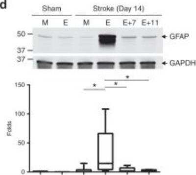
Western Blot: GAPDH Antibody (6C5cc) [DyLight 680] [NB600-502FR] - Effective targeting of EcoHIV diminishes post-ischemic stroke inflammation. Mice were infected, treated with ART-7 & ART-11, & subjected to ischemic stroke as in Fig. 6b, c. mRNA levels of cellular activation markers Iba1 (a) & GFAP (b) 7 days post stroke. Protein expression levels of Iba1 (c), GFAP (d), & ICAM1 (e) were quantified by immunoblotting in sham & ischemic stroke animals at day 14 post-stroke in mock (M) & EcoHIV-infected (E) mice that were treated with ART-7 (E + 7) or ART-11 (E + 11). Representative blots are shown, & quantified results are illustrated on the bar graphs. mRNA levels of anti-inflammatory markers ICAM-5 (f) & FoxP3 (g), & tissue degrading enzymes MMP2 (h) & MMP9 (i) were quantified by RT-qPCR. Impact of therapy on viral DNA genome levels was evaluated in ipsilateral hemisphere (j) & spleen (k); n = 5–16 mice per group, 2 independent experiments. Whiskers-box plots represent centerline median, with interquartile range & min-max whiskers. Other graphs represent data as mean & SEM with individual data points. Source data are provided as a Source Data file. *p < 0.05 or **p < 0.01; one-way ANOVA, followed by Tukey multiple comparison test (a, b & f–k), & unpaired t test (c–e) Image collected & cropped by CiteAb from the following publication (https://pubmed.ncbi.nlm.nih.gov/31043599), licensed under a CC-BY license. Not internally tested by Novus Biologicals.![GAPDH Antibody (6C5cc) [DyLight 680]](https://resources.rndsystems.com/images/products/nb600-502fr_mouse-monoclonal-gapdh-antibody-6c5cc-dylight-680-310202416163776.jpg "Western Blot: GAPDH Antibody (6C5cc) [DyLight 680] [NB600-502FR] -")
Western Blot: GAPDH Antibody (6C5cc) [DyLight 680] [NB600-502FR] -
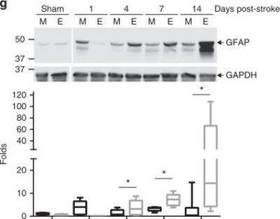
Western Blot: GAPDH Antibody (6C5cc) [DyLight 680] [NB600-502FR] - Prolonged post-ischemic stroke inflammation in EcoHIV-infected brains. Mice were infected with EcoHIV & subjected to stroke as in Fig. 1. mRNA expression of cytokines IL1 beta (a) & TNF alpha (b) chemokines CXCL1 (c) & CCL2 (d), & cellular activation markers GFAP (e) & Iba1 (f) were assessed by real-time qPCR 7 days post stroke. Sham, n = 6; Stroke, n = 12 mice per group, 3 independent experiments. Time course of GFAP (g) & Iba1 (h) protein expression levels as quantified by western blotting in sham & ischemic stroke animals at days 1, 4, 7, & 14 post-stroke in mock (M) & EcoHIV-infected (E) mice. Representative blots are shown, & quantified results are illustrated on the bar graphs. n = 5–12 mice per group, 3 independent experiments. Whiskers-box plots represent centerline median, with interquartile range & min-max whiskers. Source data are provided as a Source Data file. *p < 0.05; one-way ANOVA, followed by Tukey multiple comparison test (a–f) & unpaired t test (g, h) Image collected & cropped by CiteAb from the following publication (https://pubmed.ncbi.nlm.nih.gov/31043599), licensed under a CC-BY license. Not internally tested by Novus Biologicals.![GAPDH Antibody (6C5cc) [DyLight 680]](https://resources.rndsystems.com/images/products/nb600-502fr_mouse-monoclonal-gapdh-antibody-6c5cc-dylight-680-310202416163782.jpg "Western Blot: GAPDH Antibody (6C5cc) [DyLight 680] [NB600-502FR] -")
Western Blot: GAPDH Antibody (6C5cc) [DyLight 680] [NB600-502FR] -
Western Blot: GAPDH Antibody (6C5cc) [DyLight 680] [NB600-502FR] - Effective targeting of EcoHIV diminishes post-ischemic stroke inflammation. Mice were infected, treated with ART-7 & ART-11, & subjected to ischemic stroke as in Fig. 6b, c. mRNA levels of cellular activation markers Iba1 (a) & GFAP (b) 7 days post stroke. Protein expression levels of Iba1 (c), GFAP (d), & ICAM1 (e) were quantified by immunoblotting in sham & ischemic stroke animals at day 14 post-stroke in mock (M) & EcoHIV-infected (E) mice that were treated with ART-7 (E + 7) or ART-11 (E + 11). Representative blots are shown, & quantified results are illustrated on the bar graphs. mRNA levels of anti-inflammatory markers ICAM-5 (f) & FoxP3 (g), & tissue degrading enzymes MMP2 (h) & MMP9 (i) were quantified by RT-qPCR. Impact of therapy on viral DNA genome levels was evaluated in ipsilateral hemisphere (j) & spleen (k); n = 5–16 mice per group, 2 independent experiments. Whiskers-box plots represent centerline median, with interquartile range & min-max whiskers. Other graphs represent data as mean & SEM with individual data points. Source data are provided as a Source Data file. *p < 0.05 or **p < 0.01; one-way ANOVA, followed by Tukey multiple comparison test (a, b & f–k), & unpaired t test (c–e) Image collected & cropped by CiteAb from the following publication (https://pubmed.ncbi.nlm.nih.gov/31043599), licensed under a CC-BY license. Not internally tested by Novus Biologicals.![GAPDH Antibody (6C5cc) [DyLight 680]](https://resources.rndsystems.com/images/products/nb600-502fr_mouse-monoclonal-gapdh-antibody-6c5cc-dylight-680-310202416175249.jpg "Western Blot: GAPDH Antibody (6C5cc) [DyLight 680] [NB600-502FR] -")
Western Blot: GAPDH Antibody (6C5cc) [DyLight 680] [NB600-502FR] -
Western Blot: GAPDH Antibody (6C5cc) [DyLight 680] [NB600-502FR] - EcoHIV diminishes post-ischemic stroke NVU recovery. Mice were infected with EcoHIV & subjected to stroke as in Fig. 1. Brain sections were stained for laminin (a), ICAM-1 (b) & P-selectin (c) 24 h post stroke, & quantified for mean fluorescence index (MFI); n = 6 mice per group, 10 microvessels per mice, 2 independent experiments. d Time course of ICAM1 expression levels as quantified by western blotting in sham & ischemic stroke animals at days 1, 4, 7, & 14 post-stroke both in mock (M) & EcoHIV-infected (E) mice. Representative blots are shown, & quantified results from 5–12 samples per group are illustrated on the bar graphs. e Representative image (left) & quantified results (right) of infiltration of the infarct area by Lys6g immunoreactive cells (neutrophils) at 24 h post-ischemic stroke. The sections were also stained for MAP2 (neurons) & Hoechst (nuclei). Absence of MAP2 staining indicates infarct area. Data quantified from 6 mice per group, 2 independent experiments, 4 fields of view per mice at ×20 magnification; Z stack images. Whiskers-box plots represent centerline median, with interquartile range & min-max whiskers. Source data are provided as a Source Data file. **p < 0.01; ***p < 0.001; unpaired t test. a–c Scale bars: 40 µm; e scale bar: 320 µm Image collected & cropped by CiteAb from the following publication (https://pubmed.ncbi.nlm.nih.gov/31043599), licensed under a CC-BY license. Not internally tested by Novus Biologicals.Applications for GAPDH Antibody (6C5cc) [DyLight 680]
Application
Recommended Usage
ELISA
Optimal dilutions of this antibody should be experimentally determined.
Immunoassay
Optimal dilutions of this antibody should be experimentally determined.
Immunocytochemistry/ Immunofluorescence
Optimal dilutions of this antibody should be experimentally determined.
Immunohistochemistry
Optimal dilutions of this antibody should be experimentally determined.
Immunohistochemistry-Paraffin
Optimal dilutions of this antibody should be experimentally determined.
Immunoprecipitation
Optimal dilutions of this antibody should be experimentally determined.
Western Blot
Optimal dilutions of this antibody should be experimentally determined.
Formulation, Preparation, and Storage
Purification
Protein A purified
Formulation
50mM Sodium Borate
Preservative
0.05% Sodium Azide
Concentration
Concentrations vary lot to lot. See vial label for concentration. If unlisted please contact technical services.
Shipping
The product is shipped with polar packs. Upon receipt, store it immediately at the temperature recommended below.
Stability & Storage
Store at 4C in the dark.
Background: GAPDH
References
1) Barber RD, Harmer DW, Coleman RA, Clark BJ. (2005) GAPDH as a housekeeping gene: analysis of GAPDH mRNA expression in a panel of 72 human tissues. Physiol Genomics. 21(3):389-95. PMID: 15769908
2) Jia Y, Takimoto K. (2006) Mitogen-activated protein kinases control cardiac KChIP2 gene expression. Circ Res. 98(3):386-93. PMID: 16385079
3) Godsel LM, Hsieh SN, Amargo EV, Bass AE, Pascoe-McGillicuddy LT, Huen AC, Thorne ME, Gaudry CA, Park JK, Myung K, Goldman RD, Chew TL, Green KJ. (2005) Desmoplakin assembly dynamics in four dimensions: multiple phases differentially regulated by intermediate filaments and actin. J Cell Biol. 171(6):1045-59. PMID: 16365169
4) Sirover MA1. (1999) New insights into an old protein: the functional diversity of mammalian glyceraldehyde-3-phosphate dehydrogenase. Biochim Biophys Acta. 1432(2): 159-84. PMID: 10407139
5) Tristan C, Shahani N, Sedlak TW, Sawa A. (2011) The diverse functions of GAPDH: views from different subcellular compartments. Cell Signal. 23(2):317-23. PMID: 20727968
Long Name
Glyceraldehyde-3-phosphate Dehydrogenase
Alternate Names
G3PDH
Gene Symbol
GAPDH
UniProt
Additional GAPDH Products
Product Documents for GAPDH Antibody (6C5cc) [DyLight 680]
Certificate of Analysis
To download a Certificate of Analysis, please enter a lot or batch number in the search box below.
Product Specific Notices for GAPDH Antibody (6C5cc) [DyLight 680]
DyLight (R) is a trademark of Thermo Fisher Scientific Inc. and its subsidiaries.
This product is for research use only and is not approved for use in humans or in clinical diagnosis. Primary Antibodies are guaranteed for 1 year from date of receipt.
Related Research Areas
Citations for GAPDH Antibody (6C5cc) [DyLight 680]
 Powered by Bioz
Customer Reviews for GAPDH Antibody (6C5cc) [DyLight 680]
There are currently no reviews for this product. Be the first to review GAPDH Antibody (6C5cc) [DyLight 680] and earn rewards!
Have you used GAPDH Antibody (6C5cc) [DyLight 680]?
Submit a review and receive an Amazon gift card!
$25/€18/£15/$25CAN/¥2500 Yen for a review with an image
$10/€7/£6/$10CAN/¥1110 Yen for a review without an image
Submit a review
Protocols
Find general support by application which include: protocols, troubleshooting, illustrated assays, videos and webinars.
- Antigen Retrieval Protocol (PIER)
- Antigen Retrieval for Frozen Sections Protocol
- Appropriate Fixation of IHC/ICC Samples
- Cellular Response to Hypoxia Protocols
- Chromogenic IHC Staining of Formalin-Fixed Paraffin-Embedded (FFPE) Tissue Protocol
- Chromogenic Immunohistochemistry Staining of Frozen Tissue
- ClariTSA™ Fluorophore Kits
- Detection & Visualization of Antibody Binding
- ELISA Sample Preparation & Collection Guide
- ELISA Troubleshooting Guide
- Fluorescent IHC Staining of Frozen Tissue Protocol
- Graphic Protocol for Heat-induced Epitope Retrieval
- Graphic Protocol for the Preparation and Fluorescent IHC Staining of Frozen Tissue Sections
- Graphic Protocol for the Preparation and Fluorescent IHC Staining of Paraffin-embedded Tissue Sections
- Graphic Protocol for the Preparation of Gelatin-coated Slides for Histological Tissue Sections
- How to Run an R&D Systems DuoSet ELISA
- How to Run an R&D Systems Quantikine ELISA
- How to Run an R&D Systems Quantikine™ QuicKit™ ELISA
- ICC Cell Smear Protocol for Suspension Cells
- ICC Immunocytochemistry Protocol Videos
- ICC for Adherent Cells
- IHC Sample Preparation (Frozen sections vs Paraffin)
- Immunocytochemistry (ICC) Protocol
- Immunocytochemistry Troubleshooting
- Immunofluorescence of Organoids Embedded in Cultrex Basement Membrane Extract
- Immunofluorescent IHC Staining of Formalin-Fixed Paraffin-Embedded (FFPE) Tissue Protocol
- Immunohistochemistry (IHC) and Immunocytochemistry (ICC) Protocols
- Immunohistochemistry Frozen Troubleshooting
- Immunohistochemistry Paraffin Troubleshooting
- Immunoprecipitation Protocol
- Preparing Samples for IHC/ICC Experiments
- Preventing Non-Specific Staining (Non-Specific Binding)
- Primary Antibody Selection & Optimization
- Protocol for Heat-Induced Epitope Retrieval (HIER)
- Protocol for Making a 4% Formaldehyde Solution in PBS
- Protocol for VisUCyte™ HRP Polymer Detection Reagent
- Protocol for the Fluorescent ICC Staining of Cell Smears - Graphic
- Protocol for the Fluorescent ICC Staining of Cultured Cells on Coverslips - Graphic
- Protocol for the Preparation & Fixation of Cells on Coverslips
- Protocol for the Preparation and Chromogenic IHC Staining of Frozen Tissue Sections
- Protocol for the Preparation and Chromogenic IHC Staining of Frozen Tissue Sections - Graphic
- Protocol for the Preparation and Chromogenic IHC Staining of Paraffin-embedded Tissue Sections
- Protocol for the Preparation and Chromogenic IHC Staining of Paraffin-embedded Tissue Sections - Graphic
- Protocol for the Preparation and Fluorescent ICC Staining of Cells on Coverslips
- Protocol for the Preparation and Fluorescent ICC Staining of Non-adherent Cells
- Protocol for the Preparation and Fluorescent ICC Staining of Stem Cells on Coverslips
- Protocol for the Preparation and Fluorescent IHC Staining of Frozen Tissue Sections
- Protocol for the Preparation and Fluorescent IHC Staining of Paraffin-embedded Tissue Sections
- Protocol for the Preparation of Gelatin-coated Slides for Histological Tissue Sections
- Protocol for the Preparation of a Cell Smear for Non-adherent Cell ICC - Graphic
- Quantikine HS ELISA Kit Assay Principle, Alkaline Phosphatase
- Quantikine HS ELISA Kit Principle, Streptavidin-HRP Polymer
- R&D Systems Quality Control Western Blot Protocol
- Sandwich ELISA (Colorimetric) – Biotin/Streptavidin Detection Protocol
- Sandwich ELISA (Colorimetric) – Direct Detection Protocol
- TUNEL and Active Caspase-3 Detection by IHC/ICC Protocol
- The Importance of IHC/ICC Controls
- Troubleshooting Guide: ELISA
- Troubleshooting Guide: Immunohistochemistry
- Troubleshooting Guide: Western Blot Figures
- Western Blot Conditions
- Western Blot Protocol
- Western Blot Protocol for Cell Lysates
- Western Blot Troubleshooting
- Western Blot Troubleshooting Guide
- View all Protocols, Troubleshooting, Illustrated assays and Webinars
FAQs for GAPDH Antibody (6C5cc) [DyLight 680]
Showing
1
-
5 of
8 FAQs
Showing All
-
Q: Do you offer a smaller size for the GAPDH antibodies?
A: We offer sample sizes for many of our GAPDH loading control antibodies including NB300-221.
-
Q: I need a suitable loading control antibody that I can use in western blot of rat nerve extracts, which do you recommend?
A: This GAPDH antibody (NB300-221) is a good choice for a loading control antibody and has been cited in over 180 publications and used on rat lysates with good results.
-
Q: I want to know why GAPDH is used as a loading control antibody in WB techniques.
A: Our GAPDH antibody makes a good loading control because GAPDH is a housekeeping gene essential to metabolism that is globally expressed in most tissue types and cell lines.
-
Q: I wanted to know which of the two housekeeping genes B-actin or GAPDH can be used for best results. I intend to do an experiment to determine expression of IL-6 and TNF-alpha in liver and kidney tissues.
A: For homogenized tissue, beta-actin and GAPDH are both fine.
-
Q: I'm looking for a loading control between 24kDa - 65kDa for use in simple western, we tried an actin antibody we already had but it didn't work. What can you recommend?
A: Our GAPDH antibody NB300-221 has been validated in simple western and detects around 35 kDa so I think it would be a good choice for a simple western loading control antibody based on your size requirements.
-
Q: We need a loading control antibody for WB working with HeLa cell line, will this GAPDH antibody work for that?
A: GAPDH is expressed in HeLa cell lines but whether or not it is the best option for a loading control antibody depends on the size of the protein you are interested in. GAPDH antibody is a good choice for low to mid MW targets, as GAPDH detects around 35 kDa.
-
Q: We received GAPDH antibody NB600-502 and there was only about 15µl of antibody in the tube. I was under the impression this should have been 100µl.
A: The unit size of this product is 0.1 mg with a concentration of 6.7 mg/ml. Hence the volume should be no less than 14.9 ul.
-
Q: What secondary antibody should I use for visualization of the GAPDH antibody?
A: This GAPDH antibody is raised in mouse so you would want to use an anti-mouse secondary with the dye of your choice.
-
Q: Do you offer a smaller size for the GAPDH antibodies?
A: We offer sample sizes for many of our GAPDH loading control antibodies including NB300-221.
-
Q: I need a suitable loading control antibody that I can use in western blot of rat nerve extracts, which do you recommend?
A: This GAPDH antibody (NB300-221) is a good choice for a loading control antibody and has been cited in over 180 publications and used on rat lysates with good results.
-
Q: I want to know why GAPDH is used as a loading control antibody in WB techniques.
A: Our GAPDH antibody makes a good loading control because GAPDH is a housekeeping gene essential to metabolism that is globally expressed in most tissue types and cell lines.
-
Q: I wanted to know which of the two housekeeping genes B-actin or GAPDH can be used for best results. I intend to do an experiment to determine expression of IL-6 and TNF-alpha in liver and kidney tissues.
A: For homogenized tissue, beta-actin and GAPDH are both fine.
-
Q: I'm looking for a loading control between 24kDa - 65kDa for use in simple western, we tried an actin antibody we already had but it didn't work. What can you recommend?
A: Our GAPDH antibody NB300-221 has been validated in simple western and detects around 35 kDa so I think it would be a good choice for a simple western loading control antibody based on your size requirements.
-
Q: We need a loading control antibody for WB working with HeLa cell line, will this GAPDH antibody work for that?
A: GAPDH is expressed in HeLa cell lines but whether or not it is the best option for a loading control antibody depends on the size of the protein you are interested in. GAPDH antibody is a good choice for low to mid MW targets, as GAPDH detects around 35 kDa.
-
Q: We received GAPDH antibody NB600-502 and there was only about 15µl of antibody in the tube. I was under the impression this should have been 100µl.
A: The unit size of this product is 0.1 mg with a concentration of 6.7 mg/ml. Hence the volume should be no less than 14.9 ul.
-
Q: What secondary antibody should I use for visualization of the GAPDH antibody?
A: This GAPDH antibody is raised in mouse so you would want to use an anti-mouse secondary with the dye of your choice.
Loading...