Key Product Details
Species Reactivity
Validated:
Human, Mouse, Rat, Porcine, Alpaca, Bovine, Canine, Chicken, Equine, Feline, Rabbit
Cited:
Human, Mouse, Rat, Primate
Applications
Validated:
Immunohistochemistry, Immunohistochemistry-Paraffin, Immunohistochemistry-Frozen, Western Blot, Flow Cytometry, Immunocytochemistry/ Immunofluorescence, Simple Western
Cited:
Immunohistochemistry, Immunohistochemistry-Paraffin, Immunohistochemistry-Frozen, Western Blot, Flow Cytometry, Immunocytochemistry/ Immunofluorescence, IF/IHC
Label
Unconjugated
Antibody Source
Monoclonal Mouse IgG1 kappa Clone # GA-5
Loading...
Product Specifications
Immunogen
GFAP isolated from pig spinal cord (Uniprot: P14136)
Reactivity Notes
Alpaca, Feline, Canine, and Equine reactivity reported from a verified customer review.
Localization
Cytoplasmic
Marker
Astrocyte & Neural Stem Cell Marker
Specificity
This monoclonal antibody recognizes a protein of ~50kDa which is identified as Glial Fibrillary Acidic Protein (GFAP). It shows no cross-reaction with other intermediate filament proteins. GFAP is specifically found in astroglia. GFAP is a very popular marker for localizing benign astrocyte and neoplastic cells of glial origin in the central nervous system. Antibody to GFAP is useful in differentiating primary gliomas from metastatic lesions in the brain and for documenting astrocytic differentiation in tumors outside the CNS.
Clonality
Monoclonal
Host
Mouse
Isotype
IgG1 kappa
Theoretical MW
50 kDa.
Disclaimer note: The observed molecular weight of the protein may vary from the listed predicted molecular weight due to post translational modifications, post translation cleavages, relative charges, and other experimental factors.
Disclaimer note: The observed molecular weight of the protein may vary from the listed predicted molecular weight due to post translational modifications, post translation cleavages, relative charges, and other experimental factors.
Description
200ug/ml of antibody purified from Bioreactor Concentrate by Protein A or G. Prepared in 10 mM PBS with 0.05% BSA & 0.05% azide. Also available WITHOUT BSA & azide at 1.0 mg/ml. (NBP2-33184)
Antibody with azide - store at 2 to 8C. Antibody without azide - store at -20 to -80C.
Antibody with azide - store at 2 to 8C. Antibody without azide - store at -20 to -80C.
Scientific Data Images for GFAP Antibody (GA-5)
![Immunocytochemistry/ Immunofluorescence: GFAP Antibody (GA-5) [NBP2-29415]](https://resources.rndsystems.com/images/products/GFAP-Antibody-GA-5-Immunocytochemistry-Immunofluorescence-NBP2-29415-img0007.jpg "Immunocytochemistry/ Immunofluorescence: GFAP Antibody (GA-5) [NBP2-29415]")
Immunocytochemistry/ Immunofluorescence: GFAP Antibody (GA-5) [NBP2-29415]
Immunocytochemistry/Immunofluorescence: GFAP Antibody (GA-5) [NBP2-29415] - Cultured Rat Hippocampal Neurons. ICC/IF image submitted by a verified customer review.![Western Blot: GFAP Antibody (GA-5) [NBP2-29415]](https://resources.rndsystems.com/images/products/GFAP-Antibody-GA-5-Western-Blot-NBP2-29415-img0011.jpg "Western Blot: GFAP Antibody (GA-5) [NBP2-29415]")
Western Blot: GFAP Antibody (GA-5) [NBP2-29415]
Western Blot: GFAP Antibody (GA-5) [NBP2-29415] - Western Blot Analysis of human brain tissue lysate using GFAP Antibody (GA-5).![Immunohistochemistry-Paraffin: GFAP Antibody (GA-5) [NBP2-29415]](https://resources.rndsystems.com/images/products/GFAP-Antibody-GA-5-Immunohistochemistry-Paraffin-NBP2-29415-img0010.jpg "Immunohistochemistry-Paraffin: GFAP Antibody (GA-5) [NBP2-29415]")
Immunohistochemistry-Paraffin: GFAP Antibody (GA-5) [NBP2-29415]
Immunohistochemistry-Paraffin: GFAP Antibody (GA-5) [NBP2-29415] - Formalin-fixed, paraffin-embedded human Cerebellum stained with GFAP Antibody (GA-5).![Flow Cytometry: GFAP Antibody (GA-5) [NBP2-29415]](https://resources.rndsystems.com/images/products/GFAP-Antibody-GA-5-Flow-Cytometry-NBP2-29415-img0009.jpg "Flow Cytometry: GFAP Antibody (GA-5) [NBP2-29415]")
Flow Cytometry: GFAP Antibody (GA-5) [NBP2-29415]
Flow Cytometry: GFAP Antibody (GA-5) [NBP2-29415] - Flow Cytometric Analysis of T98G cells using GFAP Antibody (GA-5) followed by Goat anti-Mouse IgG-CF488 (Blue); Isotype Control (Red).![Western Blot: GFAP Antibody (GA-5) [NBP2-29415]](https://resources.rndsystems.com/images/products/GFAP-Antibody-GA-5-Western-Blot-NBP2-29415-img0001.jpg "Western Blot: GFAP Antibody (GA-5) [NBP2-29415]")
Western Blot: GFAP Antibody (GA-5) [NBP2-29415]
Western Blot: GFAP Antibody (GA-5) [NBP2-29415] - Analysis of GFAP in human brain lysate using GFAP (GA5) antibody at 1 ug/mL. Goat anti-mouse Ig HRP secondary antibody and PicoTect ECL substrate solution were used for this test.![Immunohistochemistry-Paraffin: GFAP Antibody (GA-5) [NBP2-29415]](https://resources.rndsystems.com/images/products/GFAP-Antibody-GA-5-Immunohistochemistry-Paraffin-NBP2-29415-img0003.jpg "Immunohistochemistry-Paraffin: GFAP Antibody (GA-5) [NBP2-29415]")
Immunohistochemistry-Paraffin: GFAP Antibody (GA-5) [NBP2-29415]
Immunohistochemistry-Paraffin: GFAP Antibody (GA-5) [NBP2-29415] - Formalin-paraffin human brain stained with GFAP Ab (GA-5). Note cytoplasmic staining.![Flow Cytometry: GFAP Antibody (GA-5) [NBP2-29415]](https://resources.rndsystems.com/images/products/GFAP-Antibody-GA-5-Flow-Cytometry-NBP2-29415-img0008.jpg "Flow Cytometry: GFAP Antibody (GA-5) [NBP2-29415]")
Flow Cytometry: GFAP Antibody (GA-5) [NBP2-29415]
Flow Cytometry: GFAP Antibody (GA-5) [NBP2-29415] - Experimental autoimmune encephalomyelitis was induced in C57BL6/J mice, and mononuclear cells were isolated from the CNS at day 10 (onset of symptoms). Cells were stained for GFAP, Neun, CX3CL1, CXCL12, CCL2, CD45 and CD11b, plus for viability to exclude dead cells. GFAP staining is shown for viable cells. Flow cytometry image submitted by a verified customer review.![Simple Western: GFAP Antibody (GA-5) [NBP2-29415]](https://resources.rndsystems.com/images/products/GFAP-Antibody-GA-5-Simple-Western-NBP2-29415-img0004.jpg "Simple Western: GFAP Antibody (GA-5) [NBP2-29415]")
Simple Western: GFAP Antibody (GA-5) [NBP2-29415]
Simple Western: GFAP Antibody (GA-5) [NBP2-29415] - Simple Western lane view shows a specific band for GFAP in 0.2 mg/mL of human brain lysate. This experiment was performed under reducing conditions using the 12-230 kDa separation system.![Simple Western: GFAP Antibody (GA-5) [NBP2-29415]](https://resources.rndsystems.com/images/products/GFAP-Antibody-GA-5-Simple-Western-NBP2-29415-img0006.jpg "Simple Western: GFAP Antibody (GA-5) [NBP2-29415]")
Simple Western: GFAP Antibody (GA-5) [NBP2-29415]
Simple Western: GFAP Antibody (GA-5) [NBP2-29415] - Electropherogram image of the corresponding Simple Western lane. GFAP antibody was used at 10 ug/mL dilution of human brain lysate. [NBP2-29415] -")
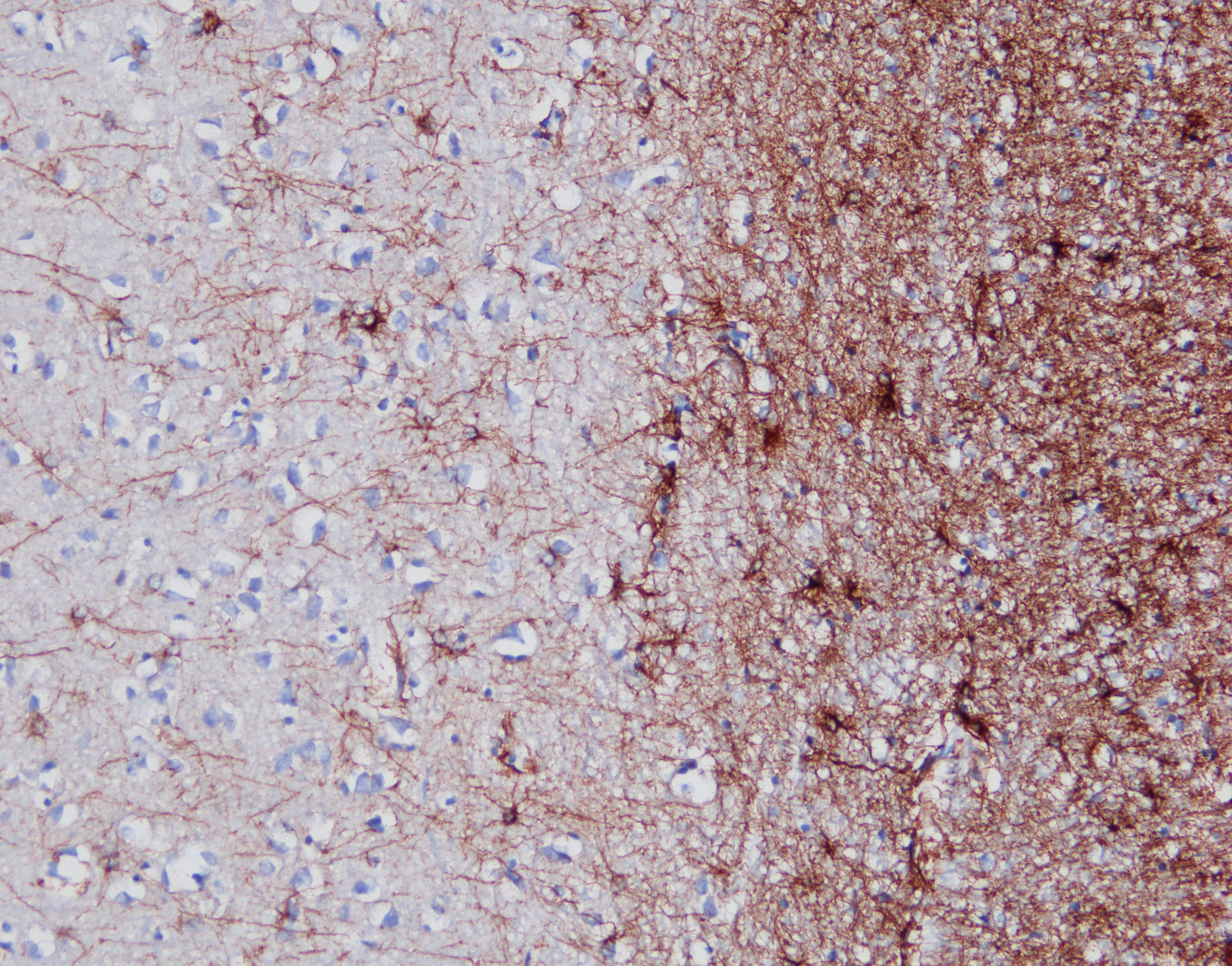
Immunohistochemistry-Paraffin: Mouse Monoclonal GFAP Antibody (GA-5) [NBP2-29415] -
Immunohistochemistry-Paraffin: Mouse Monoclonal GFAP Antibody (GA-5) [NBP2-29415] - Analysis of GFAP on adult human brain tissue. Antigen retrieval in a basic buffer x200 (1ug/mL). Image from a verified customer review. [NBP2-29415] -")
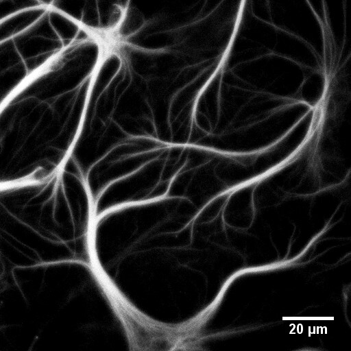
Immunocytochemistry/ Immunofluorescence: GFAP Antibody (GA-5) [NBP2-29415] -
Immunocytochemistry/ Immunofluorescence: GFAP Antibody (GA-5) [NBP2-29415] - Advancing super‐resolution microscopy for astroglial research. (A) STED images showing CA1 stratum radiatum astrocytic processes (green, whole‐cell loading with Alexa Fluor 488) adjacent to synaptic structures in organotypic slices (red; Thy1‐YFP; dendritic spines, arrows) at lower (A1) & higher (A2, area indicted by square in A1) magnification; asterisk, O‐ring structures indicating tentative cell process envelopes; adapted from (Panatier et al., 2014). (B) STED image of P2Y1 receptors (red) along a multi‐branched astrocytic process (glutamine synthase, grey) in the adult mouse hippocampus. Adapted from (Volterra et al., 2014). (C) STORM imaging of pre‐ (Bassoon; red) & postsynaptic (Homer1; green) scaffolding proteins in the mouse main olfactory bulb glomeruli imaged using conventional fluorescence imaging (C1) & STORM (C2); adapted from (Dani et al., 2010). (D, E) dSTORM images of cultured (14 DIV) mixed glial cells from rat hippocampus (ProLong Diamond in Zeiss Elyra PS.1 microscope; Fiji Plugin ThunderSTORM, 3,000 frames); unpublished data by J. Heller. (D) GFAP stained with monoclonal antibody (Novus, GA5, secondary Alexa Fluor 488 donkey anti‐mouse antibody, Life Technologies) shown at lower (D1) & higher (D2, fragment indicated in D1) magnification. (E) GLT‐1 (polyclonal, Millipore) visualized with Alexa Fluor 568 goat anti‐guinea pig antibody (Life Technologies) shown at lower (E1) & higher (E2, fragment indicated in E1) magnification. Note that the cells were permeabilized, & therefore GLT‐1 was stained throughout cellular compartments. Image collected & cropped by CiteAb from the following publication (https://pubmed.ncbi.nlm.nih.gov/25782611), licensed under a CC-BY license. Not internally tested by Novus Biologicals. [NBP2-29415]")
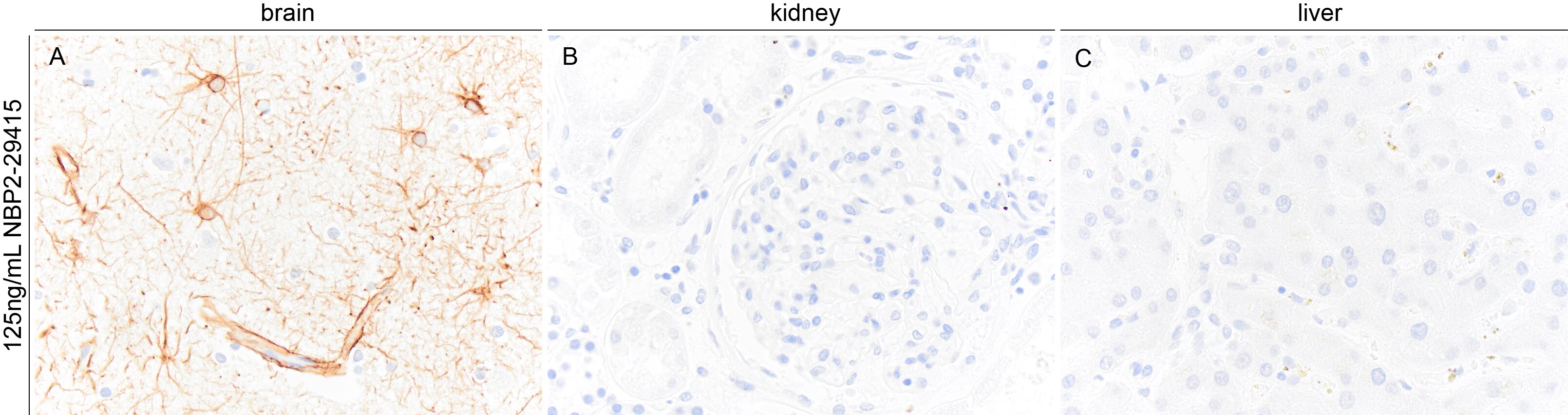
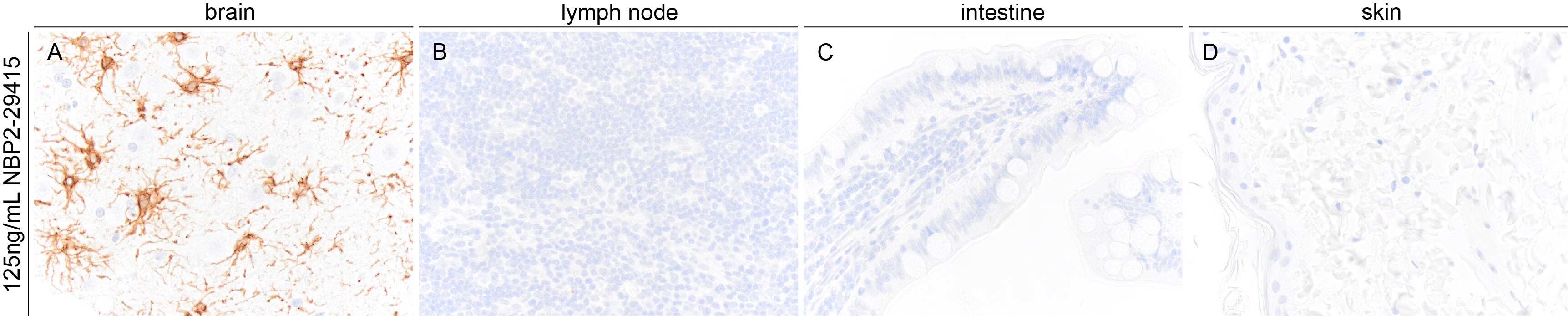
Immunohistochemistry-Paraffin - Mouse Monoclonal GFAP Antibody (GA-5) [NBP2-29415]
Images demonstrating GFAP immunoreactivity in a variety of human FFPE tissue sections. NBP2-29415 was used at a concentration of 125ng/mL and was left on tissue sections for 30m at room temperature. Heat induced epitope retrieval with a citrate-based buffer was used. Image from a verified customer review. [NBP2-29415]")
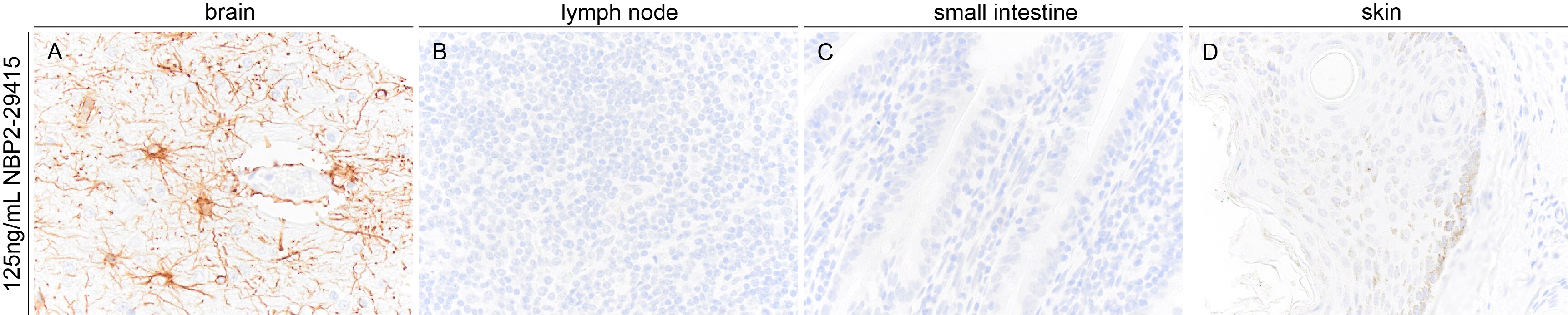
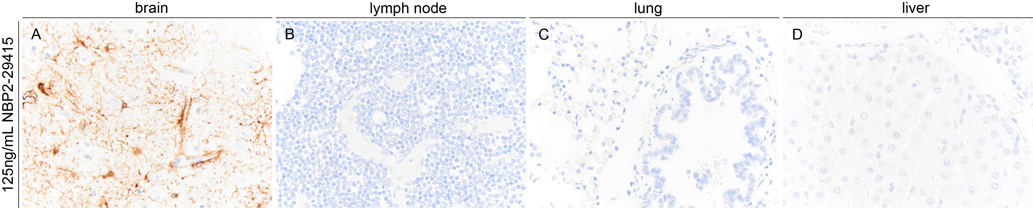
Immunohistochemistry-Paraffin: Mouse Monoclonal GFAP Antibody (GA-5) [NBP2-29415]
Images demonstrating GFAP immunoreactivity in a variety of horse FFPE tissue sections. NBP2-29415 was used at a concentration of 125ng/mL and was left on tissue sections for 30m at room temperature. Heat induced epitope retrieval with a citrate-based buffer was used. Image from a verified customer review. [NBP2-29415]")
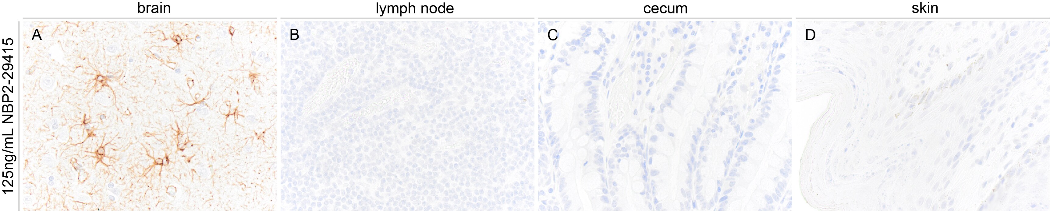
Immunohistochemistry-Paraffin: Mouse Monoclonal GFAP Antibody (GA-5) [NBP2-29415]
Images demonstrating GFAP immunoreactivity in a variety of dog FFPE tissue sections. NBP2-29415 was used at a concentration of 125ng/mL and was left on tissue sections for 30m at room temperature. Heat induced epitope retrieval with a citrate-based buffer was used. Image from a verified customer review. [NBP2-29415]")
Immunohistochemistry-Paraffin: Mouse Monoclonal GFAP Antibody (GA-5) [NBP2-29415]
Images demonstrating GFAP immunoreactivity in a variety of cat FFPE tissue sections. NBP2-29415 was used at a concentration of 125ng/mL and was left on tissue sections for 30m at room temperature. Heat induced epitope retrieval with a citrate-based buffer was used. Image from a verified customer review. [NBP2-29415]")
Immunohistochemistry-Paraffin: Mouse Monoclonal GFAP Antibody (GA-5) [NBP2-29415]
Images demonstrating GFAP immunoreactivity in a variety of alpaca FFPE tissue sections. NBP2-29415 was used at a concentration of 125ng/mL and was left on tissue sections for 30m at room temperature. Image from a verified customer review. [NBP2-29415]")
Immunohistochemistry-Paraffin: Mouse Monoclonal GFAP Antibody (GA-5) [NBP2-29415]
GFAP immunoreactivity in an FFPE section of cow brain. NBP2-29415 was diluted to 125ng/mL and was left on tissue sections for 30min at room temperature. Image from a verified customer review.Applications for GFAP Antibody (GA-5)
Application
Recommended Usage
Flow Cytometry
1-2 ug/million cells
Immunocytochemistry/ Immunofluorescence
1-2 ug/ml
Immunohistochemistry-Frozen
0.5 - 1 ug/mL
Immunohistochemistry-Paraffin
1-2 ug/ml
Simple Western
10 ug/mL
Western Blot
1-2 ug/ml
Application Notes
Immunohistochemistry (Formalin-fixed): 1-2 ug/mL for 30 minutes at RT. Staining of formalin-fixed tissues requires heating tissue sections in 10 mM Tris with 1mM EDTA, pH 9.0, for 45 min at 95C followed by cooling at RT for 20 minutes.
Optimal dilution for a specific application should be determined.
Immunohistochemistry (cryosections): see Tobin et. al. for details.
In Simple Western only 10 - 15 uL of the recommended dilution is used per data point.
See Simple Western Antibody Database for Simple Western validation: Tested in h. Brain lysate(s), separated by Size, antibody dilution of 10 ug/mL, apparent MW was 54 kDa
Reviewed Applications
Read 9 reviews rated 4.8 using NBP2-29415 in the following applications:
Flow Cytometry Panel Builder
Bio-Techne Knows Flow Cytometry
Save time and reduce costly mistakes by quickly finding compatible reagents using the Panel Builder Tool.
Advanced Features
- Spectra Viewer - Custom analysis of spectra from multiple fluorochromes
- Spillover Popups - Visualize the spectra of individual fluorochromes
- Antigen Density Selector - Match fluorochrome brightness with antigen density

Formulation, Preparation, and Storage
Purification
Protein A or G purified
Formulation
10 mM PBS with 0.05% BSA
Preservative
0.05% Sodium Azide
Concentration
0.2 mg/ml
Shipping
The product is shipped with polar packs. Upon receipt, store it immediately at the temperature recommended below.
Stability & Storage
Store at 4C.
Background: GFAP
An increase in GFAP levels is often associated with neuroinflammation which results in the activation and proliferation of astroglia cell population (1,2). GFAP expression is also observed in brains of patients with neurodegenerative diseases including Alzheimer's and Parkinson's, epilepsy disorders, and brain injuries (1-4). Lesion sites associated with neurodegeneration can exhibit an array of gliosis characteristics from glial scarring with reduced astrocyte proliferation to activated, GFAP-positive astrocytes surrounding amyloid plaques (2). Furthermore, the GFAP gene is a target of single nucleotide polymorphisms in the coding region, considered a gain-of-function mutation, characterized by astrocytic inclusions, termed Rosenthal fibers, resulting in Alexander Disease (1-4). GFAP is also a center of many post-translational modifications, such as phosphorylation, which can alter various aspects of filament assembly (1,4).
References
1. Yang, Z., & Wang, K. K. (2015). Glial fibrillary acidic protein: from intermediate filament assembly and gliosis to neurobiomarker. Trends in Neurosciences. https://doi.org/10.1016/j.tins.2015.04.003
2. Hol, E. M., & Capetanaki, Y. (2017). Type III Intermediate Filaments Desmin, Glial Fibrillary Acidic Protein (GFAP), Vimentin, and Peripherin. Cold Spring Harbor Perspectives in Biology. https://doi.org/10.1101/cshperspect.a021642
3. Potokar, M., Morita, M., Wiche, G., & Jorgacevski, J. (2020). The Diversity of Intermediate Filaments in Astrocytes. Cells. https://doi.org/10.3390/cells9071604
4. Viedma-Poyatos, a., Pajares, M. A., & Perez-Sala, D. (2020). Type III intermediate filaments as targets and effectors of electrophiles and oxidants. Redox Biology. https://doi.org/10.1016/j.redox.2020.101582
Long Name
Glial Fibrillary Acidic Protein
Alternate Names
ALXDRD, FLJ45472, GFAP, GFAP astrocytes, glial fibrillary acidic protein, GFAP elisa, GFAP flow cytometry
Gene Symbol
GFAP
UniProt
Additional GFAP Products
Product Documents for GFAP Antibody (GA-5)
Certificate of Analysis
To download a Certificate of Analysis, please enter a lot or batch number in the search box below.
Product Specific Notices for GFAP Antibody (GA-5)
This product is for research use only and is not approved for use in humans or in clinical diagnosis. Primary Antibodies are guaranteed for 1 year from date of receipt.
Related Research Areas
Citations for GFAP Antibody (GA-5)
 Powered by Bioz
Customer Reviews for GFAP Antibody (GA-5) (9)
4.8 out of 5
9 Customer Ratings
Have you used GFAP Antibody (GA-5)?
Submit a review and receive an Amazon gift card!
$25/€18/£15/$25CAN/¥2500 Yen for a review with an image
$10/€7/£6/$10CAN/¥1110 Yen for a review without an image
Submit a review
Customer Images
Showing
1
-
5 of
9 reviews
Showing All
Filter By:
-
Application: Immunohistochemistry-ParaffinSample Tested: BrainSpecies: CowVerified Customer | Posted 07/02/2025GFAP immunoreactivity in an FFPE section of cow brain. NBP2-29415 was diluted to 125ng/mL and was left on tissue sections for 30min at room temperature.

-
Application: Immunohistochemistry-ParaffinSample Tested: Multiple tissuesSpecies: HumanVerified Customer | Posted 06/03/2025Images demonstrating GFAP immunoreactivity in a variety of human FFPE tissue sections. NBP2-29415 was used at a concentration of 125ng/mL and was left on tissue sections for 30m at room temperature.Heat induced epitope retrieval with a citrate-based buffer was used.
-
Application: Immunohistochemistry-ParaffinSample Tested: Multiple tissuesSpecies: HorseVerified Customer | Posted 06/03/2025Images demonstrating GFAP immunoreactivity in a variety of horse FFPE tissue sections. NBP2-29415 was used at a concentration of 125ng/mL and was left on tissue sections for 30m at room temperature.Heat induced epitope retrieval with a citrate-based buffer was used.
Bio-Techne ResponseThis review reflects a new species or application tested on a primary antibody.
-
Application: Immunohistochemistry-ParaffinSample Tested: Multiple tissuesSpecies: DogVerified Customer | Posted 06/03/2025Images demonstrating GFAP immunoreactivity in a variety of dog FFPE tissue sections. NBP2-29415 was used at a concentration of 125ng/mL and was left on tissue sections for 30m at room temperature.Heat induced epitope retrieval with a citrate-based buffer was used.
Bio-Techne ResponseThis review reflects a new species or application tested on a primary antibody.
-
Application: Immunohistochemistry-ParaffinSample Tested: Multiple tissuesSpecies: CatVerified Customer | Posted 06/03/2025Images demonstrating GFAP immunoreactivity in a variety of cat FFPE tissue sections. NBP2-29415 was used at a concentration of 125ng/mL and was left on tissue sections for 30m at room temperature.Heat induced epitope retrieval with a citrate-based buffer was used.
Bio-Techne ResponseThis review reflects a new species or application tested on a primary antibody.
-
Application: Immunohistochemistry-ParaffinSample Tested: Multiple tissuesSpecies: AlpacaVerified Customer | Posted 06/03/2025Images demonstrating GFAP immunoreactivity in a variety of alpaca FFPE tissue sections. NBP2-29415 was used at a concentration of 125ng/mL and was left on tissue sections for 30m at room temperature.
Bio-Techne ResponseThis review reflects a new species or application tested on a primary antibody.
-
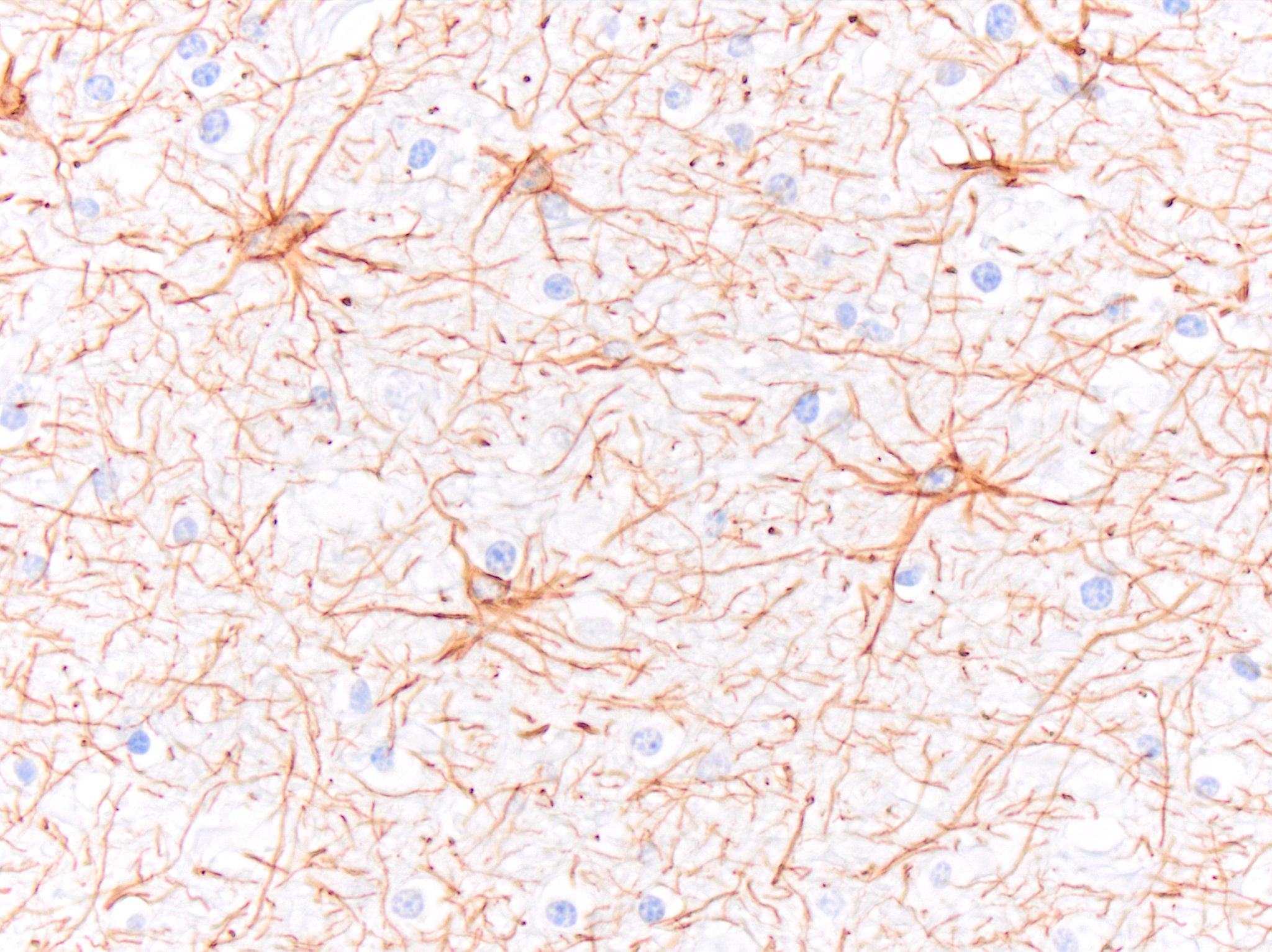
Application: Immunohistochemistry-ParaffinSample Tested: Adult brainSpecies: HumanVerified Customer | Posted 09/14/2023astrocytes were positive for GFAP (GA-5)antigen retrieval in a basic buffer x200 (1ug/mL) BOND refine kit
-
Application: ImmunocytochemistrySample Tested: Embryonic cortical glial cellsSpecies: MouseVerified Customer | Posted 09/11/2017
-
Application: ImmunocytochemistrySample Tested: Cultured rat hippocampal neuronsSpecies: RatVerified Customer | Posted 03/27/2017Fixation: 3% formaldehyde (TAAB) in PBS, 15 minutes RT Blocking: 10% donkey serum in PBS, 1 hour RT Permeabilization: 0.2% Triton-X100 Primary Antibody: 1:100 in 5% horse serum in PBS with 0.2% Triton, overnight, 4°C Wash: three times wash with PBS with 0.2% Triton, 10 min each Secondary Antibody: Invitrogen goat anti-mouse Alexa568, 1:500 in PBS with 0.2% Triton, 2 hours, RT Wash: three times wash with PBS, 10 min each Microscope: Confocal (Zeiss LSM710) The antibody labeld intermediate filaments in cultured astrocytes.
There are no reviews that match your criteria.
Protocols
Find general support by application which include: protocols, troubleshooting, illustrated assays, videos and webinars.
- 7-Amino Actinomycin D (7-AAD) Cell Viability Flow Cytometry Protocol
- Antigen Retrieval Protocol (PIER)
- Antigen Retrieval for Frozen Sections Protocol
- Appropriate Fixation of IHC/ICC Samples
- Cellular Response to Hypoxia Protocols
- Chromogenic IHC Staining of Formalin-Fixed Paraffin-Embedded (FFPE) Tissue Protocol
- Chromogenic Immunohistochemistry Staining of Frozen Tissue
- ClariTSA™ Fluorophore Kits
- Detection & Visualization of Antibody Binding
- Extracellular Membrane Flow Cytometry Protocol
- Flow Cytometry Protocol for Cell Surface Markers
- Flow Cytometry Protocol for Staining Membrane Associated Proteins
- Flow Cytometry Staining Protocols
- Flow Cytometry Troubleshooting Guide
- Fluorescent IHC Staining of Frozen Tissue Protocol
- Graphic Protocol for Heat-induced Epitope Retrieval
- Graphic Protocol for the Preparation and Fluorescent IHC Staining of Frozen Tissue Sections
- Graphic Protocol for the Preparation and Fluorescent IHC Staining of Paraffin-embedded Tissue Sections
- Graphic Protocol for the Preparation of Gelatin-coated Slides for Histological Tissue Sections
- ICC Cell Smear Protocol for Suspension Cells
- ICC Immunocytochemistry Protocol Videos
- ICC for Adherent Cells
- IHC Sample Preparation (Frozen sections vs Paraffin)
- Immunocytochemistry (ICC) Protocol
- Immunocytochemistry Troubleshooting
- Immunofluorescence of Organoids Embedded in Cultrex Basement Membrane Extract
- Immunofluorescent IHC Staining of Formalin-Fixed Paraffin-Embedded (FFPE) Tissue Protocol
- Immunohistochemistry (IHC) and Immunocytochemistry (ICC) Protocols
- Immunohistochemistry Frozen Troubleshooting
- Immunohistochemistry Paraffin Troubleshooting
- Intracellular Flow Cytometry Protocol Using Alcohol (Methanol)
- Intracellular Flow Cytometry Protocol Using Detergents
- Intracellular Nuclear Staining Flow Cytometry Protocol Using Detergents
- Intracellular Staining Flow Cytometry Protocol Using Alcohol Permeabilization
- Intracellular Staining Flow Cytometry Protocol Using Detergents to Permeabilize Cells
- Preparing Samples for IHC/ICC Experiments
- Preventing Non-Specific Staining (Non-Specific Binding)
- Primary Antibody Selection & Optimization
- Propidium Iodide Cell Viability Flow Cytometry Protocol
- Protocol for Heat-Induced Epitope Retrieval (HIER)
- Protocol for Liperfluo
- Protocol for Making a 4% Formaldehyde Solution in PBS
- Protocol for VisUCyte™ HRP Polymer Detection Reagent
- Protocol for the Characterization of Human Th22 Cells
- Protocol for the Characterization of Human Th9 Cells
- Protocol for the Fluorescent ICC Staining of Cell Smears - Graphic
- Protocol for the Fluorescent ICC Staining of Cultured Cells on Coverslips - Graphic
- Protocol for the Preparation & Fixation of Cells on Coverslips
- Protocol for the Preparation and Chromogenic IHC Staining of Frozen Tissue Sections
- Protocol for the Preparation and Chromogenic IHC Staining of Frozen Tissue Sections - Graphic
- Protocol for the Preparation and Chromogenic IHC Staining of Paraffin-embedded Tissue Sections
- Protocol for the Preparation and Chromogenic IHC Staining of Paraffin-embedded Tissue Sections - Graphic
- Protocol for the Preparation and Fluorescent ICC Staining of Cells on Coverslips
- Protocol for the Preparation and Fluorescent ICC Staining of Non-adherent Cells
- Protocol for the Preparation and Fluorescent ICC Staining of Stem Cells on Coverslips
- Protocol for the Preparation and Fluorescent IHC Staining of Frozen Tissue Sections
- Protocol for the Preparation and Fluorescent IHC Staining of Paraffin-embedded Tissue Sections
- Protocol for the Preparation of Gelatin-coated Slides for Histological Tissue Sections
- Protocol for the Preparation of a Cell Smear for Non-adherent Cell ICC - Graphic
- Protocol: Annexin V and PI Staining by Flow Cytometry
- Protocol: Annexin V and PI Staining for Apoptosis by Flow Cytometry
- R&D Systems Quality Control Western Blot Protocol
- TUNEL and Active Caspase-3 Detection by IHC/ICC Protocol
- The Importance of IHC/ICC Controls
- Troubleshooting Guide: Fluorokine Flow Cytometry Kits
- Troubleshooting Guide: Immunohistochemistry
- Troubleshooting Guide: Western Blot Figures
- Western Blot Conditions
- Western Blot Protocol
- Western Blot Protocol for Cell Lysates
- Western Blot Troubleshooting
- Western Blot Troubleshooting Guide
- View all Protocols, Troubleshooting, Illustrated assays and Webinars
Loading...
Associated Pathways
