Glucosylceramidase/GBA Antibody (JM10-76)
Novus Biologicals | Catalog # NBP2-66871
Recombinant Monoclonal Antibody

Loading...
Key Product Details
Validated by
Knockout/Knockdown, Biological Validation
Species Reactivity
Human, Mouse, Rat
Applications
Knockout Validated, Immunohistochemistry, Immunohistochemistry-Paraffin, Immunohistochemistry-Frozen, Western Blot, Flow Cytometry, Immunocytochemistry/ Immunofluorescence
Label
Unconjugated
Antibody Source
Recombinant Monoclonal Rabbit IgG Clone # JM10-76 expressed in HEK293
Loading...
Product Specifications
Immunogen
Synthetic peptide within Human Glucosylceramidase/GBA aa 477-534 / 536. (SwissProt: P04062 Human; SwissProt: P17439 Mouse; Entrez Gene:684536 Rat)
Localization
Lysosome membrane.
Clonality
Monoclonal
Host
Rabbit
Isotype
IgG
Scientific Data Images for Glucosylceramidase/GBA Antibody (JM10-76)
Western Blot Shows Glucosylceramidase/GBA Specificity Using Knockout Cell Line.
Western blot shows lysates of HAP1 parental cell line and Glucosylceramidase/GBA knockout HAP1 cell line (KO). Nitrocellulose membrane was probed with Glucosylceramidase/GBA Antibody (JM10-76) (Catalog # NBP2-66871) followed by HRP-conjugated secondary antibody. A specific band was detected for Glucosylceramidase/GBA at approximately 59.7 kDa (as indicated) in the parental HAP1 cell line, but is not detectable in knockout HAP1 cell line. Primary antibody dilution used: 1/500. The Ponceau stained transfer of the blot is shown. This experiment was conducted under reducing conditions. Image, protocol, and testing courtesy of YCharOS Inc. See ycharos.com for additional details.![Western Blot: Glucosylceramidase/GBA Antibody (JM10-76) [NBP2-66871]](https://resources.rndsystems.com/images/products/Glucosylceramidase-GBA-Antibody-JM10-76-Western-Blot-NBP2-66871-img0011.jpg "Western Blot: Glucosylceramidase/GBA Antibody (JM10-76) [NBP2-66871]")
Western Blot: Glucosylceramidase/GBA Antibody (JM10-76) [NBP2-66871]
Western Blot: Glucosylceramidase/GBA Antibody (JM10-76) [NBP2-66871] - All lanes: Western blot analysis of GBA with anti-GBA antibody [JM10-76 at 1:1,000 dilution.Lane 1: Wild-type HEK293T whole cell lysate (20 ug).Lane 2: GBA knockout HEK293T whole cell lysate (20 ug). NBP2-66871was shown to specifically react with GBA in wild-type HEK293T cells. No band was observed when GBA knockout sample was tested. Wild-type and GBA knockout samples were subjected to SDS-PAGE. Proteins were transferred to a PVDF membrane and blocked with 5% NFDM in TBST for 1 hour at room temperature. The primary antibody (1/1,000) and Loading control antibody (Rabbit anti-GAPDH, 1/10,000) was used in 5% BSA at room temperature for 2 hours. Goat Anti-Rabbit IgG-HRP Secondary Antibody at 1:200,000 dilution was used for 1 hour at room temperature.![Immunohistochemistry-Paraffin: Glucosylceramidase/GBA Antibody (JM10-76) [NBP2-66871]](https://resources.rndsystems.com/images/products/Glucosylceramidase-GBA-Antibody-JM10-76-Immunohistochemistry-Paraffin-NBP2-66871-img0015.jpg "Immunohistochemistry-Paraffin: Glucosylceramidase/GBA Antibody (JM10-76) [NBP2-66871]")
Immunohistochemistry-Paraffin: Glucosylceramidase/GBA Antibody (JM10-76) [NBP2-66871]
Immunohistochemistry-Paraffin: Glucosylceramidase/GBA Antibody (JM10-76) [NBP2-66871] - Analysis of paraffin-embedded mouse pancreas tissue using anti-GBA antibody. The section was pre-treated using heat mediated antigen retrieval with Tris-EDTA buffer (pH 8.0-8.4) for 20 minutes.The tissues were blocked in 5% BSA for 30 minutes at room temperature, washed with ddH2O and PBS, and then probed with the primary antibody (1/50) for 30 minutes at room temperature. The detection was performed using an HRP conjugated compact polymer system. DAB was used as the chromogen. Tissues were counterstained with hematoxylin and mounted with DPX.![Western Blot: Glucosylceramidase/GBA Antibody (JM10-76) [NBP2-66871]](https://resources.rndsystems.com/images/products/Glucosylceramidase-GBA-Antibody-JM10-76-Western-Blot-NBP2-66871-img0009.jpg "Western Blot: Glucosylceramidase/GBA Antibody (JM10-76) [NBP2-66871]")
Western Blot: Glucosylceramidase/GBA Antibody (JM10-76) [NBP2-66871]
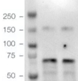
Western Blot: Glucosylceramidase/GBA Antibody (JM10-76) [NBP2-66871] - Western blot analysis of Glucosylceramidase/GBA on different lysates. Proteins were transferred to a PVDF membrane and blocked with 5% BSA in PBS for 1 hour at room temperature. The primary antibody (1/500) was used in 5% BSA at room temperature for 2 hours. Goat Anti-Rabbit IgG - HRP Secondary Antibody (HA1001) at 1:5,000 dilution was used for 1 hour at room temperature. Positive control: Lane 1: SK-Br-3 cell lysate Lane 2: A549 cell lysate Lane 3: MCF-7 cell lysate![Immunohistochemistry-Paraffin: Glucosylceramidase/GBA Antibody (JM10-76) [NBP2-66871]](https://resources.rndsystems.com/images/products/Glucosylceramidase-GBA-Antibody-JM10-76-Immunohistochemistry-Paraffin-NBP2-66871-img0001.jpg "Immunohistochemistry-Paraffin: Glucosylceramidase/GBA Antibody (JM10-76) [NBP2-66871]")
Immunohistochemistry-Paraffin: Glucosylceramidase/GBA Antibody (JM10-76) [NBP2-66871]
Immunohistochemistry-Paraffin: Glucosylceramidase/GBA Antibody (JM10-76) [NBP2-66871] - Analysis of paraffin-embedded human breast carcinoma tissue using anti-GBA antibody. Counter stained with hematoxylin.![Immunohistochemistry-Paraffin: Glucosylceramidase/GBA Antibody (JM10-76) [NBP2-66871]](https://resources.rndsystems.com/images/products/Glucosylceramidase-GBA-Antibody-JM10-76-Immunohistochemistry-Paraffin-NBP2-66871-img0003.jpg "Immunohistochemistry-Paraffin: Glucosylceramidase/GBA Antibody (JM10-76) [NBP2-66871]")
Immunohistochemistry-Paraffin: Glucosylceramidase/GBA Antibody (JM10-76) [NBP2-66871]
Immunohistochemistry-Paraffin: Glucosylceramidase/GBA Antibody (JM10-76) [NBP2-66871] - Analysis of paraffin-embedded human liver tissue using anti-GBA antibody. Counter stained with hematoxylin.![Immunohistochemistry-Paraffin: Glucosylceramidase/GBA Antibody (JM10-76) [NBP2-66871]](https://resources.rndsystems.com/images/products/Glucosylceramidase-GBA-Antibody-JM10-76-Immunohistochemistry-Paraffin-NBP2-66871-img0004.jpg "Immunohistochemistry-Paraffin: Glucosylceramidase/GBA Antibody (JM10-76) [NBP2-66871]")
Immunohistochemistry-Paraffin: Glucosylceramidase/GBA Antibody (JM10-76) [NBP2-66871]
Immunohistochemistry-Paraffin: Glucosylceramidase/GBA Antibody (JM10-76) [NBP2-66871] - Analysis of paraffin-embedded human pancreas tissue using anti-GBA antibody. Counter stained with hematoxylin.![Immunohistochemistry-Paraffin: Glucosylceramidase/GBA Antibody (JM10-76) [NBP2-66871]](https://resources.rndsystems.com/images/products/Glucosylceramidase-GBA-Antibody-JM10-76-Immunohistochemistry-Paraffin-NBP2-66871-img0012.jpg "Immunohistochemistry-Paraffin: Glucosylceramidase/GBA Antibody (JM10-76) [NBP2-66871]")
Immunohistochemistry-Paraffin: Glucosylceramidase/GBA Antibody (JM10-76) [NBP2-66871]
Immunohistochemistry-Paraffin: Glucosylceramidase/GBA Antibody (JM10-76) [NBP2-66871] - Analysis of paraffin-embedded human kidney tissue using anti-GBA antibody. The section was pre-treated using heat mediated antigen retrieval with Tris-EDTA buffer (pH 8.0-8.4) for 20 minutes.The tissues were blocked in 5% BSA for 30 minutes at room temperature, washed with ddH2O and PBS, and then probed with the primary antibody 1/50) for 30 minutes at room temperature. The detection was performed using an HRP conjugated compact polymer system. DAB was used as the chromogen. Tissues were counterstained with hematoxylin and mounted with DPX.![Immunohistochemistry-Paraffin: Glucosylceramidase/GBA Antibody (JM10-76) [NBP2-66871]](https://resources.rndsystems.com/images/products/Glucosylceramidase-GBA-Antibody-JM10-76-Immunohistochemistry-Paraffin-NBP2-66871-img0013.jpg "Immunohistochemistry-Paraffin: Glucosylceramidase/GBA Antibody (JM10-76) [NBP2-66871]")
Immunohistochemistry-Paraffin: Glucosylceramidase/GBA Antibody (JM10-76) [NBP2-66871]
Immunohistochemistry-Paraffin: Glucosylceramidase/GBA Antibody (JM10-76) [NBP2-66871] - Analysis of paraffin-embedded rat liver tissue using anti-GBA antibody. The section was pre-treated using heat mediated antigen retrieval with Tris-EDTA buffer (pH 8.0-8.4) for 20 minutes.The tissues were blocked in 5% BSA for 30 minutes at room temperature, washed with ddH2O and PBS, and then probed with the primary antibody (1/50) for 30 minutes at room temperature. The detection was performed using an HRP conjugated compact polymer system. DAB was used as the chromogen. Tissues were counterstained with hematoxylin and mounted with DPX.![Immunohistochemistry-Paraffin: Glucosylceramidase/GBA Antibody (JM10-76) [NBP2-66871]](https://resources.rndsystems.com/images/products/Glucosylceramidase-GBA-Antibody-JM10-76-Immunohistochemistry-Paraffin-NBP2-66871-img0014.jpg "Immunohistochemistry-Paraffin: Glucosylceramidase/GBA Antibody (JM10-76) [NBP2-66871]")
Immunohistochemistry-Paraffin: Glucosylceramidase/GBA Antibody (JM10-76) [NBP2-66871]
Immunohistochemistry-Paraffin: Glucosylceramidase/GBA Antibody (JM10-76) [NBP2-66871] - Analysis of paraffin-embedded rat kidney tissue using anti-GBA antibody. The section was pre-treated using heat mediated antigen retrieval with Tris-EDTA buffer (pH 8.0-8.4) for 20 minutes.The tissues were blocked in 5% BSA for 30 minutes at room temperature, washed with ddH2O and PBS, and then probed with the primary antibody (1/50) for 30 minutes at room temperature. The detection was performed using an HRP conjugated compact polymer system. DAB was used as the chromogen. Tissues were counterstained with hematoxylin and mounted with DPX.Applications for Glucosylceramidase/GBA Antibody (JM10-76)
Application
Recommended Usage
Flow Cytometry
1:500
Immunocytochemistry/ Immunofluorescence
1:100
Immunohistochemistry-Paraffin
1:50-1:200
Knockout Validated
Knockout validated from YCharOS Inc. (ycharos.com)
Western Blot
1:500-1:2000
Reviewed Applications
Read 1 review rated 5 using NBP2-66871 in the following applications:
Flow Cytometry Panel Builder
Bio-Techne Knows Flow Cytometry
Save time and reduce costly mistakes by quickly finding compatible reagents using the Panel Builder Tool.
Advanced Features
- Spectra Viewer - Custom analysis of spectra from multiple fluorochromes
- Spillover Popups - Visualize the spectra of individual fluorochromes
- Antigen Density Selector - Match fluorochrome brightness with antigen density

Formulation, Preparation, and Storage
Purification
Protein A purified
Formulation
TBS (pH7.4), 0.05% BSA, 40% Glycerol
Preservative
0.05% Sodium Azide
Concentration
1 mg/ml
Shipping
The product is shipped with polar packs. Upon receipt, store it immediately at the temperature recommended below.
Stability & Storage
Store at 4C short term. Aliquot and store at -20C long term. Avoid freeze-thaw cycles.
Background: Glucosylceramidase/GBA
Alternate Names
Alglucerase, GBA, GBA1, Imiglucerase
Gene Symbol
GBA1
Additional Glucosylceramidase/GBA Products
Product Documents for Glucosylceramidase/GBA Antibody (JM10-76)
Certificate of Analysis
To download a Certificate of Analysis, please enter a lot or batch number in the search box below.
Product Specific Notices for Glucosylceramidase/GBA Antibody (JM10-76)
This product is for research use only and is not approved for use in humans or in clinical diagnosis. Primary Antibodies are guaranteed for 1 year from date of receipt.
Related Research Areas
Citations for Glucosylceramidase/GBA Antibody (JM10-76)
 Powered by Bioz
Customer Reviews for Glucosylceramidase/GBA Antibody (JM10-76) (1)
5 out of 5
1 Customer Rating
Have you used Glucosylceramidase/GBA Antibody (JM10-76)?
Submit a review and receive an Amazon gift card!
$25/€18/£15/$25CAN/¥2500 Yen for a review with an image
$10/€7/£6/$10CAN/¥1110 Yen for a review without an image
Submit a review
Customer Images
Showing
1
-
1 of
1 review
Showing All
Filter By:
-
Application: Western BlotSample Tested: Colon tissueVerified Customer | Posted 10/02/2024Dual pink protein marker10 mg loading 1:1000 dilution HRP detection

There are no reviews that match your criteria.
Protocols
Find general support by application which include: protocols, troubleshooting, illustrated assays, videos and webinars.
- 7-Amino Actinomycin D (7-AAD) Cell Viability Flow Cytometry Protocol
- Antigen Retrieval Protocol (PIER)
- Antigen Retrieval for Frozen Sections Protocol
- Appropriate Fixation of IHC/ICC Samples
- Cellular Response to Hypoxia Protocols
- Chromogenic IHC Staining of Formalin-Fixed Paraffin-Embedded (FFPE) Tissue Protocol
- Chromogenic Immunohistochemistry Staining of Frozen Tissue
- ClariTSA™ Fluorophore Kits
- Detection & Visualization of Antibody Binding
- Extracellular Membrane Flow Cytometry Protocol
- Flow Cytometry Protocol for Cell Surface Markers
- Flow Cytometry Protocol for Staining Membrane Associated Proteins
- Flow Cytometry Staining Protocols
- Flow Cytometry Troubleshooting Guide
- Fluorescent IHC Staining of Frozen Tissue Protocol
- Graphic Protocol for Heat-induced Epitope Retrieval
- Graphic Protocol for the Preparation and Fluorescent IHC Staining of Frozen Tissue Sections
- Graphic Protocol for the Preparation and Fluorescent IHC Staining of Paraffin-embedded Tissue Sections
- Graphic Protocol for the Preparation of Gelatin-coated Slides for Histological Tissue Sections
- ICC Cell Smear Protocol for Suspension Cells
- ICC Immunocytochemistry Protocol Videos
- ICC for Adherent Cells
- IHC Sample Preparation (Frozen sections vs Paraffin)
- Immunocytochemistry (ICC) Protocol
- Immunocytochemistry Troubleshooting
- Immunofluorescence of Organoids Embedded in Cultrex Basement Membrane Extract
- Immunofluorescent IHC Staining of Formalin-Fixed Paraffin-Embedded (FFPE) Tissue Protocol
- Immunohistochemistry (IHC) and Immunocytochemistry (ICC) Protocols
- Immunohistochemistry Frozen Troubleshooting
- Immunohistochemistry Paraffin Troubleshooting
- Intracellular Flow Cytometry Protocol Using Alcohol (Methanol)
- Intracellular Flow Cytometry Protocol Using Detergents
- Intracellular Nuclear Staining Flow Cytometry Protocol Using Detergents
- Intracellular Staining Flow Cytometry Protocol Using Alcohol Permeabilization
- Intracellular Staining Flow Cytometry Protocol Using Detergents to Permeabilize Cells
- Preparing Samples for IHC/ICC Experiments
- Preventing Non-Specific Staining (Non-Specific Binding)
- Primary Antibody Selection & Optimization
- Propidium Iodide Cell Viability Flow Cytometry Protocol
- Protocol for Heat-Induced Epitope Retrieval (HIER)
- Protocol for Liperfluo
- Protocol for Making a 4% Formaldehyde Solution in PBS
- Protocol for VisUCyte™ HRP Polymer Detection Reagent
- Protocol for the Characterization of Human Th22 Cells
- Protocol for the Characterization of Human Th9 Cells
- Protocol for the Fluorescent ICC Staining of Cell Smears - Graphic
- Protocol for the Fluorescent ICC Staining of Cultured Cells on Coverslips - Graphic
- Protocol for the Preparation & Fixation of Cells on Coverslips
- Protocol for the Preparation and Chromogenic IHC Staining of Frozen Tissue Sections
- Protocol for the Preparation and Chromogenic IHC Staining of Frozen Tissue Sections - Graphic
- Protocol for the Preparation and Chromogenic IHC Staining of Paraffin-embedded Tissue Sections
- Protocol for the Preparation and Chromogenic IHC Staining of Paraffin-embedded Tissue Sections - Graphic
- Protocol for the Preparation and Fluorescent ICC Staining of Cells on Coverslips
- Protocol for the Preparation and Fluorescent ICC Staining of Non-adherent Cells
- Protocol for the Preparation and Fluorescent ICC Staining of Stem Cells on Coverslips
- Protocol for the Preparation and Fluorescent IHC Staining of Frozen Tissue Sections
- Protocol for the Preparation and Fluorescent IHC Staining of Paraffin-embedded Tissue Sections
- Protocol for the Preparation of Gelatin-coated Slides for Histological Tissue Sections
- Protocol for the Preparation of a Cell Smear for Non-adherent Cell ICC - Graphic
- Protocol: Annexin V and PI Staining by Flow Cytometry
- Protocol: Annexin V and PI Staining for Apoptosis by Flow Cytometry
- R&D Systems Quality Control Western Blot Protocol
- TUNEL and Active Caspase-3 Detection by IHC/ICC Protocol
- The Importance of IHC/ICC Controls
- Troubleshooting Guide: Fluorokine Flow Cytometry Kits
- Troubleshooting Guide: Immunohistochemistry
- Troubleshooting Guide: Western Blot Figures
- Western Blot Conditions
- Western Blot Protocol
- Western Blot Protocol for Cell Lysates
- Western Blot Troubleshooting
- Western Blot Troubleshooting Guide
- View all Protocols, Troubleshooting, Illustrated assays and Webinars
Loading...