Glyoxalase I Antibody (Glo1a) - BSA Free
Novus Biologicals | Catalog # NBP1-19015
Key Product Details
Species Reactivity
Validated:
Human, Mouse
Cited:
Human, Mouse
Applications
Validated:
Immunohistochemistry-Paraffin, Western Blot, ELISA, Immunocytochemistry/ Immunofluorescence, Simple Western, Immunoprecipitation
Cited:
Immunohistochemistry-Paraffin, Western Blot, Immunoprecipitation
Label
Unconjugated
Antibody Source
Monoclonal Mouse IgG1 kappa Clone # Glo1a
Format
BSA Free
Loading...
Product Specifications
Immunogen
Full length human GLO1 protein [Swiss-Prot #Q04760].
Clonality
Monoclonal
Host
Mouse
Isotype
IgG1 kappa
Theoretical MW
21 kDa.
Disclaimer note: The observed molecular weight of the protein may vary from the listed predicted molecular weight due to post translational modifications, post translation cleavages, relative charges, and other experimental factors.
Disclaimer note: The observed molecular weight of the protein may vary from the listed predicted molecular weight due to post translational modifications, post translation cleavages, relative charges, and other experimental factors.
Scientific Data Images for Glyoxalase I Antibody (Glo1a) - BSA Free
![Western Blot: Glyoxalase I Antibody (Glo1a) [NBP1-19015]](https://resources.rndsystems.com/images/products/Glyoxalase-I-Antibody-Glo1a-Western-Blot-NBP1-19015-img0007.jpg "Western Blot: Glyoxalase I Antibody (Glo1a) [NBP1-19015]")
Western Blot: Glyoxalase I Antibody (Glo1a) [NBP1-19015]
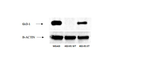
Western Blot: Glyoxalase I Antibody (Glo1a) [NBP1-19015] - Detection of GLO-1 in human cell lines. Lane 1,2 and 3: MG-63 osteosarcoma cell line (positive control), 402-91 liposarcoma cell line and 402-91 liposarcoma cell line resistant to trabectedine. Image from verified customer review.![Immunocytochemistry/ Immunofluorescence: Glyoxalase I Antibody (Glo1a) [NBP1-19015]](https://resources.rndsystems.com/images/products/Glyoxalase-I-Antibody-Glo1a-Immunocytochemistry-Immunofluorescence-NBP1-19015-img0004.jpg "Immunocytochemistry/ Immunofluorescence: Glyoxalase I Antibody (Glo1a) [NBP1-19015]")
Immunocytochemistry/ Immunofluorescence: Glyoxalase I Antibody (Glo1a) [NBP1-19015]
Immunocytochemistry/Immunofluorescence: Glyoxalase I Antibody (Glo1a) [NBP1-19015] - Immunostaining of human retinal endothelial cells. Immunoreactivity for hGlx-1 was detected in human retinal endothelial cells stained with Glo1a mAb using anti mouse IgG Texas Red as secondary Ab. The cells were counter stained with phalloidin (green) and DAPI (blue), nuclear stain. No reactivity was detected in control cells, stained without Go1a mAb. Figure is a representative of 3 independent experiments. Photo courtesy of Dr. Nagaraj, Case Western Reserve University.![Western Blot: Glyoxalase I Antibody (Glo1a) [NBP1-19015]](https://resources.rndsystems.com/images/products/Glyoxalase-I-Antibody-Glo1a-Western-Blot-NBP1-19015-img0003.jpg "Western Blot: Glyoxalase I Antibody (Glo1a) [NBP1-19015]")
Western Blot: Glyoxalase I Antibody (Glo1a) [NBP1-19015]
Western Blot: Glyoxalase I Antibody (Glo1a) [NBP1-19015] - HeLa whole cell extracts.![Simple Western: Glyoxalase I Antibody (Glo1a) [NBP1-19015]](https://resources.rndsystems.com/images/products/Glyoxalase-I-Antibody-Glo1a-Simple-Western-NBP1-19015-img0006.jpg "Simple Western: Glyoxalase I Antibody (Glo1a) [NBP1-19015]")
Simple Western: Glyoxalase I Antibody (Glo1a) [NBP1-19015]
Simple Western: Glyoxalase I Antibody (Glo1a) [NBP1-19015] - Simple Western lane view shows a specific band for GLO1 in 0.5 mg/ml of HeLa lysate. This experiment was performed under reducing conditions using the 12-230kDa separation system. * Non-specific interaction with the 230 kDa Simple Western standard may be seen with this antibody.![Simple Western: Glyoxalase I Antibody (Glo1a) [NBP1-19015]](https://resources.rndsystems.com/images/products/Glyoxalase-I-Antibody-Glo1a-Simple-Western-NBP1-19015-img0005.jpg "Simple Western: Glyoxalase I Antibody (Glo1a) [NBP1-19015]")
Simple Western: Glyoxalase I Antibody (Glo1a) [NBP1-19015]
Simple Western: Glyoxalase I Antibody (Glo1a) [NBP1-19015] - Simple Western lane view shows a specific band detected for GLO1 in HeLa lysate. This experiment was performed under reducing conditions using the Wes or Sally Sue separation system 12-230kDa (or 66-440kDa).Applications for Glyoxalase I Antibody (Glo1a) - BSA Free
Application
Recommended Usage
ELISA
0.15 ng / 1 ug protein
Immunocytochemistry/ Immunofluorescence
10 ug/ml
Immunohistochemistry-Paraffin
reported in scientific literature (10.3892/ol.2021.12808)
Immunoprecipitation
reported in scientific literature
Simple Western
1:50
Application Notes
This GLO1 antibody is useful for ELISA, Immunocytochemistry/Immunofluorescence and Western blot, where a band is seen approx. 21 kDa.
In Simple Western only 10 - 15 uL of the recommended dilution is used per data point.
See Simple Western Antibody Database for Simple Western validation: Tested in HeLa lysate 0.5 mg/mL, separated by Size, antibody dilution of 1:50, apparent MW was 32 kDa. Separated by Size-Wes, Sally Sue/Peggy Sue.
The observed molecular weight of the protein may vary from the listed predicted molecular weight due to post translational modifications, post translation cleavages, relative charges, and other experimental factors.
Reviewed Applications
Read 1 review rated 5 using NBP1-19015 in the following applications:
Formulation, Preparation, and Storage
Purification
Protein G purified
Formulation
Tris-Glycine and 0.15M NaCl
Format
BSA Free
Preservative
0.05% Sodium Azide
Concentration
1 mg/ml
Shipping
The product is shipped with polar packs. Upon receipt, store it immediately at the temperature recommended below.
Stability & Storage
Store at -20C. Avoid freeze-thaw cycles.
Background: Glyoxalase I
Alternate Names
Aldoketomutase, GLO1, GLOD1, Glx I, GLYI, Methylglyoxalase
Gene Symbol
GLO1
Additional Glyoxalase I Products
Product Documents for Glyoxalase I Antibody (Glo1a) - BSA Free
Certificate of Analysis
To download a Certificate of Analysis, please enter a lot or batch number in the search box below.
Product Specific Notices for Glyoxalase I Antibody (Glo1a) - BSA Free
This product is for research use only and is not approved for use in humans or in clinical diagnosis. Primary Antibodies are guaranteed for 1 year from date of receipt.
Citations for Glyoxalase I Antibody (Glo1a) - BSA Free
 Powered by Bioz
Customer Reviews for Glyoxalase I Antibody (Glo1a) - BSA Free (1)
5 out of 5
1 Customer Rating
Have you used Glyoxalase I Antibody (Glo1a) - BSA Free?
Submit a review and receive an Amazon gift card!
$25/€18/£15/$25CAN/¥2500 Yen for a review with an image
$10/€7/£6/$10CAN/¥1110 Yen for a review without an image
Submit a review
Customer Images
Showing
1
-
1 of
1 review
Showing All
Filter By:
-
Application: Western BlotSample Tested: Human osteosarcoma cells – MG63 and Human liposarcoma cells- 402-91Species: HumanVerified Customer | Posted 03/20/2017Western Blot: GLO1 Antibody [NBP1-19015] - Detection of GLO-1 in human cell lines. Lane 1,2 and 3: MG-63 osteosarcoma cell line (positive control), 402-91 liposarcoma cell line and 402-91 liposarcoma cell line resistant to trabectedine.

There are no reviews that match your criteria.
Protocols
View specific protocols for Glyoxalase I Antibody (Glo1a) - BSA Free (NBP1-19015):
Glyoxalase I Antibody (Glo1a):
Immunocytochemistry Protocol
1. Cells are cultured one day prior to the experiment.
2. After washing twice with PBS and they are fixed with 4% paraformaldehyde in PBS at ?20C for 15 min.
3. Followed by two washes with PBS, they are permeabilized with 0.1%Triton X-100 in PBS at ?20C for 5 min.
4. To remove the detergent the cells are washed5 times with PBS and then blocked with 2.5 % goat serum in PBS for 2 hr at RT.
5. Cells are then incubated with GLOI mAb (10 μg/ml) in PBS for 1 hr at RT and washed twice for 5 min with PBS.
6. Cells are incubated with secondary antibody (anti-mouseIgG) conjugated with Texas Red (1: 400 dilution in PBS) (Molecular Probes) for 1 hr at RT.
Images of lenses were acquired on a Leica DMI 6000 B inverted microscope using a 20x objective connected to a Retiga EXI camera (Q-imaging Vancouver British Columbia). Secondary Ab contribution to immune reaction was verified by staining without the primary Ab.
Glyoxalase I Antibody (Glo1a):
Western Blot Protocol
1. Perform SDS-PAGE (4-12% MOPS) on samples to be analyzed, loading 30 ug of total protein per lane.
2. Transfer proteins to Nitrocellulose according to the instructions provided by the manufacturer of the transfer apparatus.
3. Rinse membrane with dH2O and then stain the blot using Ponceau S for 1-2 minutes to access the transfer of proteins onto the nitrocellulose membrane. Rinse the blot in water to remove excess stain and mark the lane locations and locations of molecular weight markers using a pencil.
4. Rinse the blot in TBS for approximately 5 minutes.
5. Block the membrane using 5% BSA in TBS + Tween, 1 hour at RT.
6. Rinse the membrane in dH2O and then wash the membrane in wash buffer [TBS + 0.1% Tween] 3 times for 10 minutes each.
7. Dilute the rabbit anti-GLO1 primary antibody (NBP1-19015) in blocking buffer and incubate 1 hour at room temperature.
8. Rinse the membrane in dH2O and then wash the membrane in wash buffer [TBS + 0.1% Tween] 3 times for 10 minutes each.
9. Apply the diluted mouse-IgG HRP-conjugated secondary antibody in blocking buffer (as per manufacturers instructions) and incubate 1 hour at room temperature.
10. Wash the blot in wash buffer [TBS + 0.1% Tween] 3 times for 10 minutes each (this step can be repeated as required to reduce background).
11. Apply the detection reagent of choice in accordance with the manufacturers instructions (Pierce ECL).
**Note: Tween-20 can be added to the blocking or antibody dilution buffer at a final concentration of 0.05-0.2%, providedit does not interfere with antibody-antigen binding.
Find general support by application which include: protocols, troubleshooting, illustrated assays, videos and webinars.
- Antigen Retrieval Protocol (PIER)
- Antigen Retrieval for Frozen Sections Protocol
- Appropriate Fixation of IHC/ICC Samples
- Cellular Response to Hypoxia Protocols
- Chromogenic IHC Staining of Formalin-Fixed Paraffin-Embedded (FFPE) Tissue Protocol
- Chromogenic Immunohistochemistry Staining of Frozen Tissue
- ClariTSA™ Fluorophore Kits
- Detection & Visualization of Antibody Binding
- ELISA Sample Preparation & Collection Guide
- ELISA Troubleshooting Guide
- Fluorescent IHC Staining of Frozen Tissue Protocol
- Graphic Protocol for Heat-induced Epitope Retrieval
- Graphic Protocol for the Preparation and Fluorescent IHC Staining of Frozen Tissue Sections
- Graphic Protocol for the Preparation and Fluorescent IHC Staining of Paraffin-embedded Tissue Sections
- Graphic Protocol for the Preparation of Gelatin-coated Slides for Histological Tissue Sections
- How to Run an R&D Systems DuoSet ELISA
- How to Run an R&D Systems Quantikine ELISA
- How to Run an R&D Systems Quantikine™ QuicKit™ ELISA
- ICC Cell Smear Protocol for Suspension Cells
- ICC Immunocytochemistry Protocol Videos
- ICC for Adherent Cells
- IHC Sample Preparation (Frozen sections vs Paraffin)
- Immunocytochemistry (ICC) Protocol
- Immunocytochemistry Troubleshooting
- Immunofluorescence of Organoids Embedded in Cultrex Basement Membrane Extract
- Immunofluorescent IHC Staining of Formalin-Fixed Paraffin-Embedded (FFPE) Tissue Protocol
- Immunohistochemistry (IHC) and Immunocytochemistry (ICC) Protocols
- Immunohistochemistry Frozen Troubleshooting
- Immunohistochemistry Paraffin Troubleshooting
- Immunoprecipitation Protocol
- Preparing Samples for IHC/ICC Experiments
- Preventing Non-Specific Staining (Non-Specific Binding)
- Primary Antibody Selection & Optimization
- Protocol for Heat-Induced Epitope Retrieval (HIER)
- Protocol for Making a 4% Formaldehyde Solution in PBS
- Protocol for VisUCyte™ HRP Polymer Detection Reagent
- Protocol for the Fluorescent ICC Staining of Cell Smears - Graphic
- Protocol for the Fluorescent ICC Staining of Cultured Cells on Coverslips - Graphic
- Protocol for the Preparation & Fixation of Cells on Coverslips
- Protocol for the Preparation and Chromogenic IHC Staining of Frozen Tissue Sections
- Protocol for the Preparation and Chromogenic IHC Staining of Frozen Tissue Sections - Graphic
- Protocol for the Preparation and Chromogenic IHC Staining of Paraffin-embedded Tissue Sections
- Protocol for the Preparation and Chromogenic IHC Staining of Paraffin-embedded Tissue Sections - Graphic
- Protocol for the Preparation and Fluorescent ICC Staining of Cells on Coverslips
- Protocol for the Preparation and Fluorescent ICC Staining of Non-adherent Cells
- Protocol for the Preparation and Fluorescent ICC Staining of Stem Cells on Coverslips
- Protocol for the Preparation and Fluorescent IHC Staining of Frozen Tissue Sections
- Protocol for the Preparation and Fluorescent IHC Staining of Paraffin-embedded Tissue Sections
- Protocol for the Preparation of Gelatin-coated Slides for Histological Tissue Sections
- Protocol for the Preparation of a Cell Smear for Non-adherent Cell ICC - Graphic
- Quantikine HS ELISA Kit Assay Principle, Alkaline Phosphatase
- Quantikine HS ELISA Kit Principle, Streptavidin-HRP Polymer
- R&D Systems Quality Control Western Blot Protocol
- Sandwich ELISA (Colorimetric) – Biotin/Streptavidin Detection Protocol
- Sandwich ELISA (Colorimetric) – Direct Detection Protocol
- TUNEL and Active Caspase-3 Detection by IHC/ICC Protocol
- The Importance of IHC/ICC Controls
- Troubleshooting Guide: ELISA
- Troubleshooting Guide: Immunohistochemistry
- Troubleshooting Guide: Western Blot Figures
- Western Blot Conditions
- Western Blot Protocol
- Western Blot Protocol for Cell Lysates
- Western Blot Troubleshooting
- Western Blot Troubleshooting Guide
- View all Protocols, Troubleshooting, Illustrated assays and Webinars
Loading...