GST Epitope Tag Antibody - BSA Free
Novus Biologicals | Catalog # NB600-326
![Western Blot: GST Epitope Tag AntibodyBSA Free [NB600-326]](https://resources.rndsystems.com/images/products/GST-Epitope-Tag-Antibody-Western-Blot-NB600-326-img0001.jpg "Western Blot: GST Epitope Tag AntibodyBSA Free [NB600-326]")
Key Product Details
Species Reactivity
Validated:
Epitope Tag
Cited:
Rat, Bacteria
Applications
Validated:
Immunohistochemistry, Immunohistochemistry-Paraffin, Western Blot, ELISA, Immunocytochemistry/ Immunofluorescence, Simple Western, Immunoprecipitation
Cited:
Immunohistochemistry-Paraffin, Western Blot, Immunoprecipitation
Label
Unconjugated
Antibody Source
Polyclonal Rabbit IgG
Format
BSA Free
Loading...
Product Specifications
Immunogen
Rabbits were immunized with Glutathione-S-Transferase (GST) from Schistosoma japonicum. Antibody was isolated by affinity chromatography using GST immobilized on solid support.
Reactivity Notes
Rat reactivity in vitro transcription/translation systems reported in scientific literature (PMID:19412421). Human reactivity reported in in vitro transcription/translation systems scientific literature (PMID: 18614808). Bacteria reported in (PMID: 27470678).
Clonality
Polyclonal
Host
Rabbit
Isotype
IgG
Scientific Data Images for GST Epitope Tag Antibody - BSA Free
Western Blot: GST Epitope Tag AntibodyBSA Free [NB600-326]
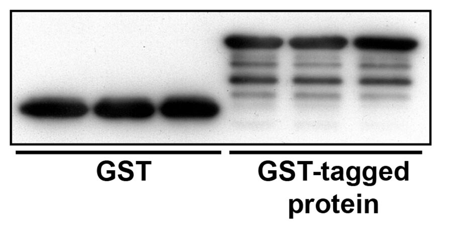
Western Blot: GST Epitope Tag Antibody [NB600-326] - 0.1, 0.3, or 1.0.ug of 293 cell lysate expressing a GST-tag fusion protein. Antibody used at 0.06 ug/ml (1:15,000).![Simple Western: GST Epitope Tag AntibodyBSA Free [NB600-326]](https://resources.rndsystems.com/images/products/GST-Epitope-Tag-Antibody-Simple-Western-NB600-326-img0004.jpg "Simple Western: GST Epitope Tag AntibodyBSA Free [NB600-326]")
Simple Western: GST Epitope Tag AntibodyBSA Free [NB600-326]
Simple Western: GST Epitope Tag Antibody [NB600-326] - Simple Western lane view shows a specific band for GST Epitope Tag in 5 ng/ml of hUbiquitn-GST (left) & hISG15-GST (right) lysate(s). This experiment was performed under reducing conditions using the 12 - 230 kDa separation system.![Simple Western: GST Epitope Tag AntibodyBSA Free [NB600-326]](https://resources.rndsystems.com/images/products/GST-Epitope-Tag-Antibody-Simple-Western-NB600-326-img0005.jpg "Simple Western: GST Epitope Tag AntibodyBSA Free [NB600-326]")
Simple Western: GST Epitope Tag AntibodyBSA Free [NB600-326]
Simple Western: GST Epitope Tag Antibody [NB600-326] - Electropherogram image(s) of corresponding Simple Western lane view. GST Epitope Tag antibody was used at 1:2000 dilution on hUbiquitn-GST (left) & hISG15-GST (right) lysate(s).![Simple Western: GST Epitope Tag AntibodyBSA Free [NB600-326]](https://resources.rndsystems.com/images/products/GST-Epitope-Tag-Antibody-Simple-Western-NB600-326-img0006.jpg "Simple Western: GST Epitope Tag AntibodyBSA Free [NB600-326]")
Simple Western: GST Epitope Tag AntibodyBSA Free [NB600-326]
Simple Western: GST Epitope Tag Antibody [NB600-326] - Simple Western lane view shows a specific band for GST Epitope Tag in 5 ng/ml of hVAV1-GST, E. coli (left) & hPARK2-GST-conditioned media (right) lysate(s). This experiment was performed under reducing conditions using the 12 - 230 kDa separation system.![Simple Western: GST Epitope Tag AntibodyBSA Free [NB600-326]](https://resources.rndsystems.com/images/products/GST-Epitope-Tag-Antibody-Simple-Western-NB600-326-img0007.jpg "Simple Western: GST Epitope Tag AntibodyBSA Free [NB600-326]")
Simple Western: GST Epitope Tag AntibodyBSA Free [NB600-326]
Simple Western: GST Epitope Tag Antibody [NB600-326] - Electropherogram image(s) of corresponding Simple Western lane view. GST Epitope Tag antibody was used at 1:2000 dilution on hVAV1-GST, E. coli (left) & hPARK2-GST-conditioned media (right) lysate(s).Applications for GST Epitope Tag Antibody - BSA Free
Application
Recommended Usage
ELISA
1:100-1:2000
Immunocytochemistry/ Immunofluorescence
1:100-1:400
Immunohistochemistry
1:10-1:500
Immunohistochemistry-Paraffin
1:100-1:400
Immunoprecipitation
1-4 ug/mg lysate
Simple Western
1:2000
Western Blot
1:1000 - 1:30000
Application Notes
In Simple Western only 10 - 15 uL of the recommended dilution is used per data point.
See Simple Western Antibody Database for Simple Western validation: Tested in hUbiquitn-GST and hISG15-GST lysates, hVAV1-GST, E. coli lysate and hPARK2-GST, conditioned media, separated by Size, antibody dilution of 1:2000, apparent MW was 39, 47, 43, 81 kDa. IHC-P reactivity reported in (PMID: 19412421).
See Simple Western Antibody Database for Simple Western validation: Tested in hUbiquitn-GST and hISG15-GST lysates, hVAV1-GST, E. coli lysate and hPARK2-GST, conditioned media, separated by Size, antibody dilution of 1:2000, apparent MW was 39, 47, 43, 81 kDa. IHC-P reactivity reported in (PMID: 19412421).
Reviewed Applications
Read 2 reviews rated 5 using NB600-326 in the following applications:
Formulation, Preparation, and Storage
Purification
Immunogen affinity purified
Formulation
PBS
Format
BSA Free
Preservative
0.09% Sodium Azide
Concentration
1.0 mg/ml
Shipping
The product is shipped with polar packs. Upon receipt, store it immediately at the temperature recommended below.
Stability & Storage
Store at 4C. Do not freeze.
Background: GST Epitope Tag
Alternate Names
EC 2.5.1.18, glutathione S-alkyltransferase, glutathione S-aralkyltransferase, glutathione S-aryltransferase, glutathione S-transferase M1, glutathione S-transferase mu 1, GST class-mu 1, GST HB subunit 4, GST1, GSTM1-1, GSTM1a-1a, GSTM1b-1b, GTH4, GTM1, H-B, HB subunit 4, MGC26563, MU, MU-1, S-(hydroxyalkyl)glutathione lyase
Gene Symbol
GSTM1
Additional GST Epitope Tag Products
Product Documents for GST Epitope Tag Antibody - BSA Free
Certificate of Analysis
To download a Certificate of Analysis, please enter a lot or batch number in the search box below.
Product Specific Notices for GST Epitope Tag Antibody - BSA Free
This product is for research use only and is not approved for use in humans or in clinical diagnosis. Primary Antibodies are guaranteed for 1 year from date of receipt.
Citations for GST Epitope Tag Antibody - BSA Free
 Powered by Bioz
Customer Reviews for GST Epitope Tag Antibody - BSA Free (2)
5 out of 5
2 Customer Ratings
Have you used GST Epitope Tag Antibody - BSA Free?
Submit a review and receive an Amazon gift card!
$25/€18/£15/$25CAN/¥2500 Yen for a review with an image
$10/€7/£6/$10CAN/¥1110 Yen for a review without an image
Submit a review
Customer Images
Showing
1
-
2 of
2 reviews
Showing All
Filter By:
-
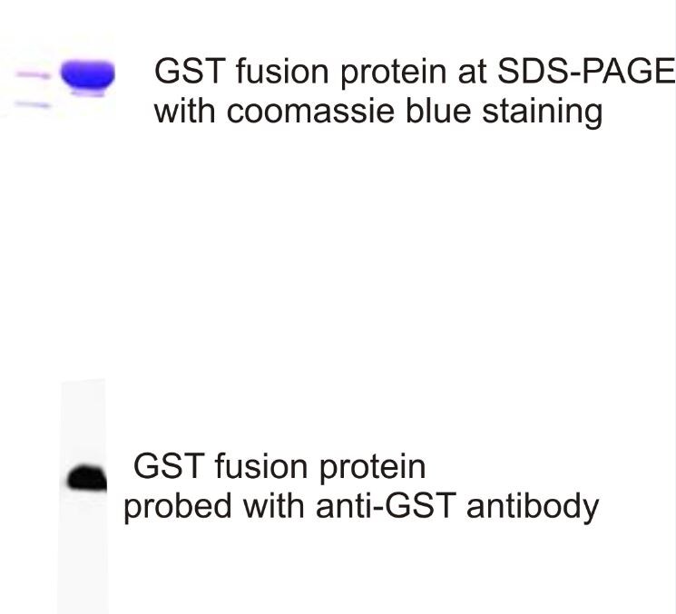
Application: Western BlotSample Tested: GST fustion proteinSpecies: OtherVerified Customer | Posted 01/07/2016GST fusion protein probed with GST antibody

-
Application: Western BlotSample Tested: GST pulldownsSpecies: OtherVerified Customer | Posted 07/24/2015GST Western Blot
There are no reviews that match your criteria.
Protocols
Find general support by application which include: protocols, troubleshooting, illustrated assays, videos and webinars.
- Antigen Retrieval Protocol (PIER)
- Antigen Retrieval for Frozen Sections Protocol
- Appropriate Fixation of IHC/ICC Samples
- Cellular Response to Hypoxia Protocols
- Chromogenic IHC Staining of Formalin-Fixed Paraffin-Embedded (FFPE) Tissue Protocol
- Chromogenic Immunohistochemistry Staining of Frozen Tissue
- ClariTSA™ Fluorophore Kits
- Detection & Visualization of Antibody Binding
- ELISA Sample Preparation & Collection Guide
- ELISA Troubleshooting Guide
- Fluorescent IHC Staining of Frozen Tissue Protocol
- Graphic Protocol for Heat-induced Epitope Retrieval
- Graphic Protocol for the Preparation and Fluorescent IHC Staining of Frozen Tissue Sections
- Graphic Protocol for the Preparation and Fluorescent IHC Staining of Paraffin-embedded Tissue Sections
- Graphic Protocol for the Preparation of Gelatin-coated Slides for Histological Tissue Sections
- How to Run an R&D Systems DuoSet ELISA
- How to Run an R&D Systems Quantikine ELISA
- How to Run an R&D Systems Quantikine™ QuicKit™ ELISA
- ICC Cell Smear Protocol for Suspension Cells
- ICC Immunocytochemistry Protocol Videos
- ICC for Adherent Cells
- IHC Sample Preparation (Frozen sections vs Paraffin)
- Immunocytochemistry (ICC) Protocol
- Immunocytochemistry Troubleshooting
- Immunofluorescence of Organoids Embedded in Cultrex Basement Membrane Extract
- Immunofluorescent IHC Staining of Formalin-Fixed Paraffin-Embedded (FFPE) Tissue Protocol
- Immunohistochemistry (IHC) and Immunocytochemistry (ICC) Protocols
- Immunohistochemistry Frozen Troubleshooting
- Immunohistochemistry Paraffin Troubleshooting
- Immunoprecipitation Protocol
- Preparing Samples for IHC/ICC Experiments
- Preventing Non-Specific Staining (Non-Specific Binding)
- Primary Antibody Selection & Optimization
- Protocol for Heat-Induced Epitope Retrieval (HIER)
- Protocol for Making a 4% Formaldehyde Solution in PBS
- Protocol for VisUCyte™ HRP Polymer Detection Reagent
- Protocol for the Fluorescent ICC Staining of Cell Smears - Graphic
- Protocol for the Fluorescent ICC Staining of Cultured Cells on Coverslips - Graphic
- Protocol for the Preparation & Fixation of Cells on Coverslips
- Protocol for the Preparation and Chromogenic IHC Staining of Frozen Tissue Sections
- Protocol for the Preparation and Chromogenic IHC Staining of Frozen Tissue Sections - Graphic
- Protocol for the Preparation and Chromogenic IHC Staining of Paraffin-embedded Tissue Sections
- Protocol for the Preparation and Chromogenic IHC Staining of Paraffin-embedded Tissue Sections - Graphic
- Protocol for the Preparation and Fluorescent ICC Staining of Cells on Coverslips
- Protocol for the Preparation and Fluorescent ICC Staining of Non-adherent Cells
- Protocol for the Preparation and Fluorescent ICC Staining of Stem Cells on Coverslips
- Protocol for the Preparation and Fluorescent IHC Staining of Frozen Tissue Sections
- Protocol for the Preparation and Fluorescent IHC Staining of Paraffin-embedded Tissue Sections
- Protocol for the Preparation of Gelatin-coated Slides for Histological Tissue Sections
- Protocol for the Preparation of a Cell Smear for Non-adherent Cell ICC - Graphic
- Quantikine HS ELISA Kit Assay Principle, Alkaline Phosphatase
- Quantikine HS ELISA Kit Principle, Streptavidin-HRP Polymer
- R&D Systems Quality Control Western Blot Protocol
- Sandwich ELISA (Colorimetric) – Biotin/Streptavidin Detection Protocol
- Sandwich ELISA (Colorimetric) – Direct Detection Protocol
- TUNEL and Active Caspase-3 Detection by IHC/ICC Protocol
- The Importance of IHC/ICC Controls
- Troubleshooting Guide: ELISA
- Troubleshooting Guide: Immunohistochemistry
- Troubleshooting Guide: Western Blot Figures
- Western Blot Conditions
- Western Blot Protocol
- Western Blot Protocol for Cell Lysates
- Western Blot Troubleshooting
- Western Blot Troubleshooting Guide
- View all Protocols, Troubleshooting, Illustrated assays and Webinars
Loading...