HA Tag Antibody - BSA Free
Novus Biologicals | Catalog # NB600-362
![Immunohistochemistry: HA Tag Antibody - BSA Free [NB600-362]](https://resources.rndsystems.com/images/products/HA-Tag-Antibody-Immunohistochemistry-NB600-362-img0011.jpg "Immunohistochemistry: HA Tag Antibody - BSA Free [NB600-362]")
Key Product Details
Validated by
Biological Validation
Species Reactivity
Validated:
Epitope Tag
Cited:
Human, Mouse, Epitope Tag
Applications
Validated:
Immunohistochemistry, Western Blot, ELISA, Immunocytochemistry/ Immunofluorescence, Immunoprecipitation
Cited:
Immunohistochemistry-Frozen, Western Blot, Immunocytochemistry/ Immunofluorescence, Immunoprecipitation, Bioassay
Label
Unconjugated
Antibody Source
Polyclonal Goat IgG
Format
BSA Free
Loading...
Product Specifications
Immunogen
This HA Tag Antibody was developed by immunizing goats with HA cleavage site (YPYDVPDYA) conjugated to KLH.
Clonality
Polyclonal
Host
Goat
Isotype
IgG
Scientific Data Images for HA Tag Antibody - BSA Free
Immunohistochemistry: HA Tag Antibody - BSA Free [NB600-362]
HA-Tag-Antibody-Immunohistochemistry-NB600-362-img0011.jpg![Western Blot: HA Tag AntibodyBSA Free [NB600-362]](https://resources.rndsystems.com/images/products/HA-Tag-Antibody-Western-Blot-NB600-362-img0006.jpg "Western Blot: HA Tag AntibodyBSA Free [NB600-362]")
Western Blot: HA Tag AntibodyBSA Free [NB600-362]
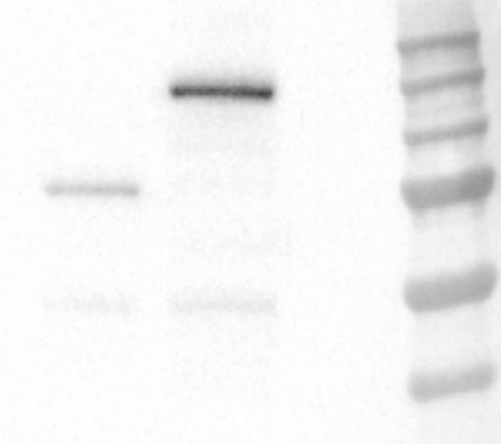
Western Blot: HA Tag Antibody [NB600-362] - Lysates of HEK293T human embryonic kidney cell line transfected with N-terminal HA-tagged MICA, N-terminal HA-tagged LGI-2, mock transfected, C-terminal HA-tagged LRRN5, and C-terminal HA-tagged Trance. PVDF membrane was probed with 1:500 dilution of 1:2500 dilution of goat anti-HA Tag polyclonal (NB600-362, Novus Biologicals), 1:5000 dilution of rabbit anti-HA Tag polyclonal (NB600-363, Novus Biologicals), followed by 1:2000 dilution of the appropriate HRP-conjugated secondary antibody, donkey anti-mouse IgG (HAF018).![Immunocytochemistry/ Immunofluorescence: HA Tag Antibody - BSA Free [NB600-362]](https://resources.rndsystems.com/images/products/HA-Tag-Antibody-Immunocytochemistry-Immunofluorescence-NB600-362-img0005.jpg "Immunocytochemistry/ Immunofluorescence: HA Tag Antibody - BSA Free [NB600-362]")
Immunocytochemistry/ Immunofluorescence: HA Tag Antibody - BSA Free [NB600-362]
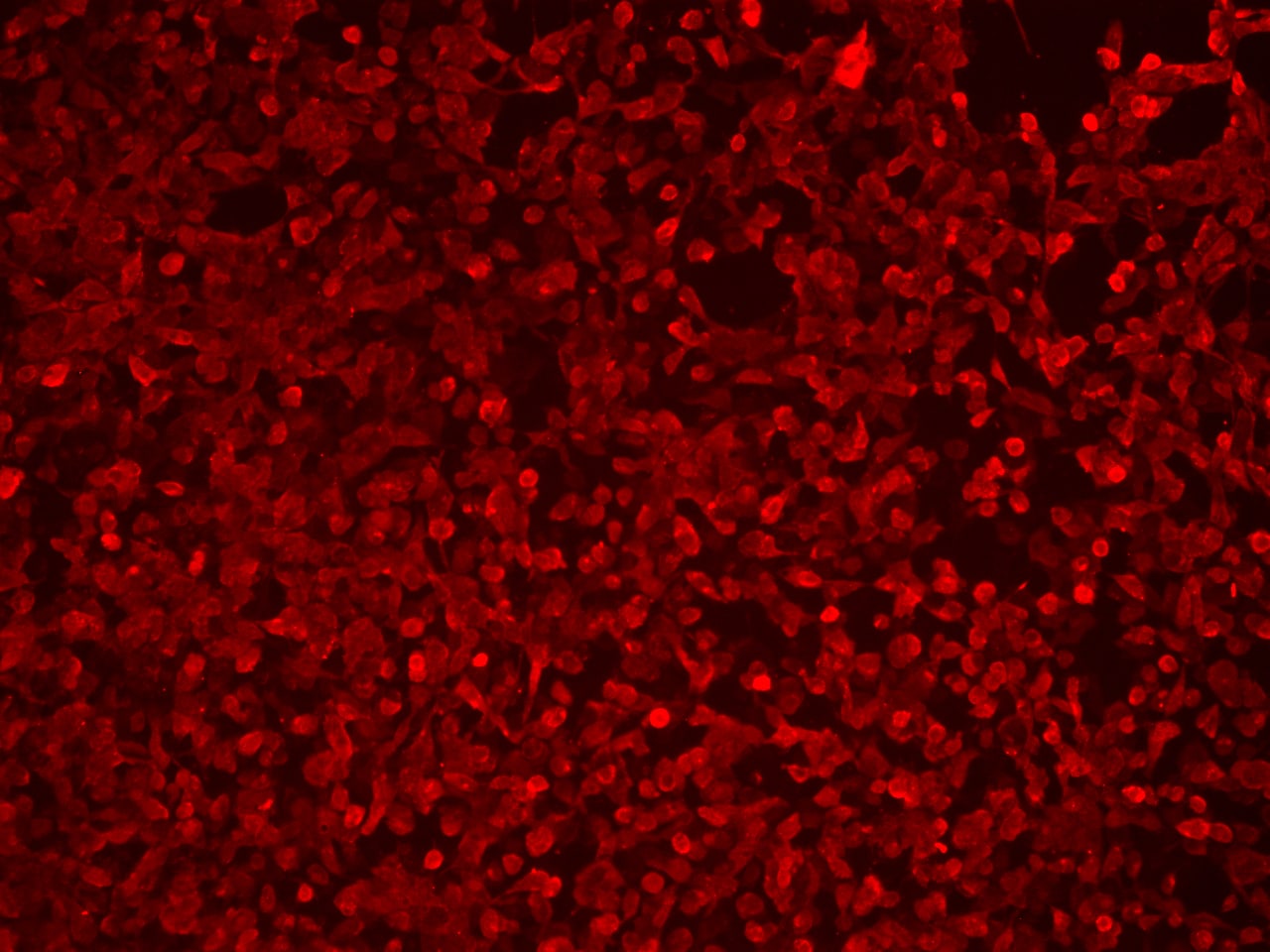
Immunocytochemistry/Immunofluorescence: HA Tag Antibody [NB600-362] - HA-tagged proteins were detected in immersion fixed HEK293 human embryonic kidney cell line transfected with HA-tagged MGAT2 using 1 ug/mL goat anti-HA Tag polyclonal (NB600-362, Novus Biologicals), 1 ug/mL rabbit anti-HA Tag polyclonal (NB600-363, Novus Biologicals). Cells were stained using the appropriate secondary antibody, donkey anti-mouse IgG-NL557 (NL007) and counterstained with DAPI (blue).![Western Blot: HA Tag AntibodyBSA Free [NB600-362]](https://resources.rndsystems.com/images/products/HA-Tag-Antibody---BSA-Free-Western-Blot-NB600-362-img0012.jpg "Western Blot: HA Tag AntibodyBSA Free [NB600-362]")
Western Blot: HA Tag AntibodyBSA Free [NB600-362]
Western Blot: HA Tag Antibody - BSA Free [NB600-362] - 200, 67, 22, 7, or 2 ng of E. coli whole cell lysateexpressing a multi-tag fusion protein. Antibodies: Affinitypurified, goat anti-HA antibody used for WBat 0.04 ug/ml (1:25,000). Detection: Chemiluminescencewith an exposure time of 10 seconds.![Immunocytochemistry/ Immunofluorescence: HA Tag Antibody - BSA Free [NB600-362]](https://resources.rndsystems.com/images/products/HA-Tag-Antibody-Immunocytochemistry-NB600-362-img0010.jpg "Immunocytochemistry/ Immunofluorescence: HA Tag Antibody - BSA Free [NB600-362]")
Immunocytochemistry/ Immunofluorescence: HA Tag Antibody - BSA Free [NB600-362]
Immunocytochemistry/Immunofluorescence: HA Tag Antibody [NB600-362] - HA tagged RFX6 overexpression in HEK293 stained with HA Tag antibody at a dilution of 1:250. ICC/IF image submitted by a verified customer review.
Immunocytochemistry/ Immunofluorescence: HA Tag Antibody - BSA Free [NB600-362] -
SUMO1 conjugates are not localized to amyloid plaques. (a) Sagittal brain sections of 24‐week‐old KI/AD & WT/AD animals were stained using anti‐HA antibodies (red), 6E10 antibodies (green) that label amyloid beta 1–42 among other amyloid beta variants (epitope lies within amino acids 3–8 of amyloid beta) & anti‐MAP2 antibodies (blue). Sections of the hippocampal subiculum are shown. Images are representatives of three independent experiments. Scale bar, 25 μm. (b) Anti‐HA signal intensity in amyloid plaques & in cell nuclei was quantified using ImageJ (N = 3, ***significance between WT/AD & KI/AD, p = .0007 in Student's t test) Image collected & cropped by CiteAb from the following publication (https://pubmed.ncbi.nlm.nih.gov/29633471), licensed under a CC-BY license. Not internally tested by Novus Biologicals.
Immunocytochemistry/ Immunofluorescence: HA Tag Antibody - BSA Free [NB600-362] -
SUMO1 conjugates remain nuclear during increased amyloid burden. (a) Western blot analysis of subcellular fractions of 36‐week‐old KI/WT & KI/AD mouse brain using anti‐HA antibody (upper two panels) & antibodies to GluN1 (a marker of the postsynaptic compartment) & Synaptophysin (a marker of the presynaptic compartment) to validate the fractionation procedure (lower two panels). H, homogenate; P1, nuclear pellet; S1, supernatant after P1 sedimentation; P2, crude synaptosomal pellet; S2, supernatant after P2 sedimentation; LP1, lysed synaptosomal membranes; LS1, supernatant after LP1 sedimentation; LP2, pellet after LS1 sedimentation, SPM, synaptic plasma membranes. Bracket indicates the anti‐HA signal representing SUMOylation, arrow indicates RanGAP1, stars indicate nonspecific signal detected by the anti‐HA antibody. (b) Brain sagittal sections of aged (36 weeks old) KI/AD (left panels) & WT/AD (right panels) mice were immunostained using antibodies directed against HA (red, labels HA‐HA conjugates), MAP2 (green, labels neuronal somata & dendrites), & Synapsin1 (Syn1, magenta, labels synapses). The images show triple‐labeled neurons of hippocampal subiculum (b), cortical layer 5 (c), & proximal apical dendrite from hippocampal CA3 (d). The white line shows the orientation of the line‐scan used to generate the image stack shown in the bottom side view. Scale bar: 10 μm. Note that the anti‐HA immunosignal is mainly located in neuronal nuclei, only background staining is observed in WT/AD mice. Little anti‐HA signal is observed along MAP2‐positive structures & does not colocalize with Synapsin1 Image collected & cropped by CiteAb from the following publication (https://pubmed.ncbi.nlm.nih.gov/29633471), licensed under a CC-BY license. Not internally tested by Novus Biologicals.Applications for HA Tag Antibody - BSA Free
Application
Recommended Usage
ELISA
1:1000 - 1:3000
Immunocytochemistry/ Immunofluorescence
1:100 - 1:400
Immunoprecipitation
2 - 10 ug / mg lysate
Western Blot
1:10000 - 1:25000
Application Notes
ICC/IF reactivity reported in scientific literature (PMID: 23050017).
Reviewed Applications
Read 3 reviews rated 4.3 using NB600-362 in the following applications:
Formulation, Preparation, and Storage
Purification
Immunogen affinity purified
Formulation
PBS
Format
BSA Free
Preservative
0.09% Sodium Azide
Concentration
1.0 mg/ml
Shipping
The product is shipped with polar packs. Upon receipt, store it immediately at the temperature recommended below.
Stability & Storage
Store at 4C. Do not freeze.
Background: HA Tag
References
1. Wilks, S., Graaf, M. D., Smith, D. J., & Burke, D. F. (2012). A review of influenza haemagglutinin receptor binding as it relates to pandemic properties. Vaccine, 30(29), 4369-4376. doi:10.1016/j.vaccine.2012.02.076
2. Wu, N. C., & Wilson, I. A. (2019). Influenza hemagglutinin structures and antibody recognition. Cold Spring Harbor Perspectives in Medicine, 10(8). doi:10.1101/cshperspect.a038778
3. Zhao, X., Li, G., & Liang, S. (2013). Several affinity tags commonly used in chromatographic purification. Journal of Analytical Methods in Chemistry, 2013, 1-8. doi:10.1155/2013/581093
4. Kimple, M. E., Brill, A. L., & Pasker, R. L. (2013). Overview of affinity tags for protein purification. Current Protocols in Protein Science, 73(1). doi:10.1002/0471140864.ps0909s73
5. Schembri, L., Dalibart, R., Tomasello, F., Legembre, P., Ichas, F., & Giorgi, F. D. (2007). The HA tag is cleaved and loses immunoreactivity during apoptosis. Nature Methods, 4(2), 107-108. doi:10.1038/nmeth0207-107
Alternate Names
HA Epitope Tag
Additional HA Tag Products
Product Documents for HA Tag Antibody - BSA Free
Certificate of Analysis
To download a Certificate of Analysis, please enter a lot or batch number in the search box below.
Product Specific Notices for HA Tag Antibody - BSA Free
This product is for research use only and is not approved for use in humans or in clinical diagnosis. Primary Antibodies are guaranteed for 1 year from date of receipt.
Related Research Areas
Citations for HA Tag Antibody - BSA Free
 Powered by Bioz
Customer Reviews for HA Tag Antibody - BSA Free (3)
4.3 out of 5
3 Customer Ratings
Have you used HA Tag Antibody - BSA Free?
Submit a review and receive an Amazon gift card!
$25/€18/£15/$25CAN/¥2500 Yen for a review with an image
$10/€7/£6/$10CAN/¥1110 Yen for a review without an image
Submit a review
Customer Images
Showing
1
-
3 of
3 reviews
Showing All
Filter By:
-
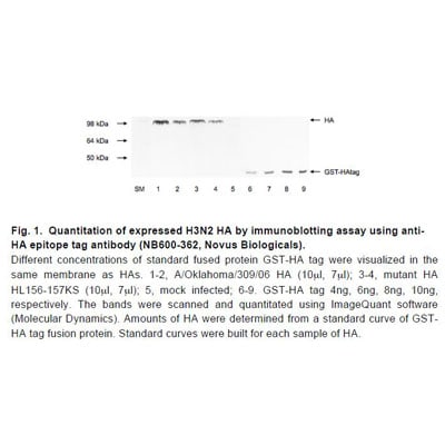
Application: Western BlotSample Tested: HEK 293T cellsSpecies: HumanVerified Customer | Posted 04/28/2017First lane is the negative control (untransfected cells) Second lane is HA-tagged WT-RFX6 of 140 kDa Third lane is HA-tagged mutant RFX6, which is a truncated protein of 70 kDa antibody dilution is 1:2000 exposure time is 1 second

-
Application: ImmunocytochemistrySample Tested: 293t HEK and U2OS cellsSpecies: HumanVerified Customer | Posted 04/28/2017HA tagged RFX6 overexpression in HEK293 stained at a dilution of 1:250
-
Application: Western BlotSample Tested: Hela, Cos whole cell lysates, serum after expression in Baculovirus systemSpecies: OtherVerified Customer | Posted 03/04/2013
There are no reviews that match your criteria.
Protocols
Find general support by application which include: protocols, troubleshooting, illustrated assays, videos and webinars.
- Antigen Retrieval Protocol (PIER)
- Antigen Retrieval for Frozen Sections Protocol
- Appropriate Fixation of IHC/ICC Samples
- Cellular Response to Hypoxia Protocols
- Chromogenic IHC Staining of Formalin-Fixed Paraffin-Embedded (FFPE) Tissue Protocol
- Chromogenic Immunohistochemistry Staining of Frozen Tissue
- ClariTSA™ Fluorophore Kits
- Detection & Visualization of Antibody Binding
- ELISA Sample Preparation & Collection Guide
- ELISA Troubleshooting Guide
- Fluorescent IHC Staining of Frozen Tissue Protocol
- Graphic Protocol for Heat-induced Epitope Retrieval
- Graphic Protocol for the Preparation and Fluorescent IHC Staining of Frozen Tissue Sections
- Graphic Protocol for the Preparation and Fluorescent IHC Staining of Paraffin-embedded Tissue Sections
- Graphic Protocol for the Preparation of Gelatin-coated Slides for Histological Tissue Sections
- How to Run an R&D Systems DuoSet ELISA
- How to Run an R&D Systems Quantikine ELISA
- How to Run an R&D Systems Quantikine™ QuicKit™ ELISA
- ICC Cell Smear Protocol for Suspension Cells
- ICC Immunocytochemistry Protocol Videos
- ICC for Adherent Cells
- IHC Sample Preparation (Frozen sections vs Paraffin)
- Immunocytochemistry (ICC) Protocol
- Immunocytochemistry Troubleshooting
- Immunofluorescence of Organoids Embedded in Cultrex Basement Membrane Extract
- Immunofluorescent IHC Staining of Formalin-Fixed Paraffin-Embedded (FFPE) Tissue Protocol
- Immunohistochemistry (IHC) and Immunocytochemistry (ICC) Protocols
- Immunohistochemistry Frozen Troubleshooting
- Immunohistochemistry Paraffin Troubleshooting
- Immunoprecipitation Protocol
- Preparing Samples for IHC/ICC Experiments
- Preventing Non-Specific Staining (Non-Specific Binding)
- Primary Antibody Selection & Optimization
- Protocol for Heat-Induced Epitope Retrieval (HIER)
- Protocol for Making a 4% Formaldehyde Solution in PBS
- Protocol for VisUCyte™ HRP Polymer Detection Reagent
- Protocol for the Fluorescent ICC Staining of Cell Smears - Graphic
- Protocol for the Fluorescent ICC Staining of Cultured Cells on Coverslips - Graphic
- Protocol for the Preparation & Fixation of Cells on Coverslips
- Protocol for the Preparation and Chromogenic IHC Staining of Frozen Tissue Sections
- Protocol for the Preparation and Chromogenic IHC Staining of Frozen Tissue Sections - Graphic
- Protocol for the Preparation and Chromogenic IHC Staining of Paraffin-embedded Tissue Sections
- Protocol for the Preparation and Chromogenic IHC Staining of Paraffin-embedded Tissue Sections - Graphic
- Protocol for the Preparation and Fluorescent ICC Staining of Cells on Coverslips
- Protocol for the Preparation and Fluorescent ICC Staining of Non-adherent Cells
- Protocol for the Preparation and Fluorescent ICC Staining of Stem Cells on Coverslips
- Protocol for the Preparation and Fluorescent IHC Staining of Frozen Tissue Sections
- Protocol for the Preparation and Fluorescent IHC Staining of Paraffin-embedded Tissue Sections
- Protocol for the Preparation of Gelatin-coated Slides for Histological Tissue Sections
- Protocol for the Preparation of a Cell Smear for Non-adherent Cell ICC - Graphic
- Quantikine HS ELISA Kit Assay Principle, Alkaline Phosphatase
- Quantikine HS ELISA Kit Principle, Streptavidin-HRP Polymer
- R&D Systems Quality Control Western Blot Protocol
- Sandwich ELISA (Colorimetric) – Biotin/Streptavidin Detection Protocol
- Sandwich ELISA (Colorimetric) – Direct Detection Protocol
- TUNEL and Active Caspase-3 Detection by IHC/ICC Protocol
- The Importance of IHC/ICC Controls
- Troubleshooting Guide: ELISA
- Troubleshooting Guide: Immunohistochemistry
- Troubleshooting Guide: Western Blot Figures
- Western Blot Conditions
- Western Blot Protocol
- Western Blot Protocol for Cell Lysates
- Western Blot Troubleshooting
- Western Blot Troubleshooting Guide
- View all Protocols, Troubleshooting, Illustrated assays and Webinars
FAQs for HA Tag Antibody - BSA Free
Showing
1
-
1 of
1 FAQ
Showing All
-
Q: We are looking for a NON-RABBIT polyclonal anti-HA (hemagglutinin - YPYDVPDYA ) antibody for immunohistochemistry that works in 4% paraformaldehyde-fixed tissue. We have a transgenic mouse expressing an HA-tagged protein and want to perform double-labeling with another antibody made in rabbit. We have had no luck using mouse or rat monoclonal anti-HA antibodies. The non-rabbit polyclonal antibody would preferably be made in goat, although if you have other species let us know. It could be unconjugated or conjugated (eg fluorochrome or HRP). However, we are only interested in a product with demonstrated effectiveness for IHC in a published paper. Do you have any such products?
A:
We do have other host species for this target and some that have been mentioned in publications. Unfortunately for our goat antibodies against this tag none of them have been validated in IHC-P, and for two we have shown they do not work. Please see this link to our goat anti-HA Epitope Tag antibodies. Since they work in ICC/IF I would assume as long as the tag is expressed in your tissues that you should still pick it up. In this case we have an Innovators Reward Program you can take advantage of for testing a new application to save some money. Here are the chicken anti-HA Epitope Tag antibodies. None of them have been tested in IHC-P as before, or that would be indicated with a positive result and the application on the datasheet or a negative results with the (-) beside the application. Another option for you as well would be going with a Rabbit polyclonal that we have available that has been well publicized and reviewed by customers (cat# NB600-363). I know you plan on double staining, but one way around this would be to use your other antibody and incubate with the secondary for detection first. After doing that you could have this one directly conjugated to your preferred detection probe and then pick up both of their signals since you would already have bound secondary to your first rabbit primary, there would not be any non-specific binding.
Loading...