HA Tag Antibody - BSA Free
Novus Biologicals | Catalog # NB600-363
![Immunocytochemistry/ Immunofluorescence: HA Tag Antibody [NB600-363]](https://resources.rndsystems.com/images/products/HA-Tag-Antibody-Immunocytochemistry-Immunofluorescence-NB600-363-img0005.jpg "Immunocytochemistry/ Immunofluorescence: HA Tag Antibody [NB600-363]")
Key Product Details
Validated by
Knockout/Knockdown
Species Reactivity
Validated:
Epitope Tag
Cited:
Human, Mouse, Rat, Epitope Tag, Non-species specific, Yeast
Applications
Validated:
Knockout Validated, Immunohistochemistry, Immunohistochemistry-Paraffin, Western Blot, ELISA, Flow Cytometry, Immunocytochemistry/ Immunofluorescence, Immunoprecipitation
Cited:
Western Blot, Block/Neutralize, Flow Cytometry, Immunocytochemistry/ Immunofluorescence, Immunoprecipitation, Chemotaxis, Gel Supershift Assay, Super-Resolution Microscopy, SDS-Page
Label
Unconjugated
Antibody Source
Polyclonal Rabbit IgG
Format
BSA Free
Loading...
Product Specifications
Immunogen
This HA Tag Antibody was developed by immunizing rabbits with HA cleavage site (YPYDVPDYA) conjugated to KLH. Antibody was isolated by affinity chromatography using the peptide immobilized on solid support.
Clonality
Polyclonal
Host
Rabbit
Isotype
IgG
Scientific Data Images for HA Tag Antibody - BSA Free
Immunocytochemistry/ Immunofluorescence: HA Tag Antibody [NB600-363]
Immunocytochemistry/Immunofluorescence: HA Tag Antibody [NB600-363] - Detection of HA-tagged Tubulin in CHO cells transfected with mutant beta-tubulin cDNA encoding an HA epitope tag. Photo contributed by R. Bhattacharya and F. Cabral, University of Texas Medical School at Houston.![Western Blot: HA Tag Antibody [NB600-363]](https://resources.rndsystems.com/images/products/HA-Tag-Antibody-Western-Blot-NB600-363-img0019.jpg "Western Blot: HA Tag Antibody [NB600-363]")
Western Blot: HA Tag Antibody [NB600-363]
Western Blot: HA Tag Antibody [NB600-363] - 200, 100, or 50 ng of E. coli whole cell lysate expressing amulti-tag fusion protein.Antibody: Affinity purified rabbitanti-HA antibody used for WB at 0.04 ug/ml(1:25,000). Detection: Chemiluminescence with anexposure time of 3 seconds.![Western Blot: HA Tag Antibody [NB600-363]](https://resources.rndsystems.com/images/products/HA-Tag-Antibody-Western-Blot-NB600-363-img0018.jpg "Western Blot: HA Tag Antibody [NB600-363]")
Western Blot: HA Tag Antibody [NB600-363]
HA-Tag-Antibody-Western-Blot-NB600-363-img0018.jpg![Western Blot: HA Tag Antibody [NB600-363]](https://resources.rndsystems.com/images/products/HA-Tag-Antibody-Western-Blot-NB600-363-img0017.jpg "Western Blot: HA Tag Antibody [NB600-363]")
Western Blot: HA Tag Antibody [NB600-363]
Western Blot: HA Tag Antibody [NB600-363] - ZFN expression levels. After co-transfection of HEK293T cells with ZFN expression vectors and pEGFP, cell lysates were probed with antibodies against the HA tag and EGFP. Amount of transfected ZFN plasmids was 75 ng, 300 ng, and 1200 ng. NT, non-transfected cells. Selection-independent generation of gene knockout mouse embryonic stem cells using zinc-finger nucleases. PLoS One (2011)![Immunocytochemistry/ Immunofluorescence: HA Tag Antibody [NB600-363]](https://resources.rndsystems.com/images/products/HA-Tag-Antibody-Immunocytochemistry-Immunofluorescence-NB600-363-img0013.jpg "Immunocytochemistry/ Immunofluorescence: HA Tag Antibody [NB600-363]")
Immunocytochemistry/ Immunofluorescence: HA Tag Antibody [NB600-363]
Immunocytochemistry/Immunofluorescence: HA Tag Antibody [NB600-363] - HA-tagged proteins were detected in immersion fixed HEK293 human embryonic kidney cell line transfected with HA-tagged MGAT2 using 1 ug/mL goat anti-HA Tag polyclonal (NB600-362, Novus Biologicals), 1 ug/mL rabbit anti-HA Tag polyclonal (NB600-363, Novus Biologicals). Cells were stained using the appropriate secondary antibody, donkey anti-mouse IgG-NL557 (NL007) and counterstained with DAPI (blue).
Western Blot: HA Tag Antibody [NB600-363] -
Western Blot: HA Tag Antibody [NB600-363] - Gene knockout in U2OS.693 cells.(A) Schematic of ZFN-mediated knockout. A ZFN pair (ZFNR & ZFNL) designed to target position 502 in the EGFP gene (E502) creates a DNA double-strand break that is sealed by the error-prone non-homologous end-joining (NHEJ) pathway & hence leads to disruption of the coding sequence. (B) Dose-dependent gene disruption in U2OS.693 cells. U2OS.693 cells that stably express a destabilized EGFP were transfected with increasing amounts of E502-specific ZFN expression vectors (75–1200 ng). The percentage of EGFP-negative cells was determined 6 days post-transfection by flow cytometry (n = 3; indicated is average & standard deviation). E502-WT, ZFN with wild-type FokI domain; E502-OH, ZFN with obligate heterodimeric FokI domain. (C) ZFN expression levels. After co-transfection of HEK293T cells with ZFN expression vectors & pEGFP, cell lysates were probed with antibodies against the HA tag & EGFP. Amount of transfected ZFN plasmids was 75 ng, 300 ng, & 1200 ng. NT, non-transfected cells. (D) Kinetics of EGFP knockout. The graph displays the percentage of EGFP-negative cells (see B) from day 1 to day 6 post-transfection for two vector amounts (n = 3; indicated is average & standard deviation). WT, E502-WT; OH, E502-OH. Image collected & cropped by CiteAb from the following publication (https://pubmed.ncbi.nlm.nih.gov/22194948), licensed under a CC-BY license. Not internally tested by Novus Biologicals.Applications for HA Tag Antibody - BSA Free
Application
Recommended Usage
ELISA
1:100-1:2000
Immunocytochemistry/ Immunofluorescence
1:100-1:400
Immunohistochemistry
1:100-1:400
Immunohistochemistry-Paraffin
1:100-1:400
Immunoprecipitation
1-4 ug/mg lysate
Western Blot
1:1000-1:10000
Application Notes
Use in Gel Super Shift Assays reported in scientific literature (PMID:34289349). Use in ChIP reported in (PMID: 30659200).. Use in IHC reported in (PMID: 22880041).. Use in FLOW reported in scientific literature (Pryce R et al).. Knockout validation (PMID: 32354171).. Recommended dilution for coating ELISA plates is 1:100 - 1:500.
Reviewed Applications
Read 3 reviews rated 4.3 using NB600-363 in the following applications:
Flow Cytometry Panel Builder
Bio-Techne Knows Flow Cytometry
Save time and reduce costly mistakes by quickly finding compatible reagents using the Panel Builder Tool.
Advanced Features
- Spectra Viewer - Custom analysis of spectra from multiple fluorochromes
- Spillover Popups - Visualize the spectra of individual fluorochromes
- Antigen Density Selector - Match fluorochrome brightness with antigen density

Formulation, Preparation, and Storage
Purification
Immunogen affinity purified
Formulation
PBS
Format
BSA Free
Preservative
0.09% Sodium Azide
Concentration
0.1 mg/ml
Shipping
The product is shipped with polar packs. Upon receipt, store it immediately at the temperature recommended below.
Stability & Storage
Store at 4C. Do not freeze.
Background: HA Tag
References
1. Wilks, S., Graaf, M. D., Smith, D. J., & Burke, D. F. (2012). A review of influenza haemagglutinin receptor binding as it relates to pandemic properties. Vaccine, 30(29), 4369-4376. doi:10.1016/j.vaccine.2012.02.076
2. Wu, N. C., & Wilson, I. A. (2019). Influenza hemagglutinin structures and antibody recognition. Cold Spring Harbor Perspectives in Medicine, 10(8). doi:10.1101/cshperspect.a038778
3. Zhao, X., Li, G., & Liang, S. (2013). Several affinity tags commonly used in chromatographic purification. Journal of Analytical Methods in Chemistry, 2013, 1-8. doi:10.1155/2013/581093
4. Kimple, M. E., Brill, A. L., & Pasker, R. L. (2013). Overview of affinity tags for protein purification. Current Protocols in Protein Science, 73(1). doi:10.1002/0471140864.ps0909s73
5. Schembri, L., Dalibart, R., Tomasello, F., Legembre, P., Ichas, F., & Giorgi, F. D. (2007). The HA tag is cleaved and loses immunoreactivity during apoptosis. Nature Methods, 4(2), 107-108. doi:10.1038/nmeth0207-107
Alternate Names
HA Epitope Tag
Additional HA Tag Products
Product Documents for HA Tag Antibody - BSA Free
Certificate of Analysis
To download a Certificate of Analysis, please enter a lot or batch number in the search box below.
Product Specific Notices for HA Tag Antibody - BSA Free
This product is for research use only and is not approved for use in humans or in clinical diagnosis. Primary Antibodies are guaranteed for 1 year from date of receipt.
Related Research Areas
Citations for HA Tag Antibody - BSA Free
 Powered by Bioz
Customer Reviews for HA Tag Antibody - BSA Free (3)
4.3 out of 5
3 Customer Ratings
Have you used HA Tag Antibody - BSA Free?
Submit a review and receive an Amazon gift card!
$25/€18/£15/$25CAN/¥2500 Yen for a review with an image
$10/€7/£6/$10CAN/¥1110 Yen for a review without an image
Submit a review
Customer Images

Showing
1
-
3 of
3 reviews
Showing All
Filter By:
-
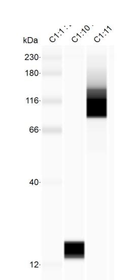
Application: Simple WesternSample Tested: Recombinant proteinsSpecies: HumanVerified Customer | Posted 06/08/2018

-
Application: Western BlotSample Tested: Hek 293T cell lysateSpecies: HumanVerified Customer | Posted 05/04/2013
-
Application: Western BlotSample Tested: Xenopus laevis inejected embryo Lysates, Sample Amount: 30ugSpecies: OtherVerified Customer | Posted 09/26/2011
There are no reviews that match your criteria.
Protocols
Find general support by application which include: protocols, troubleshooting, illustrated assays, videos and webinars.
- 7-Amino Actinomycin D (7-AAD) Cell Viability Flow Cytometry Protocol
- Antigen Retrieval Protocol (PIER)
- Antigen Retrieval for Frozen Sections Protocol
- Appropriate Fixation of IHC/ICC Samples
- Cellular Response to Hypoxia Protocols
- Chromogenic IHC Staining of Formalin-Fixed Paraffin-Embedded (FFPE) Tissue Protocol
- Chromogenic Immunohistochemistry Staining of Frozen Tissue
- ClariTSA™ Fluorophore Kits
- Detection & Visualization of Antibody Binding
- ELISA Sample Preparation & Collection Guide
- ELISA Troubleshooting Guide
- Extracellular Membrane Flow Cytometry Protocol
- Flow Cytometry Protocol for Cell Surface Markers
- Flow Cytometry Protocol for Staining Membrane Associated Proteins
- Flow Cytometry Staining Protocols
- Flow Cytometry Troubleshooting Guide
- Fluorescent IHC Staining of Frozen Tissue Protocol
- Graphic Protocol for Heat-induced Epitope Retrieval
- Graphic Protocol for the Preparation and Fluorescent IHC Staining of Frozen Tissue Sections
- Graphic Protocol for the Preparation and Fluorescent IHC Staining of Paraffin-embedded Tissue Sections
- Graphic Protocol for the Preparation of Gelatin-coated Slides for Histological Tissue Sections
- How to Run an R&D Systems DuoSet ELISA
- How to Run an R&D Systems Quantikine ELISA
- How to Run an R&D Systems Quantikine™ QuicKit™ ELISA
- ICC Cell Smear Protocol for Suspension Cells
- ICC Immunocytochemistry Protocol Videos
- ICC for Adherent Cells
- IHC Sample Preparation (Frozen sections vs Paraffin)
- Immunocytochemistry (ICC) Protocol
- Immunocytochemistry Troubleshooting
- Immunofluorescence of Organoids Embedded in Cultrex Basement Membrane Extract
- Immunofluorescent IHC Staining of Formalin-Fixed Paraffin-Embedded (FFPE) Tissue Protocol
- Immunohistochemistry (IHC) and Immunocytochemistry (ICC) Protocols
- Immunohistochemistry Frozen Troubleshooting
- Immunohistochemistry Paraffin Troubleshooting
- Immunoprecipitation Protocol
- Intracellular Flow Cytometry Protocol Using Alcohol (Methanol)
- Intracellular Flow Cytometry Protocol Using Detergents
- Intracellular Nuclear Staining Flow Cytometry Protocol Using Detergents
- Intracellular Staining Flow Cytometry Protocol Using Alcohol Permeabilization
- Intracellular Staining Flow Cytometry Protocol Using Detergents to Permeabilize Cells
- Preparing Samples for IHC/ICC Experiments
- Preventing Non-Specific Staining (Non-Specific Binding)
- Primary Antibody Selection & Optimization
- Propidium Iodide Cell Viability Flow Cytometry Protocol
- Protocol for Heat-Induced Epitope Retrieval (HIER)
- Protocol for Liperfluo
- Protocol for Making a 4% Formaldehyde Solution in PBS
- Protocol for VisUCyte™ HRP Polymer Detection Reagent
- Protocol for the Characterization of Human Th22 Cells
- Protocol for the Characterization of Human Th9 Cells
- Protocol for the Fluorescent ICC Staining of Cell Smears - Graphic
- Protocol for the Fluorescent ICC Staining of Cultured Cells on Coverslips - Graphic
- Protocol for the Preparation & Fixation of Cells on Coverslips
- Protocol for the Preparation and Chromogenic IHC Staining of Frozen Tissue Sections
- Protocol for the Preparation and Chromogenic IHC Staining of Frozen Tissue Sections - Graphic
- Protocol for the Preparation and Chromogenic IHC Staining of Paraffin-embedded Tissue Sections
- Protocol for the Preparation and Chromogenic IHC Staining of Paraffin-embedded Tissue Sections - Graphic
- Protocol for the Preparation and Fluorescent ICC Staining of Cells on Coverslips
- Protocol for the Preparation and Fluorescent ICC Staining of Non-adherent Cells
- Protocol for the Preparation and Fluorescent ICC Staining of Stem Cells on Coverslips
- Protocol for the Preparation and Fluorescent IHC Staining of Frozen Tissue Sections
- Protocol for the Preparation and Fluorescent IHC Staining of Paraffin-embedded Tissue Sections
- Protocol for the Preparation of Gelatin-coated Slides for Histological Tissue Sections
- Protocol for the Preparation of a Cell Smear for Non-adherent Cell ICC - Graphic
- Protocol: Annexin V and PI Staining by Flow Cytometry
- Protocol: Annexin V and PI Staining for Apoptosis by Flow Cytometry
- Quantikine HS ELISA Kit Assay Principle, Alkaline Phosphatase
- Quantikine HS ELISA Kit Principle, Streptavidin-HRP Polymer
- R&D Systems Quality Control Western Blot Protocol
- Sandwich ELISA (Colorimetric) – Biotin/Streptavidin Detection Protocol
- Sandwich ELISA (Colorimetric) – Direct Detection Protocol
- TUNEL and Active Caspase-3 Detection by IHC/ICC Protocol
- The Importance of IHC/ICC Controls
- Troubleshooting Guide: ELISA
- Troubleshooting Guide: Fluorokine Flow Cytometry Kits
- Troubleshooting Guide: Immunohistochemistry
- Troubleshooting Guide: Western Blot Figures
- Western Blot Conditions
- Western Blot Protocol
- Western Blot Protocol for Cell Lysates
- Western Blot Troubleshooting
- Western Blot Troubleshooting Guide
- View all Protocols, Troubleshooting, Illustrated assays and Webinars
FAQs for HA Tag Antibody - BSA Free
Showing
1
-
1 of
1 FAQ
Showing All
-
Q: We are looking for a NON-RABBIT polyclonal anti-HA (hemagglutinin - YPYDVPDYA ) antibody for immunohistochemistry that works in 4% paraformaldehyde-fixed tissue. We have a transgenic mouse expressing an HA-tagged protein and want to perform double-labeling with another antibody made in rabbit. We have had no luck using mouse or rat monoclonal anti-HA antibodies. The non-rabbit polyclonal antibody would preferably be made in goat, although if you have other species let us know. It could be unconjugated or conjugated (eg fluorochrome or HRP). However, we are only interested in a product with demonstrated effectiveness for IHC in a published paper. Do you have any such products?
A:
We do have other host species for this target and some that have been mentioned in publications. Unfortunately for our goat antibodies against this tag none of them have been validated in IHC-P, and for two we have shown they do not work. Please see this link to our goat anti-HA Epitope Tag antibodies. Since they work in ICC/IF I would assume as long as the tag is expressed in your tissues that you should still pick it up. In this case we have an Innovators Reward Program you can take advantage of for testing a new application to save some money. Here are the chicken anti-HA Epitope Tag antibodies. None of them have been tested in IHC-P as before, or that would be indicated with a positive result and the application on the datasheet or a negative results with the (-) beside the application. Another option for you as well would be going with a Rabbit polyclonal that we have available that has been well publicized and reviewed by customers (cat# NB600-363). I know you plan on double staining, but one way around this would be to use your other antibody and incubate with the secondary for detection first. After doing that you could have this one directly conjugated to your preferred detection probe and then pick up both of their signals since you would already have bound secondary to your first rabbit primary, there would not be any non-specific binding.
Loading...