Histone H2AX [p Ser139] Antibody (3F2)
Novus Biologicals | Catalog # NB100-74435

Loading...
Key Product Details
Validated by
Biological Validation
Species Reactivity
Validated:
Human, Mouse, Bovine
Cited:
Human, Mouse, Rabbit
Applications
Validated:
Immunohistochemistry, Immunohistochemistry-Paraffin, Western Blot, ELISA, Flow Cytometry, Immunocytochemistry/ Immunofluorescence, Simple Western
Cited:
Immunohistochemistry-Paraffin, Immunomicroscopy, Western Blot, Immunocytochemistry/ Immunofluorescence, Immunoprecipitation, IF/IHC
Label
Unconjugated
Antibody Source
Monoclonal Mouse IgG1 kappa Clone # 3F2
Loading...
Product Specifications
Immunogen
This Histone H2AX [p Ser139] Antibody (3F2) was developed against a synthetic peptide sequence surrounding phosphorylated Ser139.
Reactivity Notes
Bovine reactivity reported in scientific literature (PMID: 17604361). Please note that this antibody is reactive to Mouse and derived from the same host, Mouse. Additional Mouse on Mouse blocking steps may be required for IHC and ICC experiments. Please contact Technical Support for more information.
Modification
p Ser139
Marker
DNA Double-strand break marker
Specificity
In Western blot this antibody detects ~17 kDa protein representing phosphorylated H2AX in gamma irradiated HeLa cell lysate. In immunofluorescence procedures, recognizes phosphorylated H2AX in gamma irradiated HeLa cells. ELISA of phosphorylated H2AX can also be performed. Used in IHC to successfully detect H2A.X pSer140 in postnatal mouse lung section.
Clonality
Monoclonal
Host
Mouse
Isotype
IgG1 kappa
Theoretical MW
15 kDa.
Disclaimer note: The observed molecular weight of the protein may vary from the listed predicted molecular weight due to post translational modifications, post translation cleavages, relative charges, and other experimental factors.
Disclaimer note: The observed molecular weight of the protein may vary from the listed predicted molecular weight due to post translational modifications, post translation cleavages, relative charges, and other experimental factors.
Scientific Data Images for Histone H2AX [p Ser139] Antibody (3F2)
Simple Western: Histone H2AX [p Ser139] Antibody (3F2) [NB100-74435] - Simple Western lane view shows a specific band for Histone H2AX [p Ser139] in 0.2 mg/ml of Jurkat lysate(s). This experiment was performed under reducing conditions using the 12 - 230 kDa separation system.
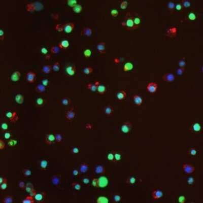
Immunocytochemistry/Immunofluorescence: Histone H2AX [p Ser139] Antibody (3F2) [NB100-74435] - Staining using NB100-74435, treatment with paraquat and iron induces MnSOD and Phosphorylation of H2AX in RAW 264.7 macrophages. Cells were treated for 20 hours with paraquat (500 uM) and iron (200 ug/ml) and stained with anti-Phospho-H2AX antibody.
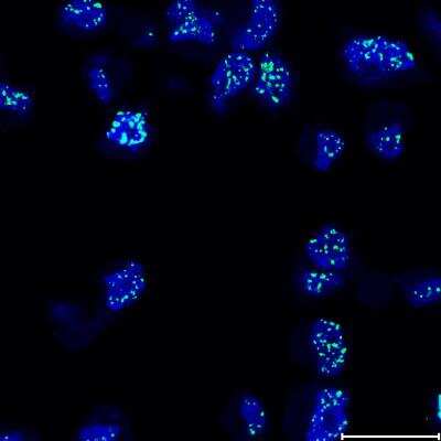
Immunocytochemistry/Immunofluorescence: Histone H2AX [p Ser139] Antibody (3F2) [NB100-74435] - STED image of MDA-MB-231 cells after 2 Gy irradiation. Blue channel: DAPI, window level [0 255]. Green channel: secondary antibody Alexa Fluor 488, window level [140 255]. Scale bar: 20 um. Image from verified customer review.
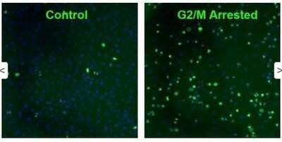
Immunocytochemistry/Immunofluorescence: Histone H2AX [p Ser139] Antibody (3F2) [NB100-74435] - Immunofluorescence analysis using Histone H2AX [p Ser139] antibody (3F2).
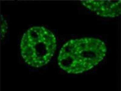
Immunocytochemistry/Immunofluorescence: Histone H2AX [p Ser139] Antibody (3F2) [NB100-74435] - IF image of phosphorylated H2AX in gamma irradiated Hela cells.
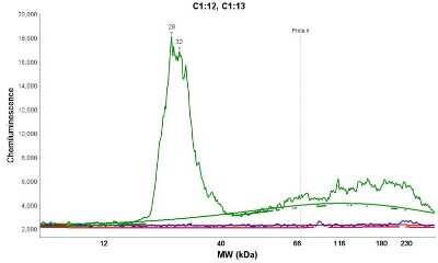
Simple Western: Histone H2AX [p Ser139] Antibody (3F2) [NB100-74435] - Electropherogram image(s) of corresponding Simple Western lane view. Histone H2AX [p Ser139] antibody (3F2) was used at 10 ug/ml dilution on Jurkat lysate(s).
![Histone H2AX [p Ser139] Antibody (3F2)](https://resources.rndsystems.com/images/products/nb100-74435_mouse-monoclonal-histone-h2ax-p-ser139-antibody-3f2-31020241534396.jpg "Immunocytochemistry/ Immunofluorescence: Histone H2AX [p Ser139] Antibody (3F2) [NB100-74435] -")
Immunocytochemistry/ Immunofluorescence: Histone H2AX [p Ser139] Antibody (3F2) [NB100-74435] -
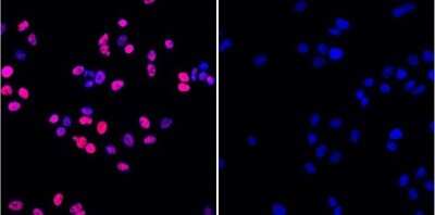
Immunocytochemistry/ Immunofluorescence: Histone H2AX [p Ser139] Antibody (3F2) [NB100-74435] - Evidence of DNA damage & oxidative stress in islets from HFD treated mice. (a) Representative immunofluorescent staining of gamma H2AX, insulin, & DAPI for islets from each treatment group. (b) Representative immunofluorescent staining of 4-hydroxynonenal (4-HNE), insulin, & DAPI for islets from each treatment group. (c) Representative immunofluorescent staining of nitrotyrosine, insulin, & DAPI for islets from each treatment group. (d–g) qRT-PCR analysis of total islet RNA for antioxidant genes, including Gpx1, SOD1, Nrf2, & Ppargc1 alpha. (n = 3) *p < 0.05, **p < 0.01. Image collected & cropped by CiteAb from the following publication (https://www.nature.com/articles/s41598-017-03869-5), licensed under a CC-BY license. Not internally tested by Novus Biologicals.Applications for Histone H2AX [p Ser139] Antibody (3F2)
Application
Recommended Usage
ELISA
1:100 - 1:2000
Flow Cytometry
1 ug/million cells
Immunocytochemistry/ Immunofluorescence
2 - 4 ug/ml
Immunohistochemistry
1:10 - 1:500
Immunohistochemistry-Paraffin
1:10 - 1:500
Simple Western
10 ug/ml
Western Blot
1 ug/ml
Application Notes
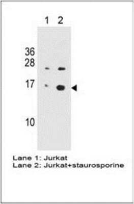
In WB: Detects an approx. 17 kDa protein representing phosphorylated H2AX in gamma irradiate Hela cell lysate.
In Simple Western only 10 - 15 uL of the recommended dilution is used per data point.
See Simple Western Antibody Database for Simple Western validation: Tested in Jurkat lysate, separated by Size, antibody dilution of 10 ug/mL, apparent MW was 28 kDa. Separated by Size-Wes, Sally Sue/Peggy Sue.
In Simple Western only 10 - 15 uL of the recommended dilution is used per data point.
See Simple Western Antibody Database for Simple Western validation: Tested in Jurkat lysate, separated by Size, antibody dilution of 10 ug/mL, apparent MW was 28 kDa. Separated by Size-Wes, Sally Sue/Peggy Sue.
Reviewed Applications
Read 2 reviews rated 5 using NB100-74435 in the following applications:
Flow Cytometry Panel Builder
Bio-Techne Knows Flow Cytometry
Save time and reduce costly mistakes by quickly finding compatible reagents using the Panel Builder Tool.
Advanced Features
- Spectra Viewer - Custom analysis of spectra from multiple fluorochromes
- Spillover Popups - Visualize the spectra of individual fluorochromes
- Antigen Density Selector - Match fluorochrome brightness with antigen density

Formulation, Preparation, and Storage
Purification
Protein G purified
Formulation
PBS with 1 mg/ml BSA
Preservative
0.05% Sodium Azide
Concentration
1 mg/ml
Shipping
The product is shipped with polar packs. Upon receipt, store it immediately at the temperature recommended below.
Stability & Storage
Store at -20C. Avoid freeze-thaw cycles.
Background: Histone H2AX
References
1. Palla, V. V., Karaolanis, G., Katafigiotis, I., Anastasiou, I., Patapis, P., Dimitroulis, D., & Perrea, D. (2017). gamma-H2AX: Can it be established as a classical cancer prognostic factor?. Tumour biology : the journal of the International Society for Oncodevelopmental Biology and Medicine. https://doi.org/10.1177/1010428317695931
2. Kuo, L. J., & Yang, L. X. (2008). Gamma-H2AX - a novel biomarker for DNA double-strand breaks. In vivo (Athens, Greece).
3. Kinner, A., Wu, W., Staudt, C., & Iliakis, G. (2008). Gamma-H2AX in recognition and signaling of DNA double-strand breaks in the context of chromatin. Nucleic acids research. https://doi.org/10.1093/nar/gkn550
4. Redon, C. E., Weyemi, U., Parekh, P. R., Huang, D., Burrell, A. S., & Bonner, W. M. (2012). gamma-H2AX and other histone post-translational modifications in the clinic. Biochimica et biophysica acta. https://doi.org/10.1016/j.bbagrm.2012.02.021
5. H2AX: Uniprot (P16104)
Additional Histone H2AX Products
Product Documents for Histone H2AX [p Ser139] Antibody (3F2)
Certificate of Analysis
To download a Certificate of Analysis, please enter a lot or batch number in the search box below.
Product Specific Notices for Histone H2AX [p Ser139] Antibody (3F2)
Licensed to Novus Biologicals LLC under U.S. Patent Nos. 6,362,317 and 6,884,873.
This product is for research use only and is not approved for use in humans or in clinical diagnosis. Primary Antibodies are guaranteed for 1 year from date of receipt.
Related Research Areas
Citations for Histone H2AX [p Ser139] Antibody (3F2)
 Powered by Bioz
Customer Reviews for Histone H2AX [p Ser139] Antibody (3F2) (2)
5 out of 5
2 Customer Ratings
Have you used Histone H2AX [p Ser139] Antibody (3F2)?
Submit a review and receive an Amazon gift card!
$25/€18/£15/$25CAN/¥2500 Yen for a review with an image
$10/€7/£6/$10CAN/¥1110 Yen for a review without an image
Submit a review
Customer Images
![Histone H2AX [p Ser139] Antibody (3F2) NB100-74435](https://resources.rndsystems.com/images/reviews/NB100-74435_74FA4E26-5545-471A-8A83-6939ED89AA05.jpg)
![Histone H2AX [p Ser139] Antibody (3F2) NB100-74435](https://resources.rndsystems.com/images/reviews/review_nb100-74435_59031_0_0.jpg)
Showing
1
-
2 of
2 reviews
Showing All
Filter By:
-
Application: Western BlotSample Tested: Colon tissueSpecies: MouseVerified Customer | Posted 10/02/2024Dual pink protein marker10 mg loading 1:1000 dilution HRP detection
![Histone H2AX [p Ser139] Antibody (3F2) NB100-74435](data:image/png;base64,R0lGODlhAQABAAD/ACwAAAAAAQABAAACADs=)
-
Application: ImmunofluorescenceSample Tested: MDA MB 231 cellsSpecies: HumanVerified Customer | Posted 05/17/2022STED image of MDA-MB-231 cells after 2 Gy irradiation. Blue channel: DAPI, window level [0 255]. Green channel: secondary antibody Alexa Fluor 488, window level [140 255]. Scale bar: 20 μm.
There are no reviews that match your criteria.
Protocols
Find general support by application which include: protocols, troubleshooting, illustrated assays, videos and webinars.
- 7-Amino Actinomycin D (7-AAD) Cell Viability Flow Cytometry Protocol
- Antigen Retrieval Protocol (PIER)
- Antigen Retrieval for Frozen Sections Protocol
- Appropriate Fixation of IHC/ICC Samples
- Cellular Response to Hypoxia Protocols
- Chromogenic IHC Staining of Formalin-Fixed Paraffin-Embedded (FFPE) Tissue Protocol
- Chromogenic Immunohistochemistry Staining of Frozen Tissue
- ClariTSA™ Fluorophore Kits
- Detection & Visualization of Antibody Binding
- ELISA Sample Preparation & Collection Guide
- ELISA Troubleshooting Guide
- Extracellular Membrane Flow Cytometry Protocol
- Flow Cytometry Protocol for Cell Surface Markers
- Flow Cytometry Protocol for Staining Membrane Associated Proteins
- Flow Cytometry Staining Protocols
- Flow Cytometry Troubleshooting Guide
- Fluorescent IHC Staining of Frozen Tissue Protocol
- Graphic Protocol for Heat-induced Epitope Retrieval
- Graphic Protocol for the Preparation and Fluorescent IHC Staining of Frozen Tissue Sections
- Graphic Protocol for the Preparation and Fluorescent IHC Staining of Paraffin-embedded Tissue Sections
- Graphic Protocol for the Preparation of Gelatin-coated Slides for Histological Tissue Sections
- How to Run an R&D Systems DuoSet ELISA
- How to Run an R&D Systems Quantikine ELISA
- How to Run an R&D Systems Quantikine™ QuicKit™ ELISA
- ICC Cell Smear Protocol for Suspension Cells
- ICC Immunocytochemistry Protocol Videos
- ICC for Adherent Cells
- IHC Sample Preparation (Frozen sections vs Paraffin)
- Immunocytochemistry (ICC) Protocol
- Immunocytochemistry Troubleshooting
- Immunofluorescence of Organoids Embedded in Cultrex Basement Membrane Extract
- Immunofluorescent IHC Staining of Formalin-Fixed Paraffin-Embedded (FFPE) Tissue Protocol
- Immunohistochemistry (IHC) and Immunocytochemistry (ICC) Protocols
- Immunohistochemistry Frozen Troubleshooting
- Immunohistochemistry Paraffin Troubleshooting
- Intracellular Flow Cytometry Protocol Using Alcohol (Methanol)
- Intracellular Flow Cytometry Protocol Using Detergents
- Intracellular Nuclear Staining Flow Cytometry Protocol Using Detergents
- Intracellular Staining Flow Cytometry Protocol Using Alcohol Permeabilization
- Intracellular Staining Flow Cytometry Protocol Using Detergents to Permeabilize Cells
- Preparing Samples for IHC/ICC Experiments
- Preventing Non-Specific Staining (Non-Specific Binding)
- Primary Antibody Selection & Optimization
- Propidium Iodide Cell Viability Flow Cytometry Protocol
- Protocol for Heat-Induced Epitope Retrieval (HIER)
- Protocol for Liperfluo
- Protocol for Making a 4% Formaldehyde Solution in PBS
- Protocol for VisUCyte™ HRP Polymer Detection Reagent
- Protocol for the Characterization of Human Th22 Cells
- Protocol for the Characterization of Human Th9 Cells
- Protocol for the Fluorescent ICC Staining of Cell Smears - Graphic
- Protocol for the Fluorescent ICC Staining of Cultured Cells on Coverslips - Graphic
- Protocol for the Preparation & Fixation of Cells on Coverslips
- Protocol for the Preparation and Chromogenic IHC Staining of Frozen Tissue Sections
- Protocol for the Preparation and Chromogenic IHC Staining of Frozen Tissue Sections - Graphic
- Protocol for the Preparation and Chromogenic IHC Staining of Paraffin-embedded Tissue Sections
- Protocol for the Preparation and Chromogenic IHC Staining of Paraffin-embedded Tissue Sections - Graphic
- Protocol for the Preparation and Fluorescent ICC Staining of Cells on Coverslips
- Protocol for the Preparation and Fluorescent ICC Staining of Non-adherent Cells
- Protocol for the Preparation and Fluorescent ICC Staining of Stem Cells on Coverslips
- Protocol for the Preparation and Fluorescent IHC Staining of Frozen Tissue Sections
- Protocol for the Preparation and Fluorescent IHC Staining of Paraffin-embedded Tissue Sections
- Protocol for the Preparation of Gelatin-coated Slides for Histological Tissue Sections
- Protocol for the Preparation of a Cell Smear for Non-adherent Cell ICC - Graphic
- Protocol: Annexin V and PI Staining by Flow Cytometry
- Protocol: Annexin V and PI Staining for Apoptosis by Flow Cytometry
- Quantikine HS ELISA Kit Assay Principle, Alkaline Phosphatase
- Quantikine HS ELISA Kit Principle, Streptavidin-HRP Polymer
- R&D Systems Quality Control Western Blot Protocol
- Sandwich ELISA (Colorimetric) – Biotin/Streptavidin Detection Protocol
- Sandwich ELISA (Colorimetric) – Direct Detection Protocol
- TUNEL and Active Caspase-3 Detection by IHC/ICC Protocol
- The Importance of IHC/ICC Controls
- Troubleshooting Guide: ELISA
- Troubleshooting Guide: Fluorokine Flow Cytometry Kits
- Troubleshooting Guide: Immunohistochemistry
- Troubleshooting Guide: Western Blot Figures
- Western Blot Conditions
- Western Blot Protocol
- Western Blot Protocol for Cell Lysates
- Western Blot Troubleshooting
- Western Blot Troubleshooting Guide
- View all Protocols, Troubleshooting, Illustrated assays and Webinars
Loading...