HLA-DR Antibody (L243) [FITC]
Novus Biologicals | Catalog # NB100-77856
Key Product Details
Species Reactivity
Human, Baboon, Canine, Primate
Applications
Flow Cytometry, Immunocytochemistry/ Immunofluorescence
Label
FITC (Excitation = 495 nm, Emission = 519 nm)
Antibody Source
Monoclonal Mouse IgG2a Kappa Clone # L243
Loading...
Product Specifications
Immunogen
Human lymphoblastoid cell line (RPMI 8866).
Reactivity Notes
Predicted cross-reactivity with Chimpanzee, Baboon, Cynomolgus, Rhesus, Marmoset, Tamarin, Squirrel Monkey and Canine.
Clonality
Monoclonal
Host
Mouse
Isotype
IgG2a Kappa
Scientific Data Images for HLA-DR Antibody (L243) [FITC]
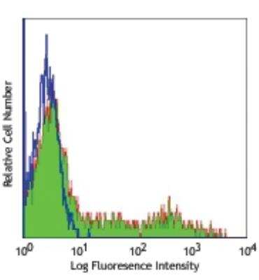
Flow Cytometry: HLA-DR Antibody (L243) [FITC] [NB100-77856] - Human peripheral blood lymphocytes stained with L243 FITC
Applications for HLA-DR Antibody (L243) [FITC]
Application
Recommended Usage
Flow Cytometry
1:10-1:1000
Application Notes
Optimal dilution of this antibody should be experimentally determined.
Spectra Viewer
Plan Your Experiments
Use our spectra viewer to interactively plan your experiments, assessing multiplexing options. View the excitation and emission spectra for our fluorescent dye range and other commonly used dyes.
Spectra Viewer
Flow Cytometry Panel Builder
Bio-Techne Knows Flow Cytometry
Save time and reduce costly mistakes by quickly finding compatible reagents using the Panel Builder Tool.
Advanced Features
- Spectra Viewer - Custom analysis of spectra from multiple fluorochromes
- Spillover Popups - Visualize the spectra of individual fluorochromes
- Antigen Density Selector - Match fluorochrome brightness with antigen density

Formulation, Preparation, and Storage
Purification
Protein A purified
Formulation
PBS
Preservative
0.05% Sodium Azide
Concentration
Please see the vial label for concentration. If unlisted please contact technical services.
Shipping
The product is shipped with polar packs. Upon receipt, store it immediately at the temperature recommended below.
Stability & Storage
Store at 4C in the dark.
Background: HLA-DR
Given the role in adaptive immunity, HLA-DR allele polymorphisms, gene misexpression, and dysfunction has been implicated in many diseases ranging from autoimmune disorders to cancer (2). HLA-DR is also a classical biomarker for disease, including sepsis where reduced expression of HLA-DR molecules on monocytes, as measured by flow cytometry, indicates diagnosis and prognosis (4,5). Immunosuppression observed with sepsis results in decreased surface expression of HLA-DR and concurrent increase in expression of programmed death 1 (PD-1), cytotoxic T-lymphocyte antigen 4 (CTLA-4), and B and T lymphocyte attenuator (BTLA) (4). This altered expression results in poor T cell response and apoptosis, along with reduced interferon-gamma (IFN-gamma) production and increased pro-inflammatory cytokine release (4). Furthermore, the decrease in HLA-DR expression is also correlated with the decrease in CD14lowCD16+ inflammatory monocytes (5). Interestingly, COVID-19 patients also exhibit a reduction in HLA-DR that correlates with disease severity and immunosuppression (5).
References
1. Andersson G. (1998). Evolution of the human HLA-DR region. Frontiers in bioscience : a journal and virtual library. https://doi.org/10.2741/a317
2. Shiina, T., Hosomichi, K., Inoko, H., & Kulski, J. K. (2009). The HLA genomic loci map: expression, interaction, diversity and disease. Journal of human genetics. https://doi.org/10.1038/jhg.2008.5
3. Stern, L. J., & Calvo-Calle, J. M. (2009). HLA-DR: molecular insights and vaccine design. Current pharmaceutical design. https://doi.org/10.2174/138161209789105171
4. Zhuang, Y., Peng, H., Chen, Y., Zhou, S., & Chen, Y. (2017). Dynamic monitoring of monocyte HLA-DR expression for the diagnosis, prognosis, and prediction of sepsis. Frontiers in bioscience (Landmark edition). https://doi.org/10.2741/4547
5. Benlyamani, I., Venet, F., Coudereau, R., Gossez, M., & Monneret, G. (2020). Monocyte HLA-DR Measurement by Flow Cytometry in COVID-19 Patients: An Interim Review. Cytometry. Part A : the journal of the International Society for Analytical Cytology. https://doi.org/10.1002/cyto.a.24249
Long Name
Major Histocompatibility Complex Class II DR
Alternate Names
HLA-DRA, HLADR, MHC Class II DR
Gene Symbol
HLA-DRA
Additional HLA-DR Products
Product Documents for HLA-DR Antibody (L243) [FITC]
Certificate of Analysis
To download a Certificate of Analysis, please enter a lot or batch number in the search box below.
Product Specific Notices for HLA-DR Antibody (L243) [FITC]
This conjugate is made on demand. Actual recovery may vary from the stated volume of this product. The volume will be greater than or equal to the unit size stated on the datasheet.
This product is for research use only and is not approved for use in humans or in clinical diagnosis. Primary Antibodies are guaranteed for 1 year from date of receipt.
Related Research Areas
Customer Reviews for HLA-DR Antibody (L243) [FITC]
There are currently no reviews for this product. Be the first to review HLA-DR Antibody (L243) [FITC] and earn rewards!
Have you used HLA-DR Antibody (L243) [FITC]?
Submit a review and receive an Amazon gift card!
$25/€18/£15/$25CAN/¥2500 Yen for a review with an image
$10/€7/£6/$10CAN/¥1110 Yen for a review without an image
Submit a review
Protocols
Find general support by application which include: protocols, troubleshooting, illustrated assays, videos and webinars.
- 7-Amino Actinomycin D (7-AAD) Cell Viability Flow Cytometry Protocol
- Appropriate Fixation of IHC/ICC Samples
- Cellular Response to Hypoxia Protocols
- ClariTSA™ Fluorophore Kits
- Detection & Visualization of Antibody Binding
- Extracellular Membrane Flow Cytometry Protocol
- Flow Cytometry Protocol for Cell Surface Markers
- Flow Cytometry Protocol for Staining Membrane Associated Proteins
- Flow Cytometry Staining Protocols
- Flow Cytometry Troubleshooting Guide
- ICC Cell Smear Protocol for Suspension Cells
- ICC Immunocytochemistry Protocol Videos
- ICC for Adherent Cells
- Immunocytochemistry (ICC) Protocol
- Immunocytochemistry Troubleshooting
- Immunofluorescence of Organoids Embedded in Cultrex Basement Membrane Extract
- Immunohistochemistry (IHC) and Immunocytochemistry (ICC) Protocols
- Intracellular Flow Cytometry Protocol Using Alcohol (Methanol)
- Intracellular Flow Cytometry Protocol Using Detergents
- Intracellular Nuclear Staining Flow Cytometry Protocol Using Detergents
- Intracellular Staining Flow Cytometry Protocol Using Alcohol Permeabilization
- Intracellular Staining Flow Cytometry Protocol Using Detergents to Permeabilize Cells
- Preparing Samples for IHC/ICC Experiments
- Preventing Non-Specific Staining (Non-Specific Binding)
- Primary Antibody Selection & Optimization
- Propidium Iodide Cell Viability Flow Cytometry Protocol
- Protocol for Liperfluo
- Protocol for VisUCyte™ HRP Polymer Detection Reagent
- Protocol for the Characterization of Human Th22 Cells
- Protocol for the Characterization of Human Th9 Cells
- Protocol for the Fluorescent ICC Staining of Cell Smears - Graphic
- Protocol for the Fluorescent ICC Staining of Cultured Cells on Coverslips - Graphic
- Protocol for the Preparation and Fluorescent ICC Staining of Cells on Coverslips
- Protocol for the Preparation and Fluorescent ICC Staining of Non-adherent Cells
- Protocol for the Preparation and Fluorescent ICC Staining of Stem Cells on Coverslips
- Protocol for the Preparation of a Cell Smear for Non-adherent Cell ICC - Graphic
- Protocol: Annexin V and PI Staining by Flow Cytometry
- Protocol: Annexin V and PI Staining for Apoptosis by Flow Cytometry
- TUNEL and Active Caspase-3 Detection by IHC/ICC Protocol
- The Importance of IHC/ICC Controls
- Troubleshooting Guide: Fluorokine Flow Cytometry Kits
- View all Protocols, Troubleshooting, Illustrated assays and Webinars


