HLA-DR Antibody (L243) - BSA Free
Novus Biologicals | Catalog # NB100-77855
Key Product Details
Validated by
Orthogonal Validation
Species Reactivity
Validated:
Human, Baboon, Canine, Primate
Cited:
Human
Applications
Validated:
Immunohistochemistry, Immunohistochemistry-Paraffin, Immunohistochemistry-Frozen, Western Blot, ELISA, Block/Neutralize, Flow Cytometry, Flow (Cell Surface), Dual RNAscope ISH-IHC, Immunocytochemistry/ Immunofluorescence, Immunoprecipitation, Functional, In vitro assay, Electron Microscopy, CyTOF-ready
Cited:
Immunohistochemistry-Frozen, Western Blot, ELISA, Fluorophore-linked immunosorbent assay, Block/Neutralize, Flow Cytometry, Immunoprecipitation, In vitro assay, Functional Assay, ICC/F, IF/IHC, Electron Microscopy
Label
Unconjugated
Antibody Source
Monoclonal Mouse IgG2a Kappa Clone # L243
Format
BSA Free
Loading...
Product Specifications
Immunogen
Human lymphoblastoid cell line (RPMI 8866).
Reactivity Notes
Predicted cross-reactivity with Chimpanzee, Baboon, Cynomolgus, Rhesus, Marmoset, Tamarin, Squirrel Monkey
Clonality
Monoclonal
Host
Mouse
Isotype
IgG2a Kappa
Theoretical MW
28 kDa.
Disclaimer note: The observed molecular weight of the protein may vary from the listed predicted molecular weight due to post translational modifications, post translation cleavages, relative charges, and other experimental factors.
Disclaimer note: The observed molecular weight of the protein may vary from the listed predicted molecular weight due to post translational modifications, post translation cleavages, relative charges, and other experimental factors.
Scientific Data Images for HLA-DR Antibody (L243) - BSA Free
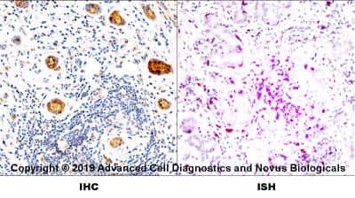
Dual RNAscope ISH-IHC: HLA-DR Antibody (L243) [NB100-77855] - Formalin-fixed paraffin-embedded tissue sections of human metastatic tonsil were probed for HLA-DR mRNA (ACD RNAScope probe, catalog # 475891; Fast Red chromogen, ACD catalog # 322500). Adjacent tissue section was processes for immunohistochemistry using mouse monoclonal (Novus catalog # NB100-77855) at 1:50 dilution for 1 hour at room temperature followed by incubation with the anti-mouse IgG VisUCyte HRP Polymer Antibody (Catalog # VC001) and DAB chromogen (yellow-brown). Tissue was counterstained with hematoxylin (blue).
![Western Blot: HLA-DR Antibody (L243)BSA Free [NB100-77855]](https://resources.rndsystems.com/images/products/HLA-DR-Antibody-L243-Western-Blot-NB100-77855-img0002.jpg "Western Blot: HLA-DR Antibody (L243)BSA Free [NB100-77855]")
Western Blot: HLA-DR Antibody (L243)BSA Free [NB100-77855]
Western Blot: HLA-DR Antibody (L243) [NB100-77855] - Analysis of human spleen (A) and human tonsil (B) tissue using HLA-DR antibody at 2 ug/mL.![Immunohistochemistry-Paraffin: HLA-DR Antibody (L243) - BSA Free [NB100-77855]](https://resources.rndsystems.com/images/products/HLA-DR-Antibody-L243-Immunohistochemistry-Paraffin-NB100-77855-img0003.jpg "Immunohistochemistry-Paraffin: HLA-DR Antibody (L243) - BSA Free [NB100-77855]")
Immunohistochemistry-Paraffin: HLA-DR Antibody (L243) - BSA Free [NB100-77855]
Immunohistochemistry-Paraffin: HLA-DR Antibody (L243) [NB100-77855] - Staining of HLA-DR in human spleen using DAB with hematoxylin counterstain.![Flow Cytometry: HLA-DR Antibody (L243) - BSA Free [NB100-77855]](https://resources.rndsystems.com/images/products/HLA-DR-Antibody-L243-Flow-Cytometry-NB100-77855-img0015.jpg "Flow Cytometry: HLA-DR Antibody (L243) - BSA Free [NB100-77855]")
Flow Cytometry: HLA-DR Antibody (L243) - BSA Free [NB100-77855]
Flow Cytometry: HLA-DR Antibody (L243) [NB100-77855] - Analysis using the FITC conjugate of NB100-77855. Staining of Human peripheral blood lymphocytes with L243 FITC.![Flow Cytometry: HLA-DR Antibody (L243) - BSA Free [NB100-77855]](https://resources.rndsystems.com/images/products/HLA-DR-Antibody-L243-Flow-Cytometry-NB100-77855-img0001.jpg "Flow Cytometry: HLA-DR Antibody (L243) - BSA Free [NB100-77855]")
Flow Cytometry: HLA-DR Antibody (L243) - BSA Free [NB100-77855]
Flow Cytometry: HLA-DR Antibody (L243) [NB100-77855] - Human peripheral blood lymphocytes stained with L243 PE![Flow (Cell Surface): HLA-DR Antibody (L243) - BSA Free [NB100-77855]](https://resources.rndsystems.com/images/products/HLA-DR-Antibody-L243-Flow-Cell-Surface-NB100-77855-img0010.jpg "Flow (Cell Surface): HLA-DR Antibody (L243) - BSA Free [NB100-77855]")
Flow (Cell Surface): HLA-DR Antibody (L243) - BSA Free [NB100-77855]
Flow (Cell Surface): HLA-DR Antibody (L243) [NB100-77855] - Analysis using the Azide Free version of NB100-77855. Staining of HLA-DR expression by 5x10^5 BDCM cells using 0.5 ug of (red) and 0.5 ug of isotype control (green). This antibody and an anti-mouse IgG PE conjugated secondary antibody were used.![Flow Cytometry: HLA-DR Antibody (L243) - BSA Free [NB100-77855]](https://resources.rndsystems.com/images/products/HLA-DR-Antibody-L243-Flow-Cytometry-NB100-77855-img0011.jpg "Flow Cytometry: HLA-DR Antibody (L243) - BSA Free [NB100-77855]")
Flow Cytometry: HLA-DR Antibody (L243) - BSA Free [NB100-77855]
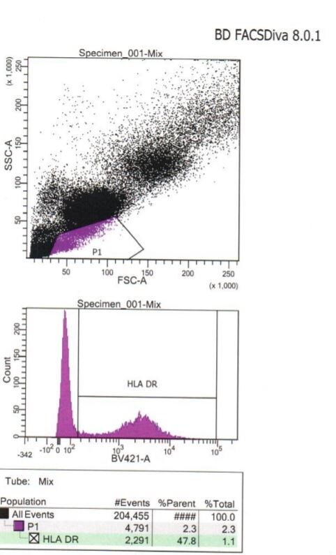
Flow Cytometry: HLA-DR Antibody (L243) [NB100-77855] - Analysis using the PerCP/Cy5.5 conjugate of NB100-77855. Staining of HLA-DR expression by 5x10^5 human PBMC using 10 uL (0.25 ug) of this antibody. Cell surface flow kit was used for this test. Left panel shows the monocyte gate, right panel shows the lymphocyte gate.![Flow Cytometry: HLA-DR Antibody (L243) - BSA Free [NB100-77855]](https://resources.rndsystems.com/images/products/HLA-DR-Antibody-L243-Flow-Cytometry-NB100-77855-img0012.jpg "Flow Cytometry: HLA-DR Antibody (L243) - BSA Free [NB100-77855]")
Flow Cytometry: HLA-DR Antibody (L243) - BSA Free [NB100-77855]
Flow Cytometry: HLA-DR Antibody (L243) [NB100-77855] - Analysis using the Alexa Fluor (R) 700 conjugate of NB100-77855. Staining of HLA-DR protein in human PBMCs using HLA-DR antibody [clone L243; conjugated to Alexa Fluor (R) 700] at 1:100 with 25 minutes incubation at 4C in 2% human serum, 0.5 mM EDTA in DPBS. Image from verified customer review.![Flow Cytometry: HLA-DR Antibody (L243) - BSA Free [NB100-77855]](https://resources.rndsystems.com/images/products/HLA-DR-Antibody-L243-Flow-Cytometry-NB100-77855-img0013.jpg "Flow Cytometry: HLA-DR Antibody (L243) - BSA Free [NB100-77855]")
Flow Cytometry: HLA-DR Antibody (L243) - BSA Free [NB100-77855]
Flow Cytometry: HLA-DR Antibody (L243) [NB100-77855] - Analysis using the Biotin conjugate of NB100-77855. Staining of Human peripheral blood lymphocytes with biotinylated L243, followed by Sav-PE.![Flow Cytometry: HLA-DR Antibody (L243) - BSA Free [NB100-77855]](https://resources.rndsystems.com/images/products/HLA-DR-Antibody-L243-Flow-Cytometry-NB100-77855-img0014.jpg "Flow Cytometry: HLA-DR Antibody (L243) - BSA Free [NB100-77855]")
Flow Cytometry: HLA-DR Antibody (L243) - BSA Free [NB100-77855]
Flow Cytometry: HLA-DR Antibody (L243) [NB100-77855] - Analysis using the DyLight 405 conjugate of NB100-77855. Staining of HLA-DR in human PBMCs using anti-HLA-DR antibody. Image from verified customer review.Applications for HLA-DR Antibody (L243) - BSA Free
Application
Recommended Usage
Block/Neutralize
reported in scientific literature (PMID 12874328)
ELISA
reported in scientific literature (PMID 11070170)
Electron Microscopy
reported in scientific literature (PMID 21660937)
Flow Cytometry
< 0.5 ug/10^6 cells
Functional
reported in scientific literature (PMID 17900279)
Immunocytochemistry/ Immunofluorescence
reported in scientific literature (PMID 7835916)
Immunohistochemistry
1:10 - 1:500
Immunohistochemistry-Frozen
1:10 - 1:500
Immunohistochemistry-Paraffin
1:100
Immunoprecipitation
1:10 - 1:500
In vitro assay
reported in scientific literature (PMID 19620786)
Western Blot
1:100 - 1:2000
Application Notes
It is recommended that the reagent be titrated for optimal performance for each application. This antibody is CyTOF ready.
Reviewed Applications
Read 1 review rated 5 using NB100-77855 in the following applications:
Flow Cytometry Panel Builder
Bio-Techne Knows Flow Cytometry
Save time and reduce costly mistakes by quickly finding compatible reagents using the Panel Builder Tool.
Advanced Features
- Spectra Viewer - Custom analysis of spectra from multiple fluorochromes
- Spillover Popups - Visualize the spectra of individual fluorochromes
- Antigen Density Selector - Match fluorochrome brightness with antigen density

Formulation, Preparation, and Storage
Purification
Protein A purified
Formulation
PBS
Format
BSA Free
Preservative
0.05% Sodium Azide
Concentration
1.0 mg/ml
Shipping
The product is shipped with polar packs. Upon receipt, store it immediately at the temperature recommended below.
Stability & Storage
Store at 4C short term. Aliquot and store at -20C long term. Avoid freeze-thaw cycles.
Background: HLA-DR
Given the role in adaptive immunity, HLA-DR allele polymorphisms, gene misexpression, and dysfunction has been implicated in many diseases ranging from autoimmune disorders to cancer (2). HLA-DR is also a classical biomarker for disease, including sepsis where reduced expression of HLA-DR molecules on monocytes, as measured by flow cytometry, indicates diagnosis and prognosis (4,5). Immunosuppression observed with sepsis results in decreased surface expression of HLA-DR and concurrent increase in expression of programmed death 1 (PD-1), cytotoxic T-lymphocyte antigen 4 (CTLA-4), and B and T lymphocyte attenuator (BTLA) (4). This altered expression results in poor T cell response and apoptosis, along with reduced interferon-gamma (IFN-gamma) production and increased pro-inflammatory cytokine release (4). Furthermore, the decrease in HLA-DR expression is also correlated with the decrease in CD14lowCD16+ inflammatory monocytes (5). Interestingly, COVID-19 patients also exhibit a reduction in HLA-DR that correlates with disease severity and immunosuppression (5).
References
1. Andersson G. (1998). Evolution of the human HLA-DR region. Frontiers in bioscience : a journal and virtual library. https://doi.org/10.2741/a317
2. Shiina, T., Hosomichi, K., Inoko, H., & Kulski, J. K. (2009). The HLA genomic loci map: expression, interaction, diversity and disease. Journal of human genetics. https://doi.org/10.1038/jhg.2008.5
3. Stern, L. J., & Calvo-Calle, J. M. (2009). HLA-DR: molecular insights and vaccine design. Current pharmaceutical design. https://doi.org/10.2174/138161209789105171
4. Zhuang, Y., Peng, H., Chen, Y., Zhou, S., & Chen, Y. (2017). Dynamic monitoring of monocyte HLA-DR expression for the diagnosis, prognosis, and prediction of sepsis. Frontiers in bioscience (Landmark edition). https://doi.org/10.2741/4547
5. Benlyamani, I., Venet, F., Coudereau, R., Gossez, M., & Monneret, G. (2020). Monocyte HLA-DR Measurement by Flow Cytometry in COVID-19 Patients: An Interim Review. Cytometry. Part A : the journal of the International Society for Analytical Cytology. https://doi.org/10.1002/cyto.a.24249
Long Name
Major Histocompatibility Complex Class II DR
Alternate Names
HLA-DRA, HLADR, MHC Class II DR
Gene Symbol
HLA-DRA
Additional HLA-DR Products
Product Documents for HLA-DR Antibody (L243) - BSA Free
Certificate of Analysis
To download a Certificate of Analysis, please enter a lot or batch number in the search box below.
Product Specific Notices for HLA-DR Antibody (L243) - BSA Free
This product is for research use only and is not approved for use in humans or in clinical diagnosis. Primary Antibodies are guaranteed for 1 year from date of receipt.
Related Research Areas
Citations for HLA-DR Antibody (L243) - BSA Free
 Powered by Bioz
Customer Reviews for HLA-DR Antibody (L243) - BSA Free (1)
5 out of 5
1 Customer Rating
Have you used HLA-DR Antibody (L243) - BSA Free?
Submit a review and receive an Amazon gift card!
$25/€18/£15/$25CAN/¥2500 Yen for a review with an image
$10/€7/£6/$10CAN/¥1110 Yen for a review without an image
Submit a review
Customer Images
Showing
1
-
1 of
1 review
Showing All
Filter By:
-
Application: Flow CytometrySample Tested: Whole blood cellsSpecies: CanineVerified Customer | Posted 05/18/2018Canine IP.The whole blood cells were lysed to get rid of the RBC. The washed cell were then incubated with the Antibody. They were washed and then incubated with a conjugated secondary. Allowed to sit for 20mins and then washed again and read on the BD Fortessa.

There are no reviews that match your criteria.
Protocols
Find general support by application which include: protocols, troubleshooting, illustrated assays, videos and webinars.
- 7-Amino Actinomycin D (7-AAD) Cell Viability Flow Cytometry Protocol
- Antigen Retrieval Protocol (PIER)
- Antigen Retrieval for Frozen Sections Protocol
- Appropriate Fixation of IHC/ICC Samples
- Cellular Response to Hypoxia Protocols
- Chromogenic IHC Staining of Formalin-Fixed Paraffin-Embedded (FFPE) Tissue Protocol
- Chromogenic Immunohistochemistry Staining of Frozen Tissue
- ClariTSA™ Fluorophore Kits
- Detection & Visualization of Antibody Binding
- ELISA Sample Preparation & Collection Guide
- ELISA Troubleshooting Guide
- Extracellular Membrane Flow Cytometry Protocol
- Flow Cytometry Protocol for Cell Surface Markers
- Flow Cytometry Protocol for Staining Membrane Associated Proteins
- Flow Cytometry Staining Protocols
- Flow Cytometry Troubleshooting Guide
- Fluorescent IHC Staining of Frozen Tissue Protocol
- Graphic Protocol for Heat-induced Epitope Retrieval
- Graphic Protocol for the Preparation and Fluorescent IHC Staining of Frozen Tissue Sections
- Graphic Protocol for the Preparation and Fluorescent IHC Staining of Paraffin-embedded Tissue Sections
- Graphic Protocol for the Preparation of Gelatin-coated Slides for Histological Tissue Sections
- How to Run an R&D Systems DuoSet ELISA
- How to Run an R&D Systems Quantikine ELISA
- How to Run an R&D Systems Quantikine™ QuicKit™ ELISA
- ICC Cell Smear Protocol for Suspension Cells
- ICC Immunocytochemistry Protocol Videos
- ICC for Adherent Cells
- IHC Sample Preparation (Frozen sections vs Paraffin)
- ISH-IHC Protocol for Chromogenic Detection on Formalin Fixed Paraffin Embedded (FFPE) Tissue
- Immunocytochemistry (ICC) Protocol
- Immunocytochemistry Troubleshooting
- Immunofluorescence of Organoids Embedded in Cultrex Basement Membrane Extract
- Immunofluorescent IHC Staining of Formalin-Fixed Paraffin-Embedded (FFPE) Tissue Protocol
- Immunohistochemistry (IHC) and Immunocytochemistry (ICC) Protocols
- Immunohistochemistry Frozen Troubleshooting
- Immunohistochemistry Paraffin Troubleshooting
- Immunoprecipitation Protocol
- Intracellular Flow Cytometry Protocol Using Alcohol (Methanol)
- Intracellular Flow Cytometry Protocol Using Detergents
- Intracellular Nuclear Staining Flow Cytometry Protocol Using Detergents
- Intracellular Staining Flow Cytometry Protocol Using Alcohol Permeabilization
- Intracellular Staining Flow Cytometry Protocol Using Detergents to Permeabilize Cells
- Preparing Samples for IHC/ICC Experiments
- Preventing Non-Specific Staining (Non-Specific Binding)
- Primary Antibody Selection & Optimization
- Propidium Iodide Cell Viability Flow Cytometry Protocol
- Protocol for Heat-Induced Epitope Retrieval (HIER)
- Protocol for Liperfluo
- Protocol for Making a 4% Formaldehyde Solution in PBS
- Protocol for VisUCyte™ HRP Polymer Detection Reagent
- Protocol for the Characterization of Human Th22 Cells
- Protocol for the Characterization of Human Th9 Cells
- Protocol for the Fluorescent ICC Staining of Cell Smears - Graphic
- Protocol for the Fluorescent ICC Staining of Cultured Cells on Coverslips - Graphic
- Protocol for the Preparation & Fixation of Cells on Coverslips
- Protocol for the Preparation and Chromogenic IHC Staining of Frozen Tissue Sections
- Protocol for the Preparation and Chromogenic IHC Staining of Frozen Tissue Sections - Graphic
- Protocol for the Preparation and Chromogenic IHC Staining of Paraffin-embedded Tissue Sections
- Protocol for the Preparation and Chromogenic IHC Staining of Paraffin-embedded Tissue Sections - Graphic
- Protocol for the Preparation and Fluorescent ICC Staining of Cells on Coverslips
- Protocol for the Preparation and Fluorescent ICC Staining of Non-adherent Cells
- Protocol for the Preparation and Fluorescent ICC Staining of Stem Cells on Coverslips
- Protocol for the Preparation and Fluorescent IHC Staining of Frozen Tissue Sections
- Protocol for the Preparation and Fluorescent IHC Staining of Paraffin-embedded Tissue Sections
- Protocol for the Preparation of Gelatin-coated Slides for Histological Tissue Sections
- Protocol for the Preparation of a Cell Smear for Non-adherent Cell ICC - Graphic
- Protocol: Annexin V and PI Staining by Flow Cytometry
- Protocol: Annexin V and PI Staining for Apoptosis by Flow Cytometry
- Quantikine HS ELISA Kit Assay Principle, Alkaline Phosphatase
- Quantikine HS ELISA Kit Principle, Streptavidin-HRP Polymer
- R&D Systems Quality Control Western Blot Protocol
- Sandwich ELISA (Colorimetric) – Biotin/Streptavidin Detection Protocol
- Sandwich ELISA (Colorimetric) – Direct Detection Protocol
- TUNEL and Active Caspase-3 Detection by IHC/ICC Protocol
- The Importance of IHC/ICC Controls
- Troubleshooting Guide: ELISA
- Troubleshooting Guide: Fluorokine Flow Cytometry Kits
- Troubleshooting Guide: Immunohistochemistry
- Troubleshooting Guide: Western Blot Figures
- Western Blot Conditions
- Western Blot Protocol
- Western Blot Protocol for Cell Lysates
- Western Blot Troubleshooting
- Western Blot Troubleshooting Guide
- View all Protocols, Troubleshooting, Illustrated assays and Webinars
FAQs for HLA-DR Antibody (L243) - BSA Free
Showing
1
-
1 of
1 FAQ
Showing All
-
Q: We have an inquiry of your product Cat. ID # NB100-2735, NB600-1185, NB100-77758, NB100-78016, NB100-77855. We are interested in this product and would like to make sure that there is no any other protein added in the buffer. I saw the storage buffer in datasheet doesn't has any other protein but still would like to confirm with you again. Would you please help us to check and provide the purification method and buffer of the antibody?
A: Thank you for your inquiry. Catalog numbers NB100-77758, NB100-78016 and NB100-77855 are in a buffer containing PBS and sodium azide. NB600-1185 is in a buffer containing PBS, 150mM NaCl and sodium azide. NB100-2735 is whole tissue culture supernatant and may therefore contain other proteins.


