HMGB1/HMG-1 Antibody - BSA Free
Novus Biologicals | Catalog # NB100-2322
![Knockdown Validated: HMGB1/HMG-1 Antibody [NB100-2322]](https://resources.rndsystems.com/images/products/HMGB1-HMG-1-Antibody-Knockdown-Validated-NB100-2322-img0025.jpg "Western Blot: HMGB1/HMG-1 Antibody [NB100-2322]")
Key Product Details
Validated by
Species Reactivity
Validated:
Cited:
Predicted:
Applications
Validated:
Cited:
Label
Antibody Source
Format
Product Specifications
Immunogen
Reactivity Notes
Localization
Clonality
Host
Isotype
Theoretical MW
Disclaimer note: The observed molecular weight of the protein may vary from the listed predicted molecular weight due to post translational modifications, post translation cleavages, relative charges, and other experimental factors.
Scientific Data Images for HMGB1/HMG-1 Antibody - BSA Free
Western Blot: HMGB1/HMG-1 Antibody [NB100-2322]
Western Blot: HMGB1/HMG-1 Antibody [NB100-2322] - Western blot shows lysates of HEK293T human embryonic kidney parental cell line and HMGB1 knockout (KO) HEK293T cell line. PVDF membrane was probed with 1.0 ug/ml of Rabbit Anti-Human HMGB1 Polyclonal Antibody (Catalog # NB100-2322) followed by HRP-conjugated Anti-Rabbit IgG Secondary Antibody (Catalog #HAF008). Specific band was detected for HMGB1 at approximately 30 kDa (as indicated) in the parental HEK293T cell line, but is not detectable in the knockout HEK293T cell line. This experiment was conducted under reducing conditions.![Western Blot: HMGB1/HMG-1 Antibody [NB100-2322]](https://resources.rndsystems.com/images/products/HMGB1-HMG-1-Antibody-Western-Blot-NB100-2322-img0017.jpg "Western Blot: HMGB1/HMG-1 Antibody [NB100-2322]")
Western Blot: HMGB1/HMG-1 Antibody [NB100-2322]
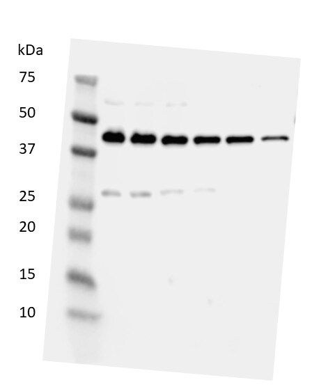
Western Blot: HMGB1/HMG-1 Antibody [NB100-2322] - Total protein from SHSY-5Y, MCF7, Neuro2A and HeLa was separated on a 12% gel by SDS-PAGE, transferred to PVDF membrane and blocked in 5% non-fat milk in TBST. The membrane was probed with 2.0 ug/mL anti-HMGB1 in 1% non-fat milk in TBST and detected with an anti-rabbit HRP secondary antibody using chemiluminescence.![Immunohistochemistry-Paraffin: HMGB1/HMG-1 Antibody [NB100-2322]](https://resources.rndsystems.com/images/products/HMGB1-HMG-1-Antibody-Immunohistochemistry-Paraffin-NB100-2322-img0011.jpg "Immunohistochemistry-Paraffin: HMGB1/HMG-1 Antibody [NB100-2322]")
Immunohistochemistry-Paraffin: HMGB1/HMG-1 Antibody [NB100-2322]
Immunohistochemistry-Paraffin: HMGB1/HMG-1 Antibody [NB100-2322] - Staining of HMGB1 in mouse liver using NB100-2322.![Simple Western: HMGB1/HMG-1 Antibody [NB100-2322]](https://resources.rndsystems.com/images/products/HMGB1-HMG-1-Antibody-Simple-Western-NB100-2322-img0014.jpg "Simple Western: HMGB1/HMG-1 Antibody [NB100-2322]")
Simple Western: HMGB1/HMG-1 Antibody [NB100-2322]
Simple Western: HMGB1/HMG-1 Antibody [NB100-2322] - Image shows a specific band for HMGB1 in 0.05 mg/mL of Jurkat lysate. This experiment was performed under reducing conditions using the 12-230 kDa separation system.![Immunocytochemistry/ Immunofluorescence: HMGB1/HMG-1 Antibody [NB100-2322]](https://resources.rndsystems.com/images/products/HMGB1-HMG-1-Antibody-Immunocytochemistry-Immunofluorescence-NB100-2322-img0027.jpg "Immunocytochemistry/ Immunofluorescence: HMGB1/HMG-1 Antibody [NB100-2322]")
Immunocytochemistry/ Immunofluorescence: HMGB1/HMG-1 Antibody [NB100-2322]
Immunocytochemistry/Immunofluorescence: HMGB1/HMG-1 Antibody [NB100-2322] - MCF7 cells were fixed in 4% paraformaldehyde for 10 minutes and permeabilized in 0.5% Triton X-100 in PBS for 5 minutes. The cells were incubated with anti-HMGB1/HMG-1 Antibody NB100-2322 at 1 ug/ml for overnight at 4C and detected with an anti-rabbit Dylight 488 (Green) at a 1:1000 dilution for 60 minutes. Nuclei were counterstained with DAPI (Blue). Cells were imaged using a 100X objective and digitally deconvolved.![Immunocytochemistry/ Immunofluorescence: HMGB1/HMG-1 Antibody [NB100-2322]](https://resources.rndsystems.com/images/products/HMGB1-HMG-1-Antibody-Immunocytochemistry-Immunofluorescence-NB100-2322-img0018.jpg "Immunocytochemistry/ Immunofluorescence: HMGB1/HMG-1 Antibody [NB100-2322]")
Immunocytochemistry/ Immunofluorescence: HMGB1/HMG-1 Antibody [NB100-2322]
Immunocytochemistry/Immunofluorescence: HMGB1/HMG-1 Antibody [NB100-2322] - Neuro2a cells were fixed for 10 minutes using 10% formalin and then permeabilized for 5 minutes using 1X TBS + 0.5% Triton-X100. The cells were incubated with anti-HMGB1 at 5 ug/ml overnight at 4C and detected with an anti-rabbit Dylight 488 (Green) at a 1:500 dilution. Alpha tubulin (DM1A) NB100-690 was used as a co-stain at a 1:1000 dilution and detected with an anti-mouse Dylight 550 (Red) at a 1:500 dilution. Nuclei were counterstained with DAPI (Blue). Cells were imaged using a 40X objective.![Immunohistochemistry-Paraffin: HMGB1/HMG-1 Antibody [NB100-2322]](https://resources.rndsystems.com/images/products/HMGB1-HMG-1-Antibody-Immunohistochemistry-Paraffin-NB100-2322-img0022.jpg "Immunohistochemistry-Paraffin: HMGB1/HMG-1 Antibody [NB100-2322]")
Immunohistochemistry-Paraffin: HMGB1/HMG-1 Antibody [NB100-2322]
Immunohistochemistry-Paraffin: HMGB1/HMG-1 Antibody [NB100-2322] - Rabbit blood vessel. Image from verified customer review.![Western Blot: HMGB1/HMG-1 Antibody [NB100-2322]](https://resources.rndsystems.com/images/products/HMGB1-HMG-1-Antibody-Western-Blot-NB100-2322-img0026.jpg "Western Blot: HMGB1/HMG-1 Antibody [NB100-2322]")
Western Blot: HMGB1/HMG-1 Antibody [NB100-2322]
HMGB1-HMG-1-Antibody-Western-Blot-NB100-2322-img0026.jpg![Immunocytochemistry/ Immunofluorescence: HMGB1/HMG-1 Antibody [NB100-2322]](https://resources.rndsystems.com/images/products/HMGB1-HMG-1-Antibody-Immunocytochemistry-Immunofluorescence-NB100-2322-img0028.jpg "Immunocytochemistry/ Immunofluorescence: HMGB1/HMG-1 Antibody [NB100-2322]")
Immunocytochemistry/ Immunofluorescence: HMGB1/HMG-1 Antibody [NB100-2322]
Immunocytochemistry/Immunofluorescence: HMGB1/HMG-1 Antibody [NB100-2322] - NIH3T3 cells were fixed in 4% paraformaldehyde for 10 minutes and permeabilized in 0.5% Triton X-100 in PBS for 5 minutes. The cells were incubated with anti-HMGB1/HMG-1 Antibody NB100-2322 at 1 ug/ml for overnight at 4C and detected with an anti-rabbit Dylight 488 (Green) at a 1:1000 dilution for 60 minutes. Nuclei were counterstained with DAPI (Blue). Cells were imaged using a 100X objective and digitally deconvolved.![Flow Cytometry: HMGB1/HMG-1 Antibody [NB100-2322]](https://resources.rndsystems.com/images/products/HMGB1-HMG-1-Antibody-Flow-Cytometry-NB100-2322-img0029.jpg "Flow Cytometry: HMGB1/HMG-1 Antibody [NB100-2322]")
Flow Cytometry: HMGB1/HMG-1 Antibody [NB100-2322]
Flow Cytometry: HMGB1/HMG-1 Antibody [NB100-2322] - An intracellular stain was performed on HeLa cells with HMGB1/HMG-1 Antibody NB100-2322AF488 (blue) and a matched isotype control (orange). Cells were fixed with 4% PFA and then permeabilized with 0.1% saponin. Cells were incubated in an antibody dilution of 5 ug/mL for 30 minutes at room temperature. Both antibodies were conjugated to Alexa Fluor 488.![Western Blot: HMGB1/HMG-1 Antibody [NB100-2322]](https://resources.rndsystems.com/images/products/HMGB1-HMG-1-Antibody-Western-Blot-NB100-2322-img0013.jpg "Western Blot: HMGB1/HMG-1 Antibody [NB100-2322]")
Western Blot: HMGB1/HMG-1 Antibody [NB100-2322]
Western Blot: HMGB1/HMG-1 Antibody [NB100-2322] - Hepatocyte protein lysate at 1:1000 4C overnight. Image from verfified customer review.![Immunocytochemistry/ Immunofluorescence: HMGB1/HMG-1 Antibody [NB100-2322]](https://resources.rndsystems.com/images/products/HMGB1-HMG-1-Antibody-Immunocytochemistry-Immunofluorescence-NB100-2322-img0015.jpg "Immunocytochemistry/ Immunofluorescence: HMGB1/HMG-1 Antibody [NB100-2322]")
Immunocytochemistry/ Immunofluorescence: HMGB1/HMG-1 Antibody [NB100-2322]
Immunocytochemistry/Immunofluorescence: HMGB1/HMG-1 Antibody [NB100-2322] - HeLa cells were fixed for 10 minutes using 10% formalin and then permeabilized for 5 minutes using 1X TBS + 0.5% Triton-X100. The cells were incubated with anti-HMGB1 [NB100-2322] at a 1:200 dilution overnight at 4C and detected with an anti-rabbit Dylight 488 (Green) at a 1:500 dilution. Alpha tubulin (DM1A) [NB100-690] was used as a co-stain at a 1:1000 dilution and detected with an anti-mouse Dylight 550 (Red) at a 1:500 dilution. Nuclei were counterstained with DAPI (Blue). Cells were imaged using a 40X objective.![Flow (Intracellular): HMGB1/HMG-1 Antibody [NB100-2322]](https://resources.rndsystems.com/images/products/HMGB1-HMG-1-Antibody-Flow-Intracellular-NB100-2322-img0021.jpg "Flow (Intracellular): HMGB1/HMG-1 Antibody [NB100-2322]")
Flow (Intracellular): HMGB1/HMG-1 Antibody [NB100-2322]
Flow (Intracellular): HMGB1/HMG-1 Antibody [NB100-2322] - An intracellular stain was performed on HeLa with NB100-2322 and a matched isotype control. Cells were fixed with 4% PFA and then permeabilized with 0.1% saponin. Cells were incubated in an antibody dilution of 1 ug/mL for 30 minutes at room temperature, followed by Rabbit IgG (H+L) Cross-Adsorbed Secondary Antibody.![Flow Cytometry: HMGB1/HMG-1 Antibody [NB100-2322]](https://resources.rndsystems.com/images/products/HMGB1-HMG-1-Antibody-Flow-Cytometry-NB100-2322-img0024.jpg "Flow Cytometry: HMGB1/HMG-1 Antibody [NB100-2322]")
Flow Cytometry: HMGB1/HMG-1 Antibody [NB100-2322]
Flow Cytometry: HMGB1/HMG-1 Antibody [NB100-2322] - An intracellular stain was performed on RH-30 cells with NB100-2322F (blue) and a matched isotype control (orange). Cells were fixed with 4% PFA and then permeabilized with 0.1% saponin. Cells were incubated in an antibody dilution of 10 ug/mL for 30 minutes at room temperature. Both antibodies were conjugated to FITC.![ELISA: HMGB1/HMG-1 Antibody [NB100-2322]](https://resources.rndsystems.com/images/products/HMGB1-HMG-1-Antibody-ELISA-NB100-2322-img0023.jpg "ELISA: HMGB1/HMG-1 Antibody [NB100-2322]")
ELISA: HMGB1/HMG-1 Antibody [NB100-2322]
ELISA: HMGB1/HMG-1 Antibody [NB100-2322] - A dose-dependent titration of the HRP conjugated anti-HMGB1 antibody on recombinant human HMGB1 protein. Image from verified customer review. Image using the HRP format of this antibody.
Western Blot: HMGB1/HMG-1 Antibody [NB100-2322] -
Expression of endogenous ligands for TLR2. (A) Expression of biglycan in AD-TLR2KO mice increased significantly compared with that in WT, AD, and TLR2KO mice (p<0.05). (B) HMGB1 in the four groups did not show a significant difference (p>0.05).
Western Blot: HMGB1/HMG-1 Antibody [NB100-2322] -
Western Blot: HMGB1/HMG-1 Antibody [NB100-2322] - Expression of endogenous ligands for TLR2. (A) Expression of biglycan in AD-TLR2KO mice increased significantly compared with that in WT, AD, & TLR2KO mice (p<0.05). (B) HMGB1 in the four groups did not show a significant difference (p>0.05). Image collected & cropped by CiteAb from the following publication (https://pubmed.ncbi.nlm.nih.gov/31509519), licensed under a CC-BY license. Not internally tested by Novus Biologicals.Applications for HMGB1/HMG-1 Antibody - BSA Free
Flow Cytometry
Immunocytochemistry/ Immunofluorescence
Immunohistochemistry
Immunohistochemistry-Paraffin
Simple Western
Western Blot
In Simple Western only 10 - 15 uL of the recommended dilution is used per data point.
See Simple Western Antibody Database for Simple Western validation: Tested in Jurkat lysate 0.05 mg/mL, separated by Size, antibody dilution of 1:2000. Separated by Size-Wes, Sally Sue/Peggy Sue.
The observed molecular weight of the protein may vary from the listed predicted molecular weight due to post translational modifications, post translation cleavages, relative charges, and other experimental factors.
Reviewed Applications
Read 5 reviews rated 4.4 using NB100-2322 in the following applications:
Flow Cytometry Panel Builder
Bio-Techne Knows Flow Cytometry
Save time and reduce costly mistakes by quickly finding compatible reagents using the Panel Builder Tool.
Advanced Features
- Spectra Viewer - Custom analysis of spectra from multiple fluorochromes
- Spillover Popups - Visualize the spectra of individual fluorochromes
- Antigen Density Selector - Match fluorochrome brightness with antigen density

Formulation, Preparation, and Storage
Purification
Formulation
Format
Preservative
Concentration
Shipping
Stability & Storage
Background: HMGB1/HMG-1
Long Name
Alternate Names
Gene Symbol
Additional HMGB1/HMG-1 Products
Product Documents for HMGB1/HMG-1 Antibody - BSA Free
Certificate of Analysis
To download a Certificate of Analysis, please enter a lot or batch number in the search box below.
Product Specific Notices for HMGB1/HMG-1 Antibody - BSA Free
This product is for research use only and is not approved for use in humans or in clinical diagnosis. Primary Antibodies are guaranteed for 1 year from date of receipt.
Related Research Areas
Citations for HMGB1/HMG-1 Antibody - BSA Free
 Powered by Bioz
Customer Reviews for HMGB1/HMG-1 Antibody - BSA Free (5)
Have you used HMGB1/HMG-1 Antibody - BSA Free?
Submit a review and receive an Amazon gift card!
$25/€18/£15/$25CAN/¥2500 Yen for a review with an image
$10/€7/£6/$10CAN/¥1110 Yen for a review without an image
Submit a review
Customer Images

-(01-ml)_NB100-2322_8456.jpg)
-
Application: Immunohistochemistry-ParaffinSample Tested: veeselSpecies: RabbitVerified Customer | Posted 04/24/2018

-
Application: Western BlotSample Tested: Brain homogenateSpecies: RatVerified Customer | Posted 03/28/2018Lane 1: protein ladder. Lanes 2-7 decreasing protein load from 30 ug to 5 ug protein per well. Upper bands are B-actin, lower bands are HMGB1. The HMGB1 antibody exhibited excellent linearity with an r2 of 0.959.Blocked in 5% milk + 1% BSA in TBS for 2 h, incubated overnight in 1:500 HMGB1 antibody at 4 degrees C and then 2 h at room temperature with HRP-conjugated secondary.
-
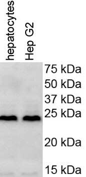
Application: Western BlotSample Tested: mouse hepatocytes and HepG2 cellsSpecies: HumanVerified Customer | Posted 11/25/2017
-
Application: Simple WesternSample Tested: mouse serumSpecies: MouseVerified Customer | Posted 12/30/2015HMGB1 in mouse serum protein extract
-
Application: Western BlotSample Tested:Species: MouseVerified Customer | Posted 06/26/2014
There are no reviews that match your criteria.
Protocols
View specific protocols for HMGB1/HMG-1 Antibody - BSA Free (NB100-2322):
Immunohistochemistry-paraffin embedded sections
Antigen Unmasking
Bring slides to a boil in 10 mM sodium citrate buffer pH 6.0 then maintain at a sub-boiling temperature for 10 minutes. Cool slides on bench top for 30 minutes.
Staining
1. Wash sections in dH2O three times for 5 minutes each.
2. Wash section in wash buffer (1X PBS/0.1% Tween-20 (1X PBST)) for 5 minutes.
3. Block each section with 100-400 ul blocking solution (1X PBST, 5% goat serum) for 1 hour at room temperature.
4. Remove blocking solution and add 100-400 ul primary antibody diluted in 1X PBST, 5% goat serum to each section. Incubate overnight at 4C.
5. Remove antibody solution and wash sections in wash buffer three times for 5 minutes each.
6. Add 100-400 ul biotinylated secondary antibody, diluted in 1X PBST, 5% goat serum. Incubate 30 minutes at room temperature.
7. Remove secondary antibody solution and wash sections three times with wash buffer for 5 minutes each.
8. Add 100-400 ul Striptavidin-HRP reagent to each section and incubate for 30 minutes at room temperature.
9. Wash sections three times in wash buffer for 5 minutes each.
10. Add 100-400 ul DAB substrate to each section and monitor staining closely.
11. As soon as the sections develop, immerse slides in dH2O.
12. Counterstain sections in hematoxylin.
13. Wash sections in dH2O two times for 5 minutes each.
14. Dehydrate sections.
15. Mount coverslips.
Western Blot Protocol
1. Perform SDS-PAGE on samples to be analyzed, loading 10-25 ug of total protein per lane.
2. Transfer proteins to PVDF membrane according to the instructions provided by the manufacturer of the membrane and transfer apparatus.
3. Stain the membrane with Ponceau S (or similar product) to assess transfer success, and mark molecular weight standards where appropriate.
4. Rinse the blot TBS -0.05% Tween 20 (TBST).
5. Block the membrane in 5% Non-fat milk in TBST (blocking buffer) for at least 1 hour.
6. Wash the membrane in TBST three times for 10 minutes each.
7. Dilute primary antibody in blocking buffer and incubate overnight at 4C with gentle rocking.
8. Wash the membrane in TBST three times for 10 minutes each.
9. Incubate the membrane in diluted HRP conjugated secondary antibody in blocking buffer (as per manufacturer's instructions) for 1 hour at room temperature.
10. Wash the blot in TBST three times for 10 minutes each (this step can be repeated as required to reduce background).
11. Apply the detection reagent of choice in accordance with the manufacturers instructions.
Find general support by application which include: protocols, troubleshooting, illustrated assays, videos and webinars.
- 7-Amino Actinomycin D (7-AAD) Cell Viability Flow Cytometry Protocol
- Antigen Retrieval Protocol (PIER)
- Antigen Retrieval for Frozen Sections Protocol
- Appropriate Fixation of IHC/ICC Samples
- Cellular Response to Hypoxia Protocols
- Chromogenic IHC Staining of Formalin-Fixed Paraffin-Embedded (FFPE) Tissue Protocol
- Chromogenic Immunohistochemistry Staining of Frozen Tissue
- ClariTSA™ Fluorophore Kits
- Detection & Visualization of Antibody Binding
- ELISA Sample Preparation & Collection Guide
- ELISA Troubleshooting Guide
- Extracellular Membrane Flow Cytometry Protocol
- Flow Cytometry Protocol for Cell Surface Markers
- Flow Cytometry Protocol for Staining Membrane Associated Proteins
- Flow Cytometry Staining Protocols
- Flow Cytometry Troubleshooting Guide
- Fluorescent IHC Staining of Frozen Tissue Protocol
- Graphic Protocol for Heat-induced Epitope Retrieval
- Graphic Protocol for the Preparation and Fluorescent IHC Staining of Frozen Tissue Sections
- Graphic Protocol for the Preparation and Fluorescent IHC Staining of Paraffin-embedded Tissue Sections
- Graphic Protocol for the Preparation of Gelatin-coated Slides for Histological Tissue Sections
- How to Run an R&D Systems DuoSet ELISA
- How to Run an R&D Systems Quantikine ELISA
- How to Run an R&D Systems Quantikine™ QuicKit™ ELISA
- ICC Cell Smear Protocol for Suspension Cells
- ICC Immunocytochemistry Protocol Videos
- ICC for Adherent Cells
- IHC Sample Preparation (Frozen sections vs Paraffin)
- Immunocytochemistry (ICC) Protocol
- Immunocytochemistry Troubleshooting
- Immunofluorescence of Organoids Embedded in Cultrex Basement Membrane Extract
- Immunofluorescent IHC Staining of Formalin-Fixed Paraffin-Embedded (FFPE) Tissue Protocol
- Immunohistochemistry (IHC) and Immunocytochemistry (ICC) Protocols
- Immunohistochemistry Frozen Troubleshooting
- Immunohistochemistry Paraffin Troubleshooting
- Intracellular Flow Cytometry Protocol Using Alcohol (Methanol)
- Intracellular Flow Cytometry Protocol Using Detergents
- Intracellular Nuclear Staining Flow Cytometry Protocol Using Detergents
- Intracellular Staining Flow Cytometry Protocol Using Alcohol Permeabilization
- Intracellular Staining Flow Cytometry Protocol Using Detergents to Permeabilize Cells
- Preparing Samples for IHC/ICC Experiments
- Preventing Non-Specific Staining (Non-Specific Binding)
- Primary Antibody Selection & Optimization
- Propidium Iodide Cell Viability Flow Cytometry Protocol
- Protocol for Heat-Induced Epitope Retrieval (HIER)
- Protocol for Liperfluo
- Protocol for Making a 4% Formaldehyde Solution in PBS
- Protocol for VisUCyte™ HRP Polymer Detection Reagent
- Protocol for the Characterization of Human Th22 Cells
- Protocol for the Characterization of Human Th9 Cells
- Protocol for the Fluorescent ICC Staining of Cell Smears - Graphic
- Protocol for the Fluorescent ICC Staining of Cultured Cells on Coverslips - Graphic
- Protocol for the Preparation & Fixation of Cells on Coverslips
- Protocol for the Preparation and Chromogenic IHC Staining of Frozen Tissue Sections
- Protocol for the Preparation and Chromogenic IHC Staining of Frozen Tissue Sections - Graphic
- Protocol for the Preparation and Chromogenic IHC Staining of Paraffin-embedded Tissue Sections
- Protocol for the Preparation and Chromogenic IHC Staining of Paraffin-embedded Tissue Sections - Graphic
- Protocol for the Preparation and Fluorescent ICC Staining of Cells on Coverslips
- Protocol for the Preparation and Fluorescent ICC Staining of Non-adherent Cells
- Protocol for the Preparation and Fluorescent ICC Staining of Stem Cells on Coverslips
- Protocol for the Preparation and Fluorescent IHC Staining of Frozen Tissue Sections
- Protocol for the Preparation and Fluorescent IHC Staining of Paraffin-embedded Tissue Sections
- Protocol for the Preparation of Gelatin-coated Slides for Histological Tissue Sections
- Protocol for the Preparation of a Cell Smear for Non-adherent Cell ICC - Graphic
- Protocol: Annexin V and PI Staining by Flow Cytometry
- Protocol: Annexin V and PI Staining for Apoptosis by Flow Cytometry
- Quantikine HS ELISA Kit Assay Principle, Alkaline Phosphatase
- Quantikine HS ELISA Kit Principle, Streptavidin-HRP Polymer
- R&D Systems Quality Control Western Blot Protocol
- Sandwich ELISA (Colorimetric) – Biotin/Streptavidin Detection Protocol
- Sandwich ELISA (Colorimetric) – Direct Detection Protocol
- TUNEL and Active Caspase-3 Detection by IHC/ICC Protocol
- The Importance of IHC/ICC Controls
- Troubleshooting Guide: ELISA
- Troubleshooting Guide: Fluorokine Flow Cytometry Kits
- Troubleshooting Guide: Immunohistochemistry
- Troubleshooting Guide: Western Blot Figures
- Western Blot Conditions
- Western Blot Protocol
- Western Blot Protocol for Cell Lysates
- Western Blot Troubleshooting
- Western Blot Troubleshooting Guide
- View all Protocols, Troubleshooting, Illustrated assays and Webinars
FAQs for HMGB1/HMG-1 Antibody - BSA Free
-
Q: I'd like to use NB100-2322 for FACS. One of the images shows successful staining. Which permeabilization method and fixative was used? I've seen conflicting reports, especially for fixation.
A: This image was obtained using 10% formalin as fixative and 90% methanol for permeabilization.