HSP90 beta Antibody (H9010) - BSA Free
Novus Biologicals | Catalog # NB110-61640
![Western Blot: HSP90 beta Antibody (H9010) [NB110-61640]](https://resources.rndsystems.com/images/products/HSP90-beta-Antibody-H9010-Western-Blot-NB110-61640-img0040.jpg "Western Blot: HSP90 beta Antibody (H9010) [NB110-61640]")
Key Product Details
Species Reactivity
Validated:
Human, Mouse, Rat, Canine, Chicken, Fish, Hamster, Rabbit
Cited:
Human, Mouse
Applications
Validated:
Immunohistochemistry, Western Blot, ELISA, Immunocytochemistry/ Immunofluorescence, Immunoprecipitation, Microarray
Cited:
Western Blot, Knockdown Validated
Label
Unconjugated
Antibody Source
Monoclonal Mouse IgG2A Clone # H9010
Format
BSA Free
Loading...
Product Specifications
Immunogen
Recombinant human HSP90beta
Localization
Cytoplasm, Melanosome
Marker
ER Stress Marker
Specificity
Detects 90 kDa protein. Detects HSP90 beta in all reactive species except in Chicken, where it detects both alpha and beta isoforms.
Clonality
Monoclonal
Host
Mouse
Isotype
IgG2A
Scientific Data Images for HSP90 beta Antibody (H9010) - BSA Free
Western Blot: HSP90 beta Antibody (H9010) [NB110-61640]
Western Blot: HSP90 beta Antibody (H9010) [NB110-61640] - Western blot analysis of Human Lysates showing detection of Hsp90 protein using Mouse Anti-Hsp90 Monoclonal Antibody, Clone H9010 (NB110-61640). Primary Antibody: Mouse Anti-Hsp90 Monoclonal Antibody (NB110-61640) at 1:1000. Comparison of clone H9010 behavior with Hsp90 human beta (1) and Hsp90 human alpha (2). Courtesy of: David Toft, Mayo Clinic.![Immunocytochemistry/ Immunofluorescence: HSP90 beta Antibody (H9010) [NB110-61640]](https://resources.rndsystems.com/images/products/HSP90-beta-Antibody-H9010-Immunocytochemistry-Immunofluorescence-NB110-61640-img0034.jpg "Immunocytochemistry/ Immunofluorescence: HSP90 beta Antibody (H9010) [NB110-61640]")
Immunocytochemistry/ Immunofluorescence: HSP90 beta Antibody (H9010) [NB110-61640]
Immunocytochemistry/Immunofluorescence: HSP90 beta Antibody (H9010) [NB110-61640] - Tissue: backskin. Species: Mouse. Fixation: Bouins Fixative and paraffin-embedded. Primary Antibody: Mouse Anti-Hsp90 Monoclonal Antibody at 1:100 for 1 hour at RT. Secondary Antibody: FITC Goat Anti-Mouse (green) at 1:50 for 1 hour at RT. Localization: Epidermis.![Immunohistochemistry: HSP90 beta Antibody (H9010) [NB110-61640]](https://resources.rndsystems.com/images/products/HSP90-beta-Antibody-H9010-Immunohistochemistry-NB110-61640-img0044.jpg "Immunohistochemistry: HSP90 beta Antibody (H9010) [NB110-61640]")
Immunohistochemistry: HSP90 beta Antibody (H9010) [NB110-61640]
Immunohistochemistry: HSP90 beta Antibody (H9010) [NB110-61640] - Immunohistochemistry analysis using Mouse Anti-Hsp90 Monoclonal Antibody, Clone H9010 (NB110-61640). Tissue: inflamed colon. Species: Mouse. Fixation: Formalin. Primary Antibody: Mouse Anti-Hsp90 Monoclonal Antibody (NB110-61640) at 1:10000 for 12 hours at 4C. Secondary Antibody: Biotin Goat Anti-Mouse at 1:2000 for 1 hour at RT. Counterstain: Mayer Hematoxylin (purple/blue) nuclear stain at 200 l for 2 minutes at RT. Localization: Inflammatory cells. Magnification: 40x. This image was produced using an amplifying IHC wash buffer. The antibody has therefore been diluted more than is recommended for other applications..![Western Blot: HSP90 beta Antibody (H9010) [NB110-61640]](https://resources.rndsystems.com/images/products/HSP90-beta-Antibody-H9010-Western-Blot-NB110-61640-img0031.jpg "Western Blot: HSP90 beta Antibody (H9010) [NB110-61640]")
Western Blot: HSP90 beta Antibody (H9010) [NB110-61640]
Western Blot: HSP90 beta Antibody (H9010) [NB110-61640] - Human cell lysates from various cell lines showing detection of Hsp90 protein using Mouse Anti-Hsp90 Monoclonal Antibody, Clone H9010. 15 ug protein each lane. Block: 1.5% BSA for 30 minutes at RT. Primary antibody at 1:1000 for 2 hours at RT. Secondary antibody: Sheep Anti-Mouse IgG: HRP for 1 hour at RT.![Western Blot: HSP90 beta Antibody (H9010) [NB110-61640]](https://resources.rndsystems.com/images/products/HSP90-beta-Antibody-H9010-Western-Blot-NB110-61640-img0039.jpg "Western Blot: HSP90 beta Antibody (H9010) [NB110-61640]")
Western Blot: HSP90 beta Antibody (H9010) [NB110-61640]
Western Blot: HSP90 beta Antibody (H9010) [NB110-61640] - Western Blot analysis of Human Cervical cancer cell line (HeLa) lysate showing detection of Hsp90 protein using Mouse Anti-Hsp90 Monoclonal Antibody, Clone H9010 (NB110-61640). Primary Antibody: Mouse Anti-Hsp90 Monoclonal Antibody (NB110-61640) at 1:1000. Secondary Antibody: HRP Goat Anti-Mouse.![Immunohistochemistry: HSP90 beta Antibody (H9010) [NB110-61640]](https://resources.rndsystems.com/images/products/HSP90-beta-Antibody-H9010-Immunohistochemistry-NB110-61640-img0041.jpg "Immunohistochemistry: HSP90 beta Antibody (H9010) [NB110-61640]")
Immunohistochemistry: HSP90 beta Antibody (H9010) [NB110-61640]
Immunohistochemistry: HSP90 beta Antibody (H9010) [NB110-61640] - Immunohistochemistry analysis using Mouse Anti-Hsp90 Monoclonal Antibody, Clone H9010 (NB110-61640). Tissue: inflamed colon. Species: Mouse. Fixation: Formalin. Primary Antibody: Mouse Anti-Hsp90 Monoclonal Antibody (NB110-61640) at 1:10000 for 12 hours at 4C. Secondary Antibody: Alexa Fluor 555 Goat Anti-Mouse (red) at 1:5000 for 1 hour at RT. Localization: Inflammatory and epithelial mucosa. Magnification: 40x. Inflammatory and epithelial mucosa. This image was produced using an amplifying IHC wash buffer. The antibody has therefore been diluted more than is recommended for other applications..![Immunohistochemistry: HSP90 beta Antibody (H9010) [NB110-61640]](https://resources.rndsystems.com/images/products/HSP90-beta-Antibody-H9010-Immunohistochemistry-NB110-61640-img0042.jpg "Immunohistochemistry: HSP90 beta Antibody (H9010) [NB110-61640]")
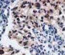
Immunohistochemistry: HSP90 beta Antibody (H9010) [NB110-61640]
Immunohistochemistry: HSP90 beta Antibody (H9010) [NB110-61640] - Immunohistochemistry analysis using Mouse Anti-Hsp90 Monoclonal Antibody, Clone H9010 (NB110-61640). Tissue: colon carcinoma. Species: Human. Fixation: Formalin. Primary Antibody: Mouse Anti-Hsp90 Monoclonal Antibody (NB110-61640) at 1:10000 for 12 hours at 4C. Secondary Antibody: Biotin Goat Anti-Mouse at 1:2000 for 1 hour at RT. Counterstain: Mayer Hematoxylin (purple/blue) nuclear stain at 200 l for 2 minutes at RT. Localization: Inflammatory cells. Magnification: 40x. This image was produced using an amplifying IHC wash buffer. The antibody has therefore been diluted more than is recommended for other applications..![Immunohistochemistry: HSP90 beta Antibody (H9010) [NB110-61640]](https://resources.rndsystems.com/images/products/HSP90-beta-Antibody-H9010-Immunohistochemistry-NB110-61640-img0043.jpg "Immunohistochemistry: HSP90 beta Antibody (H9010) [NB110-61640]")
Immunohistochemistry: HSP90 beta Antibody (H9010) [NB110-61640]
Immunohistochemistry: HSP90 beta Antibody (H9010) [NB110-61640] - Immunohistochemistry analysis using Mouse Anti-Hsp90 Monoclonal Antibody, Clone H9010 (NB110-61640). Tissue: colon carcinoma. Species: Human. Fixation: Formalin. Primary Antibody: Mouse Anti-Hsp90 Monoclonal Antibody (NB110-61640) at 1:10000 for 12 hours at 4C. Secondary Antibody: Alexa Fluor 555 Goat Anti-Mouse (red) at 1:5000 for 1 hour at RT. Magnification: 40x. This image was produced using an amplifying IHC wash buffer. The antibody has therefore been diluted more than is recommended for other applications..Applications for HSP90 beta Antibody (H9010) - BSA Free
Application
Recommended Usage
Immunohistochemistry
1:100
Western Blot
1:2500
Application Notes
1 ug/ml of HSP90 antibody was sufficient for detection of HSP90beta in 20 ug of heat shocked HeLa cell lysate by colorimetric immunoblot analysis using goat anti-mouse IgG:HRP as the secondary antibody.
Reviewed Applications
Read 5 reviews rated 4.8 using NB110-61640 in the following applications:
Formulation, Preparation, and Storage
Purification
Protein G purified
Formulation
PBS (pH 7.2), 50% Glycerol
Format
BSA Free
Preservative
0.09% Sodium Azide
Concentration
1 mg/ml
Shipping
The product is shipped with polar packs. Upon receipt, store it immediately at the temperature recommended below.
Stability & Storage
Store at 4C short term. Aliquot and store at -20C long term. Avoid freeze-thaw cycles.
Background: HSP90 beta
Long Name
Heat Shock Protein 90 beta
Alternate Names
D6S182, HSP84, HSP90AB1, HSP90B, HSPC2, HSPCB
Gene Symbol
HSP90AB1
Additional HSP90 beta Products
Product Documents for HSP90 beta Antibody (H9010) - BSA Free
Certificate of Analysis
To download a Certificate of Analysis, please enter a lot or batch number in the search box below.
Product Specific Notices for HSP90 beta Antibody (H9010) - BSA Free
This product is for research use only and is not approved for use in humans or in clinical diagnosis. Primary Antibodies are guaranteed for 1 year from date of receipt.
Related Research Areas
Citations for HSP90 beta Antibody (H9010) - BSA Free
 Powered by Bioz
Customer Reviews for HSP90 beta Antibody (H9010) - BSA Free (5)
4.8 out of 5
5 Customer Ratings
Have you used HSP90 beta Antibody (H9010) - BSA Free?
Submit a review and receive an Amazon gift card!
$25/€18/£15/$25CAN/¥2500 Yen for a review with an image
$10/€7/£6/$10CAN/¥1110 Yen for a review without an image
Submit a review
Customer Images


Showing
1
-
5 of
5 reviews
Showing All
Filter By:
-
Application: Western BlotSample Tested: Human liver cancer cell line (HuH7) and HepG2Species: HumanVerified Customer | Posted 04/04/2020

-
Application: Immunohistochemistry-ParaffinSample Tested: Breast cancer tissueSpecies: HumanVerified Customer | Posted 04/04/2020
-
Application: Immunohistochemistry-ParaffinSample Tested: Breast cancer tissueSpecies: HumanVerified Customer | Posted 02/19/2019
-
Application: Western BlotSample Tested: hela cell and 293TSpecies: HumanVerified Customer | Posted 12/19/2018
-
Application: Western BlotSample Tested: HCC827 human non-small cell lung cancer cell line and A549 cellsSpecies: HumanVerified Customer | Posted 10/17/2018
There are no reviews that match your criteria.
Protocols
Find general support by application which include: protocols, troubleshooting, illustrated assays, videos and webinars.
- Antigen Retrieval Protocol (PIER)
- Antigen Retrieval for Frozen Sections Protocol
- Appropriate Fixation of IHC/ICC Samples
- Cellular Response to Hypoxia Protocols
- Chromogenic IHC Staining of Formalin-Fixed Paraffin-Embedded (FFPE) Tissue Protocol
- Chromogenic Immunohistochemistry Staining of Frozen Tissue
- ClariTSA™ Fluorophore Kits
- Detection & Visualization of Antibody Binding
- ELISA Sample Preparation & Collection Guide
- ELISA Troubleshooting Guide
- Fluorescent IHC Staining of Frozen Tissue Protocol
- Graphic Protocol for Heat-induced Epitope Retrieval
- Graphic Protocol for the Preparation and Fluorescent IHC Staining of Frozen Tissue Sections
- Graphic Protocol for the Preparation and Fluorescent IHC Staining of Paraffin-embedded Tissue Sections
- Graphic Protocol for the Preparation of Gelatin-coated Slides for Histological Tissue Sections
- How to Run an R&D Systems DuoSet ELISA
- How to Run an R&D Systems Quantikine ELISA
- How to Run an R&D Systems Quantikine™ QuicKit™ ELISA
- ICC Cell Smear Protocol for Suspension Cells
- ICC Immunocytochemistry Protocol Videos
- ICC for Adherent Cells
- IHC Sample Preparation (Frozen sections vs Paraffin)
- Immunocytochemistry (ICC) Protocol
- Immunocytochemistry Troubleshooting
- Immunofluorescence of Organoids Embedded in Cultrex Basement Membrane Extract
- Immunofluorescent IHC Staining of Formalin-Fixed Paraffin-Embedded (FFPE) Tissue Protocol
- Immunohistochemistry (IHC) and Immunocytochemistry (ICC) Protocols
- Immunohistochemistry Frozen Troubleshooting
- Immunohistochemistry Paraffin Troubleshooting
- Immunoprecipitation Protocol
- Preparing Samples for IHC/ICC Experiments
- Preventing Non-Specific Staining (Non-Specific Binding)
- Primary Antibody Selection & Optimization
- Protocol for Heat-Induced Epitope Retrieval (HIER)
- Protocol for Making a 4% Formaldehyde Solution in PBS
- Protocol for VisUCyte™ HRP Polymer Detection Reagent
- Protocol for the Fluorescent ICC Staining of Cell Smears - Graphic
- Protocol for the Fluorescent ICC Staining of Cultured Cells on Coverslips - Graphic
- Protocol for the Preparation & Fixation of Cells on Coverslips
- Protocol for the Preparation and Chromogenic IHC Staining of Frozen Tissue Sections
- Protocol for the Preparation and Chromogenic IHC Staining of Frozen Tissue Sections - Graphic
- Protocol for the Preparation and Chromogenic IHC Staining of Paraffin-embedded Tissue Sections
- Protocol for the Preparation and Chromogenic IHC Staining of Paraffin-embedded Tissue Sections - Graphic
- Protocol for the Preparation and Fluorescent ICC Staining of Cells on Coverslips
- Protocol for the Preparation and Fluorescent ICC Staining of Non-adherent Cells
- Protocol for the Preparation and Fluorescent ICC Staining of Stem Cells on Coverslips
- Protocol for the Preparation and Fluorescent IHC Staining of Frozen Tissue Sections
- Protocol for the Preparation and Fluorescent IHC Staining of Paraffin-embedded Tissue Sections
- Protocol for the Preparation of Gelatin-coated Slides for Histological Tissue Sections
- Protocol for the Preparation of a Cell Smear for Non-adherent Cell ICC - Graphic
- Quantikine HS ELISA Kit Assay Principle, Alkaline Phosphatase
- Quantikine HS ELISA Kit Principle, Streptavidin-HRP Polymer
- R&D Systems Quality Control Western Blot Protocol
- Sandwich ELISA (Colorimetric) – Biotin/Streptavidin Detection Protocol
- Sandwich ELISA (Colorimetric) – Direct Detection Protocol
- TUNEL and Active Caspase-3 Detection by IHC/ICC Protocol
- The Importance of IHC/ICC Controls
- Troubleshooting Guide: ELISA
- Troubleshooting Guide: Immunohistochemistry
- Troubleshooting Guide: Western Blot Figures
- Western Blot Conditions
- Western Blot Protocol
- Western Blot Protocol for Cell Lysates
- Western Blot Troubleshooting
- Western Blot Troubleshooting Guide
- View all Protocols, Troubleshooting, Illustrated assays and Webinars
Loading...
Associated Pathways
