Cellular responses to bone morphogenetic proteins (BMPs) have been shown to be mediated by the formation of hetero-oligomeric complexes of the type I and type II serine/threonine kinase receptors. BMP receptor 1A (BMPR-1A), also known as activin receptor-like kinase (ALK)-3, is one of seven known type I serine/threonine kinases that are required for the signal transduction of TGF-beta family cytokines. In contrast to the TGF-beta receptor system in which the type I receptor does not bind TGF-beta in the absence of the type II receptor, type I receptors involved in BMP signaling (including BMPR-IA, BMPR-IB/ALK-6, and ActR-I/ALK-2) can independently bind the various BMP family proteins in the absence of type II receptors. Recombinant soluble BMPR-IA binds BMP-4 with high-affinity in solution and is a potent BMP-4 antagonist in vitro. BMPR-IA is ubiquitously expressed during embryogenesis. In adult tissues, BMPR-IA mRNA is also widely distributed with the highest expression levels found in skeletal muscle. The extracellular domain of BMPR-IA shares little amino acid sequence identity with the other mammalian ALK type I receptor kinases, but the cysteine residues are conserved. Human and mouse BMPR-IA are highly conserved and share 98% sequence identity.

Key Product Details
Species Reactivity
Validated:
Human
Cited:
Human, Mouse
Applications
Validated:
Immunohistochemistry, Western Blot
Cited:
Immunohistochemistry, Immunohistochemistry-Paraffin, Western Blot, Flow Cytometry, Immunocytochemistry, Immunoprecipitation, Functional Assay
Label
Unconjugated
Antibody Source
Polyclonal Goat IgG
Loading...
Product Specifications
Immunogen
Mouse myeloma cell line NS0-derived recombinant human BMPR-IA/AKL-3
Gln24-Arg152
Accession # P36894
Gln24-Arg152
Accession # P36894
Specificity
Detects human BMPR-IA in direct ELISAs and Western blots.
Clonality
Polyclonal
Host
Goat
Isotype
IgG
Scientific Data Images for Human BMPR-IA/ALK-3 Antibody
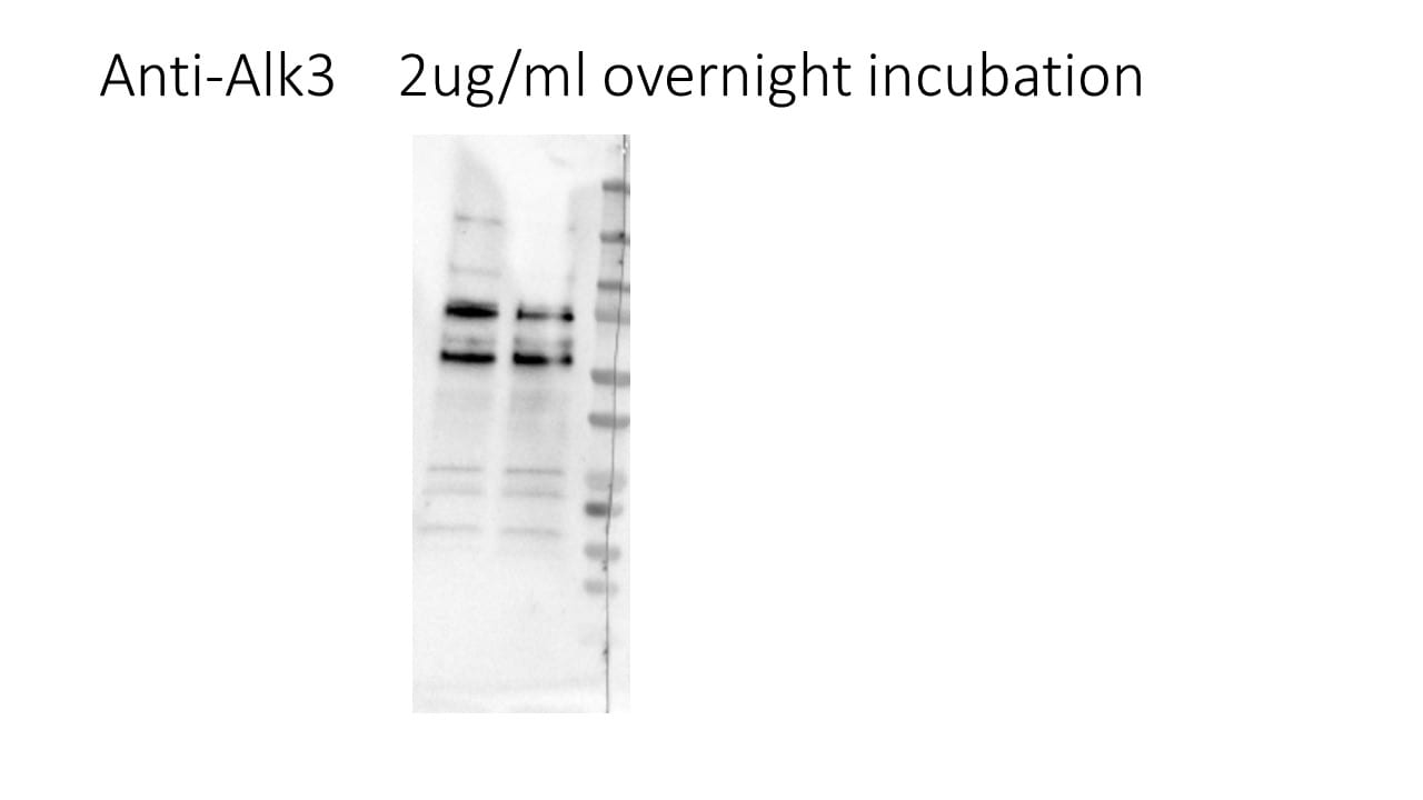
Detection of Human BMPR‑IA/ALK‑3 by Western Blot.
Western blot shows lysates of human skeletal muscle tissue. PVDF membrane was probed with 2 µg/mL of Goat Anti-Human BMPR-IA/ALK-3 Antigen Affinity-purified Polyclonal Antibody (Catalog # AF346) followed by HRP-conjugated Anti-Goat IgG Secondary Antibody (HAF017). A specific band was detected for BMPR-IA/ALK-3 at approximately 60 kDa (as indicated). This experiment was conducted under reducing conditions and using Immunoblot Buffer Group 1.
BMPR-IA/ALK-3 in Human Prostate Cancer Tissue.
BMPR‑IA/ALK‑3 was detected in immersion fixed paraffin-embedded sections of normal human prostate tissue (negative) and human prostate cancer tissue (positive) using Goat Anti-Human BMPR‑IA/ALK‑3 Antigen Affinity-purified Polyclonal Antibody (Catalog # AF346) at 1 µg/mL for 1 hour at room temperature followed by incubation with the Anti-Sheep IgG VisUCyte™ HRP Polymer Antibody (VC006). Tissue was stained using DAB (brown) and counterstained with hematoxylin (blue). Specific staining was localized to epithelial cells and stroma. Staining was performed using our IHC Staining with VisUCyte HRP Polymer Detection Reagents.
Detection of Human BMPR-IA/ALK-3 by Western Blot
cP1P-augmented cardiac differentiation in hESCs is dependent on BMP receptor-mediated SMAD1/5/8 signaling. (A) The activity of BMPR/SMAD signaling with or without cP1P treatment was evaluated by the expression levels of ALK2 and ALK3 by Western blot at the indicated time points along with GAPDH expression as the loading control. (B) Schematic representation of the treatment with BMPR inhibitor LDN during days 3 to 5 of CM differentiation coinciding with IWR1 inhibitor treatment. (C) The activity of BMPR/SMAD signaling was inhibited by the administration of LDN, as shown by the downregulation of ALK3 and p-SMAD 1/5/8, while cP1P treatment could not reverse the inhibitory effect of LDN. The effects of BMPR inhibition by LDN on cardiomyocyte differentiation, shown by (D) the downregulation of NKX2.5 at day 5. (E) Treatment of LDN between day 3 to 5 also downregulated the expression of TNNT2 and MLC2V at day 8 of CM differentiation even upon cP1P treatment, as demonstrated by Western blot. (F) Schematic representation of the treatment with LDN during days 5 to 8 of CM differentiation. (G) Immunoblots of ALK3, p-SMAD1/5/8, SMAD1/5/8, and NKX2.5 at day 5 of LDN treatment in DMSO-treated controls and cP1P-treated CMs. (H) Immunoblots of TNNT2 and MLC2V at day 8 of LDN treatment in DMSO-treated controls and cP1P-treated CMs. Image collected and cropped by CiteAb from the following open publication (https://pubmed.ncbi.nlm.nih.gov/34209900), licensed under a CC-BY license. Not internally tested by R&D Systems.
Human CCL26 / Eotaxin-3 ELISA Standard Curve
Recombinant Human CCL26/Eotaxin‑3 (Catalog # 653-E3) was serially diluted and captured by Mouse Anti-Human CCL26/Eotaxin‑3 Monoclonal Antibody (Catalog # MAB653) coated on a Clear Polystyrene Microplate (Catalog # DY990). Goat Anti-Human BMPR‑IA/ALK‑3 Antigen Affinity-purified Polyclonal Antibody (Catalog # AF346) was biotinylated and incubated with the protein captured on the plate. Detection of the standard curve was achieved by incubating Streptavidin-HRP (Catalog # DY998)Applications for Human BMPR-IA/ALK-3 Antibody
Application
Recommended Usage
Immunohistochemistry
1-15 µg/mL
Sample: Immersion fixed paraffin-embedded sections of human prostate cancer
Sample: Immersion fixed paraffin-embedded sections of human prostate cancer
Western Blot
2 µg/mL
Sample: Human skeletal muscle tissue
Sample: Human skeletal muscle tissue
Reviewed Applications
Read 1 review rated 5 using AF346 in the following applications:
Formulation, Preparation, and Storage
Purification
Antigen Affinity-purified
Reconstitution
Reconstitute at 0.2 mg/mL in sterile PBS. For liquid material, refer to CoA for concentration.
Loading...
Formulation
Lyophilized from a 0.2 μm filtered solution in PBS with Trehalose. See Certificate of Analysis for details.
*Small pack size (-SP) is supplied either lyophilized or as a 0.2 µm filtered solution in PBS.
*Small pack size (-SP) is supplied either lyophilized or as a 0.2 µm filtered solution in PBS.
Shipping
Lyophilized product is shipped at ambient temperature. Liquid small pack size (-SP) is shipped with polar packs. Upon receipt, store immediately at the temperature recommended below.
Stability & Storage
Use a manual defrost freezer and avoid repeated freeze-thaw cycles.
- 12 months from date of receipt, -20 to -70 °C as supplied.
- 1 month, 2 to 8 °C under sterile conditions after reconstitution.
- 6 months, -20 to -70 °C under sterile conditions after reconstitution.
Calculators
Background: BMPR-IA/ALK-3
References
- Kawabata, M. et al. (1998) Cytokine and Growth Factor Reviews 9:49.
- Ebendal, T. et al. (1998) J. Neuroscience Research 51:139.
Long Name
Bone Morphogenetic Protein Receptor IA/Activin Receptor-like Kinase 3
Alternate Names
ALK-3, BMPR1A, BMPRIA, CD292
Gene Symbol
BMPR1A
UniProt
Additional BMPR-IA/ALK-3 Products
Product Documents for Human BMPR-IA/ALK-3 Antibody
Certificate of Analysis
To download a Certificate of Analysis, please enter a lot or batch number in the search box below.
Note: Certificate of Analysis not available for kit components.
Product Specific Notices for Human BMPR-IA/ALK-3 Antibody
For research use only
Related Research Areas
Citations for Human BMPR-IA/ALK-3 Antibody
 Powered by Bioz
Customer Reviews for Human BMPR-IA/ALK-3 Antibody (1)
5 out of 5
1 Customer Rating
Have you used Human BMPR-IA/ALK-3 Antibody?
Submit a review and receive an Amazon gift card!
$25/€18/£15/$25CAN/¥2500 Yen for a review with an image
$10/€7/£6/$10CAN/¥1110 Yen for a review without an image
Submit a review
Customer Images
Showing
1
-
1 of
1 review
Showing All
Filter By:
-
Application: Western BlotSample Tested: A549 human lung carcinoma cell lineSpecies: HumanVerified Customer | Posted 10/26/2021

There are no reviews that match your criteria.
Protocols
Find general support by application which include: protocols, troubleshooting, illustrated assays, videos and webinars.
- Antigen Retrieval Protocol (PIER)
- Antigen Retrieval for Frozen Sections Protocol
- Appropriate Fixation of IHC/ICC Samples
- Cellular Response to Hypoxia Protocols
- Chromogenic IHC Staining of Formalin-Fixed Paraffin-Embedded (FFPE) Tissue Protocol
- Chromogenic Immunohistochemistry Staining of Frozen Tissue
- ClariTSA™ Fluorophore Kits
- Detection & Visualization of Antibody Binding
- Fluorescent IHC Staining of Frozen Tissue Protocol
- Graphic Protocol for Heat-induced Epitope Retrieval
- Graphic Protocol for the Preparation and Fluorescent IHC Staining of Frozen Tissue Sections
- Graphic Protocol for the Preparation and Fluorescent IHC Staining of Paraffin-embedded Tissue Sections
- Graphic Protocol for the Preparation of Gelatin-coated Slides for Histological Tissue Sections
- IHC Sample Preparation (Frozen sections vs Paraffin)
- Immunofluorescent IHC Staining of Formalin-Fixed Paraffin-Embedded (FFPE) Tissue Protocol
- Immunohistochemistry (IHC) and Immunocytochemistry (ICC) Protocols
- Immunohistochemistry Frozen Troubleshooting
- Immunohistochemistry Paraffin Troubleshooting
- Preparing Samples for IHC/ICC Experiments
- Preventing Non-Specific Staining (Non-Specific Binding)
- Primary Antibody Selection & Optimization
- Protocol for Heat-Induced Epitope Retrieval (HIER)
- Protocol for Making a 4% Formaldehyde Solution in PBS
- Protocol for VisUCyte™ HRP Polymer Detection Reagent
- Protocol for the Preparation & Fixation of Cells on Coverslips
- Protocol for the Preparation and Chromogenic IHC Staining of Frozen Tissue Sections
- Protocol for the Preparation and Chromogenic IHC Staining of Frozen Tissue Sections - Graphic
- Protocol for the Preparation and Chromogenic IHC Staining of Paraffin-embedded Tissue Sections
- Protocol for the Preparation and Chromogenic IHC Staining of Paraffin-embedded Tissue Sections - Graphic
- Protocol for the Preparation and Fluorescent IHC Staining of Frozen Tissue Sections
- Protocol for the Preparation and Fluorescent IHC Staining of Paraffin-embedded Tissue Sections
- Protocol for the Preparation of Gelatin-coated Slides for Histological Tissue Sections
- R&D Systems Quality Control Western Blot Protocol
- TUNEL and Active Caspase-3 Detection by IHC/ICC Protocol
- The Importance of IHC/ICC Controls
- Troubleshooting Guide: Immunohistochemistry
- Troubleshooting Guide: Western Blot Figures
- Western Blot Conditions
- Western Blot Protocol
- Western Blot Protocol for Cell Lysates
- Western Blot Troubleshooting
- Western Blot Troubleshooting Guide
- View all Protocols, Troubleshooting, Illustrated assays and Webinars
Loading...
Associated Pathways
