CD109 is a GPI-anchored member of the alpha-2-macroglobulin (A2M) and complement family of proteins (1). Mature human CD109 contains a bait region with recognition sequences for multiple proteases, an internal thioester bond, and a domain similar to the receptor binding domain of A2M (2). Cleavage of A2M family proteins within the bait region activates the thioester bond to promote covalent bonding to nucleophilic groups in adjacent molecules (3, 4). Within the region included in this recombinant protein, human CD109 shares 71-73% amino acid (aa) sequence identity with mouse and rat CD109. It shares 27-33% aa sequence identity with A2M and complement factors C3, C4, and C5. Alternate splicing of human CD109 generates two isoforms with short deletions and one that is truncated within the bait region. CD109 is expressed on activated T cells and platelets, hematopoietic stem cells, megakaryocyte precursors, vascular endothelial cells, basal and myoepithelial cells of secretory glands, and squamous cell carcinomas (2, 5-9). It is produced as a 170-180 kDa glycoprotein that is autocatalytically processed to 150 kDa and 120 kDa forms (2, 6, 10). CD109 on keratinocytes binds TGF-beta and associates with TGF-beta RI and TGF-beta RII, resulting in inhibition of TGF-beta signaling (11). Polymorphisms of CD109 include the platelet-specific Gov antigen and the blood group ABH antigens (12, 13). Alloantibodies directed against these antigens result in unsuccessful platelet transfusions, neonatal alloimmune thrombocytopenia, and posttransfusion purpura (14).

Key Product Details
Species Reactivity
Validated:
Human
Cited:
Human
Applications
Validated:
Immunohistochemistry, Western Blot, Flow Cytometry, Simple Western, CyTOF-ready
Cited:
Immunohistochemistry, Western Blot, Immunocytochemistry, Co-Immunoprecipitation
Label
Unconjugated
Antibody Source
Polyclonal Sheep IgG
Loading...
Product Specifications
Immunogen
Mouse myeloma cell line NS0-derived recombinant human CD109
Val22-Ser1268 (Tyr703Ser, Thr1241Met)
Accession # Q6YHK3
Val22-Ser1268 (Tyr703Ser, Thr1241Met)
Accession # Q6YHK3
Specificity
Detects human CD109 in direct ELISAs and Western blots.
Clonality
Polyclonal
Host
Sheep
Isotype
IgG
Scientific Data Images for Human CD109 Antibody
Detection of Human CD109 by Western Blot.
Western blot shows lysates of U‑87 MG human glioblastoma/astrocytoma cell line. PVDF membrane was probed with 0.5 µg/mL of Sheep Anti-Human CD109 Antigen Affinity-purified Polyclonal Antibody (Catalog # AF4385) followed by HRP-conjugated Anti-Sheep IgG Secondary Antibody (HAF016). A specific band was detected for CD109 at approximately 161 kDa (as indicated). This experiment was conducted under reducing conditions and using Western Blot Buffer Group 1.
Detection of CD109 in A431 Human Cell Line by Flow Cytometry.
A431 human epithelial carcinoma cell line was stained with Human CD109 Antigen Affinity‑purified Polyclonal Antibody (Catalog # AF4385, filled histogram) or isotype control antibody (Catalog # 5-001-A, open histogram), followed by NorthernLights™ 557-conjugated Anti-Sheep IgG Secondary Antibody (Catalog # NL010).
CD109 in Human Squamous Cell Carcinoma.
CD109 was detected in immersion fixed paraffin-embedded sections of human squamous cell carcinoma using 15 µg/mL Sheep Anti-Human CD109 Antigen Affinity-purified Polyclonal Antibody (Catalog # AF4385) overnight at 4 °C. Tissue was stained with the Anti-Sheep HRP-DAB Cell & Tissue Staining Kit (brown; Catalog # CTS019) and counterstained with hematoxylin (blue). View our protocol for Chromogenic IHC Staining of Paraffin-embedded Tissue Sections.
Detection of Human CD109 by Simple WesternTM.
Simple Western lane view shows lysates of U‑87 MG human glioblastoma/astrocytoma cell line, loaded at 0.2 mg/mL. A specific band was detected for CD109 at approximately 161 kDa (as indicated) using 20 µg/mL of Sheep Anti-Human CD109 Antigen Affinity-purified Polyclonal Antibody (Catalog # AF4385). This experiment was conducted under reducing conditions and using the 12-230 kDa separation system.Applications for Human CD109 Antibody
Application
Recommended Usage
CyTOF-ready
Ready to be labeled using established conjugation methods. No BSA or other carrier proteins that could interfere with conjugation.
Flow Cytometry
2.5 µg/106 cells
Sample: A431 human epithelial carcinoma cell line
Sample: A431 human epithelial carcinoma cell line
Immunohistochemistry
5-15 µg/mL
Sample: Immersion fixed paraffin-embedded sections of human squamous cell carcinoma
Sample: Immersion fixed paraffin-embedded sections of human squamous cell carcinoma
Simple Western
20 µg/mL
Sample: U‑87 MG human glioblastoma/astrocytoma cell line
Sample: U‑87 MG human glioblastoma/astrocytoma cell line
Western Blot
0.5 µg/mL
Sample: U‑87 MG human glioblastoma/astrocytoma cell line
Sample: U‑87 MG human glioblastoma/astrocytoma cell line
Reviewed Applications
Read 1 review rated 4 using AF4385 in the following applications:
Flow Cytometry Panel Builder
Bio-Techne Knows Flow Cytometry
Save time and reduce costly mistakes by quickly finding compatible reagents using the Panel Builder Tool.
Advanced Features
- Spectra Viewer - Custom analysis of spectra from multiple fluorochromes
- Spillover Popups - Visualize the spectra of individual fluorochromes
- Antigen Density Selector - Match fluorochrome brightness with antigen density

Formulation, Preparation, and Storage
Purification
Antigen Affinity-purified
Reconstitution
Reconstitute at 0.2 mg/mL in sterile PBS. For liquid material, refer to CoA for concentration.
Loading...
Formulation
Lyophilized from a 0.2 μm filtered solution in PBS with Trehalose. *Small pack size (SP) is supplied either lyophilized or as a 0.2 µm filtered solution in PBS.
Shipping
Lyophilized product is shipped at ambient temperature. Liquid small pack size (-SP) is shipped with polar packs. Upon receipt, store immediately at the temperature recommended below.
Stability & Storage
Use a manual defrost freezer and avoid repeated freeze-thaw cycles.
- 12 months from date of receipt, -20 to -70 °C as supplied.
- 1 month, 2 to 8 °C under sterile conditions after reconstitution.
- 6 months, -20 to -70 °C under sterile conditions after reconstitution.
Calculators
Background: CD109
References
- Travis, J. and G.S. Salvesen (1983) Annu. Rev. Biochem. 52:655.
- Lin, M. et al. (2002) Blood 99:1683.
- Christensen, U. and L. Sottrup-Jensen (1984) Biochemistry 23:6619.
- Wallis, R. et al. (2007) J. Biol. Chem. 282:7844.
- Murray, L.J. et al. (1999) Exp. Hematol. 27:1282.
- Haregewoin, A. et al. (1994) Cell. Immunol. 156:357.
- Hasegawa, M. et al. (2007) Pathol. Int. 57:245.
- Brashem-Stein, C. et al. (1988) J. Immunol. 140:2330.
- Hashimoto, M. et al. (2004) Oncogene 23:3716.
- Solomon, K.R. et al. (2004) Gene 327:171.
- Finnson, K.W. et al. (2006) FASEB J. 20:1525.
- Schuh, A.C. et al. (2002) Blood 99:1692.
- Kelton, J.G. et al. (1998) J. Lab. Clin. Med. 132:142.
- Rozman, P. (2002) Transpl. Immunol. 10:165.
Alternate Names
CD109, CPAMD7
Gene Symbol
CD109
UniProt
Additional CD109 Products
Product Documents for Human CD109 Antibody
Certificate of Analysis
To download a Certificate of Analysis, please enter a lot or batch number in the search box below.
Note: Certificate of Analysis not available for kit components.
Product Specific Notices for Human CD109 Antibody
For research use only
Related Research Areas
Citations for Human CD109 Antibody
 Powered by Bioz
Customer Reviews for Human CD109 Antibody (1)
4 out of 5
1 Customer Rating
Have you used Human CD109 Antibody?
Submit a review and receive an Amazon gift card!
$25/€18/£15/$25CAN/¥2500 Yen for a review with an image
$10/€7/£6/$10CAN/¥1110 Yen for a review without an image
Submit a review
Customer Images
Showing
1
-
1 of
1 review
Showing All
Filter By:
-
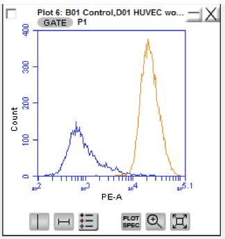
Application: Flow CytometrySample Tested: HUVEC human umbilical vein endothelial cellsSpecies: HumanVerified Customer | Posted 11/29/2016

There are no reviews that match your criteria.
Protocols
Find general support by application which include: protocols, troubleshooting, illustrated assays, videos and webinars.
- 7-Amino Actinomycin D (7-AAD) Cell Viability Flow Cytometry Protocol
- Antigen Retrieval Protocol (PIER)
- Antigen Retrieval for Frozen Sections Protocol
- Appropriate Fixation of IHC/ICC Samples
- Cellular Response to Hypoxia Protocols
- Chromogenic IHC Staining of Formalin-Fixed Paraffin-Embedded (FFPE) Tissue Protocol
- Chromogenic Immunohistochemistry Staining of Frozen Tissue
- ClariTSA™ Fluorophore Kits
- Detection & Visualization of Antibody Binding
- Extracellular Membrane Flow Cytometry Protocol
- Flow Cytometry Protocol for Cell Surface Markers
- Flow Cytometry Protocol for Staining Membrane Associated Proteins
- Flow Cytometry Staining Protocols
- Flow Cytometry Troubleshooting Guide
- Fluorescent IHC Staining of Frozen Tissue Protocol
- Graphic Protocol for Heat-induced Epitope Retrieval
- Graphic Protocol for the Preparation and Fluorescent IHC Staining of Frozen Tissue Sections
- Graphic Protocol for the Preparation and Fluorescent IHC Staining of Paraffin-embedded Tissue Sections
- Graphic Protocol for the Preparation of Gelatin-coated Slides for Histological Tissue Sections
- IHC Sample Preparation (Frozen sections vs Paraffin)
- Immunofluorescent IHC Staining of Formalin-Fixed Paraffin-Embedded (FFPE) Tissue Protocol
- Immunohistochemistry (IHC) and Immunocytochemistry (ICC) Protocols
- Immunohistochemistry Frozen Troubleshooting
- Immunohistochemistry Paraffin Troubleshooting
- Intracellular Flow Cytometry Protocol Using Alcohol (Methanol)
- Intracellular Flow Cytometry Protocol Using Detergents
- Intracellular Nuclear Staining Flow Cytometry Protocol Using Detergents
- Intracellular Staining Flow Cytometry Protocol Using Alcohol Permeabilization
- Intracellular Staining Flow Cytometry Protocol Using Detergents to Permeabilize Cells
- Preparing Samples for IHC/ICC Experiments
- Preventing Non-Specific Staining (Non-Specific Binding)
- Primary Antibody Selection & Optimization
- Propidium Iodide Cell Viability Flow Cytometry Protocol
- Protocol for Heat-Induced Epitope Retrieval (HIER)
- Protocol for Liperfluo
- Protocol for Making a 4% Formaldehyde Solution in PBS
- Protocol for VisUCyte™ HRP Polymer Detection Reagent
- Protocol for the Characterization of Human Th22 Cells
- Protocol for the Characterization of Human Th9 Cells
- Protocol for the Preparation & Fixation of Cells on Coverslips
- Protocol for the Preparation and Chromogenic IHC Staining of Frozen Tissue Sections
- Protocol for the Preparation and Chromogenic IHC Staining of Frozen Tissue Sections - Graphic
- Protocol for the Preparation and Chromogenic IHC Staining of Paraffin-embedded Tissue Sections
- Protocol for the Preparation and Chromogenic IHC Staining of Paraffin-embedded Tissue Sections - Graphic
- Protocol for the Preparation and Fluorescent IHC Staining of Frozen Tissue Sections
- Protocol for the Preparation and Fluorescent IHC Staining of Paraffin-embedded Tissue Sections
- Protocol for the Preparation of Gelatin-coated Slides for Histological Tissue Sections
- Protocol: Annexin V and PI Staining by Flow Cytometry
- Protocol: Annexin V and PI Staining for Apoptosis by Flow Cytometry
- R&D Systems Quality Control Western Blot Protocol
- TUNEL and Active Caspase-3 Detection by IHC/ICC Protocol
- The Importance of IHC/ICC Controls
- Troubleshooting Guide: Fluorokine Flow Cytometry Kits
- Troubleshooting Guide: Immunohistochemistry
- Troubleshooting Guide: Western Blot Figures
- Western Blot Conditions
- Western Blot Protocol
- Western Blot Protocol for Cell Lysates
- Western Blot Troubleshooting
- Western Blot Troubleshooting Guide
- View all Protocols, Troubleshooting, Illustrated assays and Webinars
Loading...